



Ki67 Antibody (YA6300)
(Synonyms: MKI67; Antigen KI-67)Based on 1 publication(s) in Google Scholar
Ki67 Antibody (YA6300) is a Rabbit-derived and non-conjugated IgG monoclonal antibody, targeting to Ki67.
-
Host:
Rabbit
-
Isotype:
IgG
-
Application:
WB, IHC-P, ICC/IF, ELISA
-
Reactivity :
Human, Mouse, Rat
-
Formulation:
Supplied in PBS, 50% glycerol, 0.05% Proclin 300, 0.05%BSA
-
Conjugation:
Non-conjugated


To place orders, for customer services and technical support, please contact: MedChemExpress USA
Tel: 609-228-6898 E-mail: [email protected] [email protected]



Publications Citing Use of MedChemExpress (MCE) Ki67 Antibody (YA6300)
More

Applications
| Application |
IHC-P

IHC-P: Immunohistochemistry-Paraffin
|
WB
WB: Western Blot
|
ICC/IF
ICC/IF: Immunocytochemistry/
Immunofluorescence |
ELISA
ELISA: Enzyme Linked Immunosorbent Assay
|
|---|---|---|---|---|
| Dilution Ratio | 1:200-1000 | 1:1000-5000 | 1:200-1000 | 1:5000-20000 |


Product Details
Ki67 Antibody (YA6300) is a Rabbit-derived and non-conjugated IgG monoclonal antibody, targeting to Ki67.
-
Host Rabbit

-
Clonality Monoclonal

-
Species ReactivityHuman, Mouse, Rat
-
Observed Molecular WeightObserved band size: 359 kDa
Note: Due to possible protein modifications or aggregation, the molecular weight should be confirmed by actual measurement, and the predicted value is for reference only.

-
Calculated Molecular Weight Predicted band size: 359 kDa

Protein A
Non-conjugated
Unmodified
IgG


Product Properties
-
Appearance
Solution
-
Formulation
Supplied in PBS, 50% glycerol, 0.05% Proclin 300, 0.05%BSA
-
Concentration
Batch-dependent, Please check the COA for the concentration of each lot. Check Lot Concentration
-
Shipping
Shipping with blue ice.


Publications (1)
-
Journal Impact Factor
-
Most Recent
-
Biomedicines
Engineering Bone-Mimetic Microspheres to Recapitulate the Tumor Microenvironment for In Vitro Osteosarcoma Modeling. [Abstract]2026 Apr 10;14(4):868. PMID: 42072409


Verification Images
-
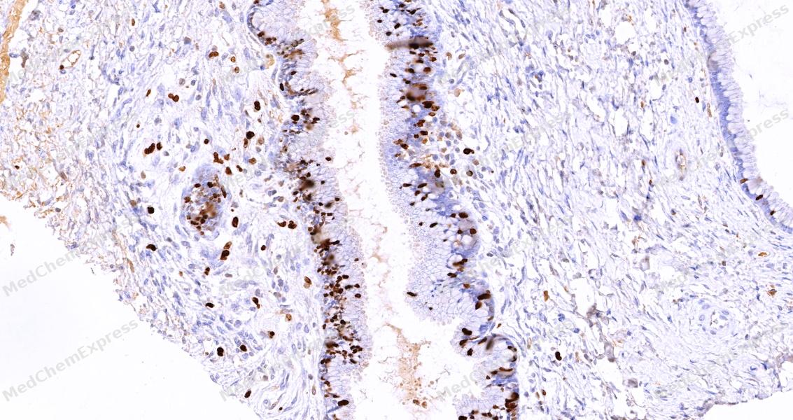 Immunohistochemical analysis of paraffin-embedded human cervix tissue using Ki67 Antibody (HY-P86608, 1/500) . The section was pretreated using heat mediated antigen retrieval with sodium citrate buffer (PH 6.0) for 8 minutes. The tissues were blocked with quick block buffer for 0.5 hours at room temperature, washed with PBS and PBST, and then incubated with the primary antibody overnight at 8℃. The detection was performed using an HRP conjugated compact polymer system. DAB was used as the chromogen. Tissues were counterstained with hematoxylin and mounted with DPX.
 -
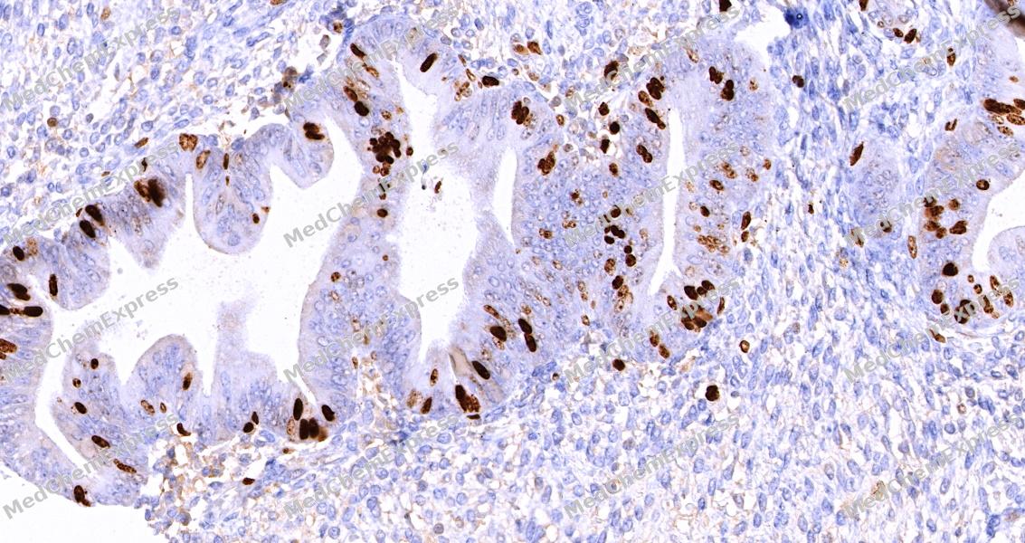 Immunohistochemical analysis of paraffin-embedded human endometrium tissue using Ki67 Antibody (HY-P86608, 1/500) . The section was pretreated using heat mediated antigen retrieval with sodium citrate buffer (PH 6.0) for 8 minutes. The tissues were blocked with quick block buffer for 0.5 hours at room temperature, washed with PBS and PBST, and then incubated with the primary antibody overnight at 8℃. The detection was performed using an HRP conjugated compact polymer system. DAB was used as the chromogen. Tissues were counterstained with hematoxylin and mounted with DPX.
 -
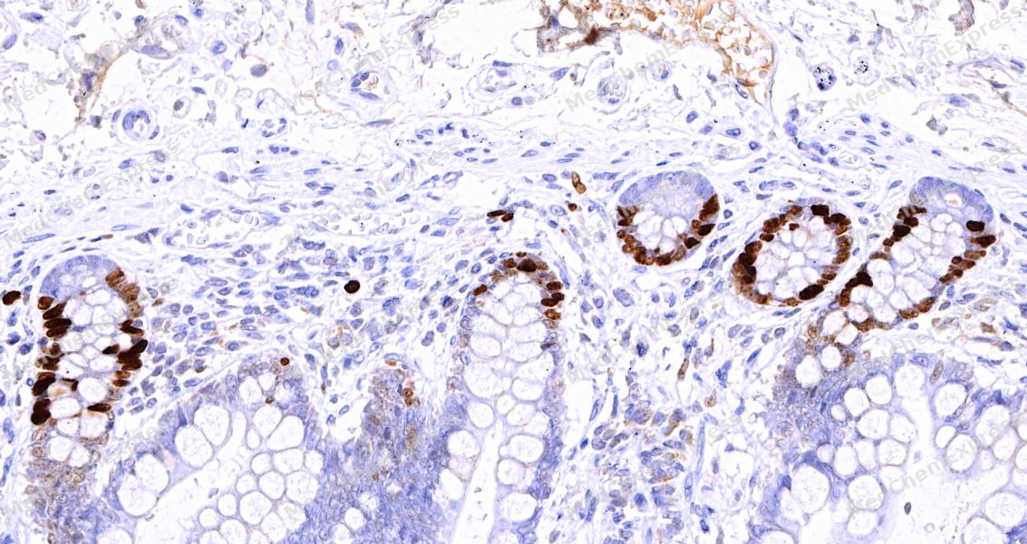 Immunohistochemical analysis of paraffin-embedded human small intestine tissue using Ki67 Antibody (HY-P86608, 1/500) . The section was pretreated using heat mediated antigen retrieval with sodium citrate buffer (PH 6.0) for 8 minutes. The tissues were blocked with quick block buffer for 0.5 hours at room temperature, washed with PBS and PBST, and then incubated with the primary antibody overnight at 8℃. The detection was performed using an HRP conjugated compact polymer system. DAB was used as the chromogen. Tissues were counterstained with hematoxylin and mounted with DPX.
 -
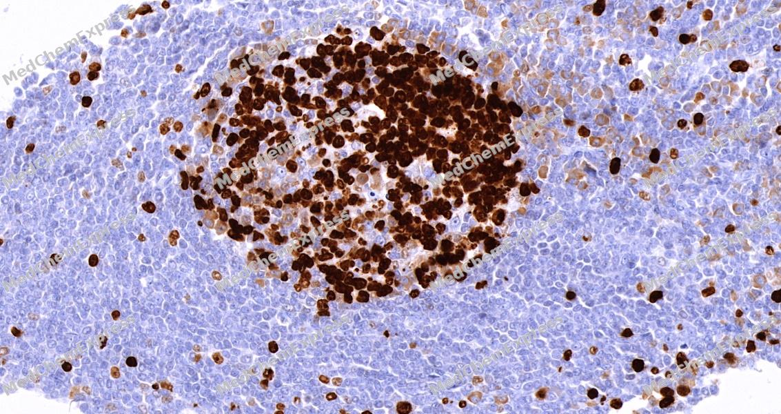 Immunohistochemical analysis of paraffin-embedded human tonsil tissue using Ki67 Antibody (HY-P86608, 1/500) . The section was pretreated using heat mediated antigen retrieval with sodium citrate buffer (PH 6.0) for 8 minutes. The tissues were blocked with quick block buffer for 0.5 hours at room temperature, washed with PBS and PBST, and then incubated with the primary antibody overnight at 8℃. The detection was performed using an HRP conjugated compact polymer system. DAB was used as the chromogen. Tissues were counterstained with hematoxylin and mounted with DPX.
 -
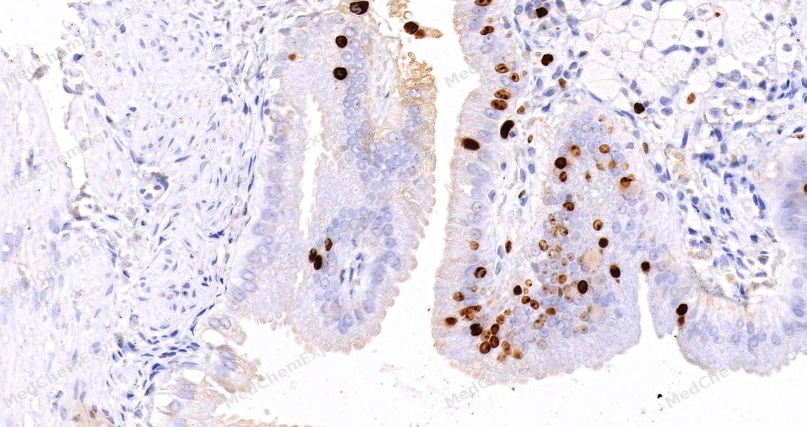 Immunohistochemical analysis of paraffin-embedded human gallbladder tissue using Ki67 Antibody (HY-P86608, 1/500) . The section was pretreated using heat mediated antigen retrieval with sodium citrate buffer (PH 6.0) for 8 minutes. The tissues were blocked with quick block buffer for 0.5 hours at room temperature, washed with PBS and PBST, and then incubated with the primary antibody overnight at 8℃. The detection was performed using an HRP conjugated compact polymer system. DAB was used as the chromogen. Tissues were counterstained with hematoxylin and mounted with DPX.
 -
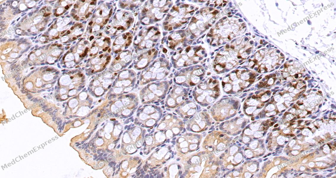 Immunohistochemical analysis of paraffin-embedded mouse colon tissue using Ki67 Antibody (HY-P86608, 1/500) . The section was pretreated using heat mediated antigen retrieval with sodium citrate buffer (PH 6.0) for 8 minutes. The tissues were blocked with quick block buffer for 0.5 hours at room temperature, washed with PBS and PBST, and then incubated with the primary antibody overnight at 8℃. The detection was performed using an HRP conjugated compact polymer system. DAB was used as the chromogen. Tissues were counterstained with hematoxylin and mounted with DPX.
 -
 Immunocytochemistry analysis of Hela cells labeling Ki67 with Ki67 Antibody (HY-P86608) at 1/300 dilution. Cells were fixed in 4% paraformaldehyde for 15 minutes at room temperature, permeabilized with 0.1% Triton X-100 in PBS for 15 minutes at room temperature, then blocked with quick block buffer for 10 minutes at room temperature. Cells were then incubated with Ki67 Antibody (HY-P86608) at 1/300 dilution in quick block buffer overnight at 4 ℃. AF488-conjugated Goat Anti-Rabbit IgG H&L (HY-P8002, Green) was used as the secondary antibody at 1/1,000 dilution. PBS instead of the primary antibody was used as the secondary antibody only control. The Nuclear counterstain was DAPI (Blue).
 -
 Immunocytochemistry analysis of Hela cells labeling Ki67 with Ki67 Antibody (HY-P86608) at 1/600 dilution. Cells were fixed in 4% paraformaldehyde for 15 minutes at room temperature, permeabilized with 0.1% Triton X-100 in PBS for 15 minutes at room temperature, then blocked with quick block buffer for 10 minutes at room temperature. Cells were then incubated with Ki67 Antibody (HY-P86608) at 1/600 dilution in quick block buffer overnight at 4 ℃. AF488-conjugated Goat Anti-Rabbit IgG H&L (HY-P8002, Green) was used as the secondary antibody at 1/1,000 dilution. PBS instead of the primary antibody was used as the secondary antibody only control. The Nuclear counterstain was DAPI (Blue).


Background
-
Function
Ki67 is a Protein that associates with the surface of mitotic chromosomes and acts both as a chromosome repellent during early mitosis and chromosome attractant during late mitosis. Required to maintain individual mitotic chromosomes dispersed in the cytoplasm following nuclear envelope disassembly. During early mitosis, relocalizes from nucleoli to the chromosome surface where it forms extended brush structures that cover a substantial fraction of the chromosome surface. The MKI67 brush structure prevents chromosomes from collapsing into a single chromatin mass by forming a steric and electrostatic charge barrier: the protein has a high net electrical charge and acts as a surfactant, dispersing chromosomes and enabling independent chromosome motility. During mitotic anaphase, the MKI67 brush structure collapses and MKI67 switches from a chromosome repellent to a chromosome attractant to promote chromosome clustering and facilitate the exclusion of large cytoplasmic particles from the future nuclear space. Mechanistically, dephosphorylation during mitotic exit and simultaneous exposure of a conserved basic patch induce the RNA-dependent formation of a liquid-like condensed phase on the chromosome surface, promoting coalescence of neighboring chromosome surfaces and clustering of chromosomes. Binds premature ribosomal RNAs during anaphase; promoting liquid-liquid phase separation. Binds DNA, with a preference for supercoiled DNA and AT-rich DNA. Does not contribute to the internal structure of mitotic chromosomes. May play a role in chromatin organization; it is however unclear whether it plays a direct role in chromatin organization or whether it is an indirect consequence of its function in mitotic chromosome[1][2][3][4][5][6][7].
-
Subcellular Localization
Chromosome; Nucleus; Nucleus, nucleolus
-
Isoforms & Post-Translational Modification
P46013 has 2 isomers: P46013-1: 358694 Da (predicted); P46013-2: 319444 Da (predicted).
Hyperphosphorylated by CDK1 in mitosis; hyperphosphorylatiom prevents undergoing liquid-liquid phase separation (PubMed:10502411, PubMed:10653604, PubMed:25012651, PubMed:39153474). Dephosphorylated by PPP1CC at the onset of anaphase (PubMed:25012651). Dephosphorylated by protein phosphatase 2A (PP2A) at the onset of anaphase (By similarity). Dephosphorylation by protein phosphatase 2A (PP2A) and simultaneous exposure of the positively charged patch (CP) during mitotic exit induce the RNA-dependent formation of a liquid-like condensed phase on the chromosome surface (PubMed:39153474);Ubiquitinated by the APC/C complex after neuronal progenitors exit mitosis during brain development, leading to clearance from constitutive heterochromatin -
Subunit
Interacts with KIF15 (PubMed:10878014). Interacts (via the FHA domain) with NIFK (PubMed:11342549, PubMed:14659764, PubMed:16244663). Interacts with PPP1CC (PubMed:24867636, PubMed:25012651). Component of a complex at least composed of ZNF335, HCFC1, CCAR2, EMSY, MKI67, RBBP5, ASH2L and WDR5; the complex is formed as a result of interactions between components of a nuclear receptor-mediated transcription complex and a histone methylation complex (PubMed:19131338). Interacts with ZNF335 (PubMed:19131338, PubMed:23178126)
-
SwissProt ID
-
Synonyms
MKI67; Antigen KI-67
Documentation
-
Data Sheet (261 KB)
-
SDS (251 KB)
- English - EN (251 KB)
- Français - FR (251 KB)
- Deutsch - DE (251 KB)
- Norwegian - NO (251 KB)
- Español - ES (251 KB)
- Swedish - SV (251 KB)
- Italian - IT (251 KB)
- Korean - KR (251 KB)
- Portuguese - PT (251 KB)
-
User Guide for Antibodies (1077 KB)
References
[1]. Cuylen S, et al. Ki-67 acts as a biological surfactant to disperse mitotic chromosomes. Nature. 2016 Jul 14;535(7611):308-12. [Content Brief]
[2]. Cuylen-Haering S, et al. Chromosome clustering by Ki-67 excludes cytoplasm during nuclear assembly. Nature. 2020 Nov;587(7833):285-290. [Content Brief]
[3]. Yamazaki H, et al. Cell cycle-specific phase separation regulated by protein charge blockiness. Nat Cell Biol. 2022 May;24(5):625-632. [Content Brief]
[4]. Hernandez-Armendariz A, et al. A liquid-like coat mediates chromosome clustering during mitotic exit. Mol Cell. 2024 Sep 5;84(17):3254-3270.e9. [Content Brief]
[5]. Hayashi Y, et al. The hierarchical structure of the perichromosomal layer comprises Ki67, ribosomal RNAs, and nucleolar proteins. Biochem Biophys Res Commun. 2017 Nov 18;493(2):1043-1049. [Content Brief]
[6]. MacCallum DE, et al. The biochemical characterization of the DNA binding activity of pKi67. J Pathol. 2000 Jul;191(3):286-98. [Content Brief]
[7]. Booth DG, et al. Ki-67 is a PP1-interacting protein that organises the mitotic chromosome periphery. Elife. 2014 May 27;3:e01641. [Content Brief]

 Powered by Bioz
