



Lactoferrin Antibody (YA2274)
(Synonyms: LTF; LF; Lactotransferrin; Lactoferrin; Talalactoferrin)
Based on 1 Customer Validation
Lactoferrin Antibody (YA2274) is a Rabbit-derived and non-conjugated IgG monoclonal antibody, targeting to Lactoferrin.
-
Host:
Rabbit
-
Isotype:
IgG
-
Application:
WB, ICC/IF, IP
-
Reactivity :
Human
-
Formulation:
Supplied in 50mM Tris-Glycine(pH 7.4), 0.15M NaCl, 40%Glycerol, 0.01% sodium azide and 0.05% BSA.
-
Conjugation:
Non-conjugated


To place orders, for customer services and technical support, please contact: MedChemExpress USA
Tel: 609-228-6898 E-mail: [email protected] [email protected]





Applications
| Application |
WB

WB: Western Blot
|
ICC/IF
ICC/IF: Immunocytochemistry/
Immunofluorescence |
IP
IP: Immunoprecipitation
|
|---|---|---|---|
| Dilution Ratio | 1:500-1:1000 | 1:50-1:200 | 1:50 |


Product Details
Lactoferrin Antibody (YA2274) is a Rabbit-derived and non-conjugated IgG monoclonal antibody, targeting to Lactoferrin.
-
Host Rabbit

-
Clonality Recombinant,Monoclonal

-
Species ReactivityHuman
-
Observed Molecular WeightObserved band size: 85 kDa
Note: Due to possible protein modifications or aggregation, the molecular weight should be confirmed by actual measurement, and the predicted value is for reference only.

-
Calculated Molecular Weight Predicted band size: 78 kDa

A synthesized peptide derived from human Lactoferrin aa100-150/710.
Endogenous
Affinity Chromatography
Non-conjugated
Unmodified
IgG


Product Properties
-
Appearance
Solution
-
Formulation
Supplied in 50mM Tris-Glycine(pH 7.4), 0.15M NaCl, 40%Glycerol, 0.01% sodium azide and 0.05% BSA.
-
Concentration
Batch-dependent, Please check the COA for the concentration of each lot. Check Lot Concentration
-
Storage & Stability
Stored at -20°C for 1 year. Avoid repeated freeze / thaw cycles.
-
Shipping
Shipping with blue ice.


Verification Images
-
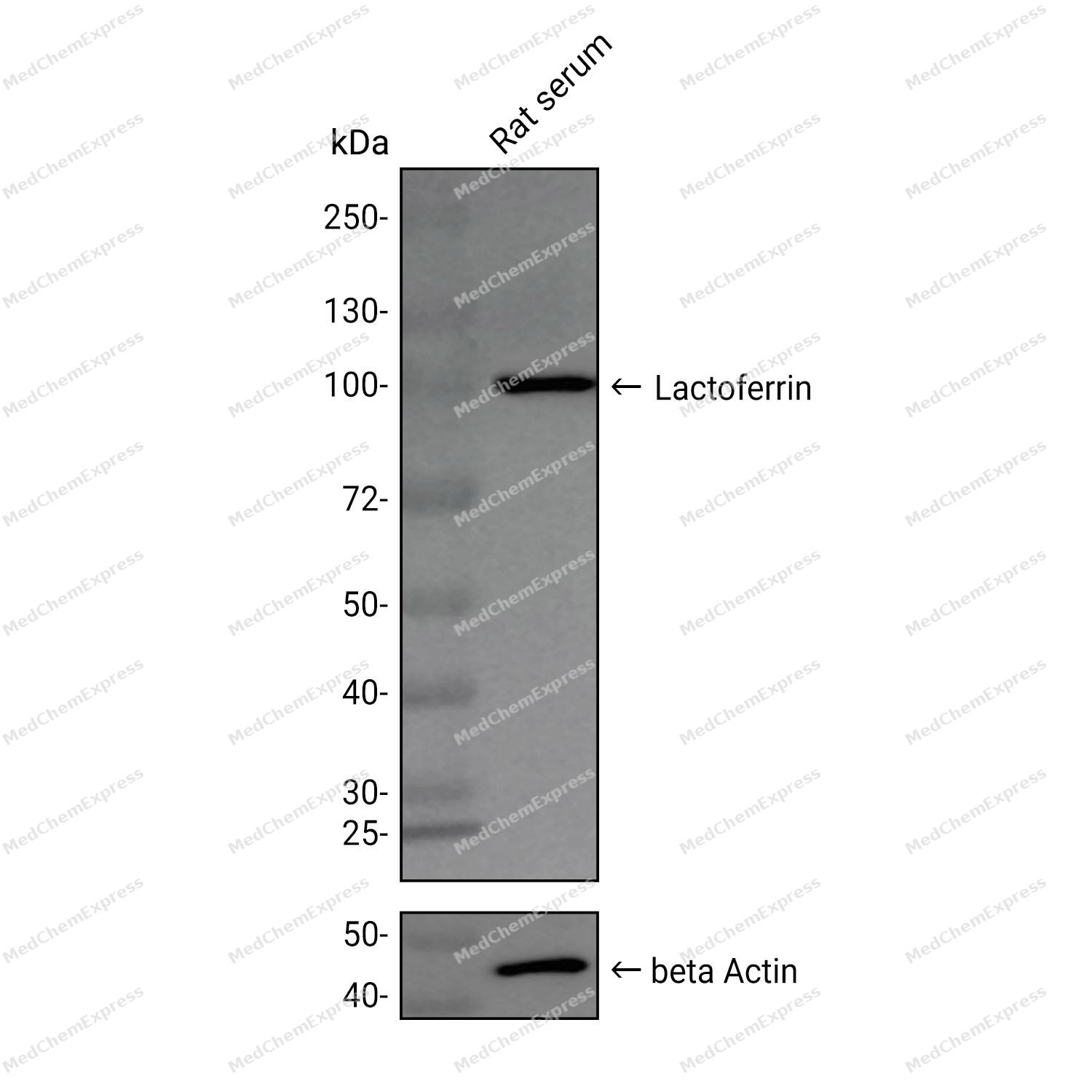 Western blot analysis was performed on extracts from Rat serum (lane 1, 15 μg) using Lactoferrin Rabbit mAb.Proteins were transferred to a PVDF membrane and blocked with 5% non-fat milk in TBST at 4°C overnight.The primary antibody (1:1000 dilution) and the loading control antibody (beta Actin, HY-P80438, 1:20000 dilution) were incubated in 5% non-fat milk in TBST for 1 hour at 37°C.Goat Anti-Rabbit IgG-HRP Secondary Antibody (1:20000 dilution) was then applied for 40 minutes at 37°C.
 -
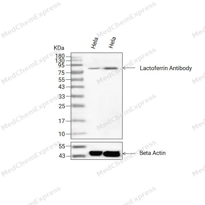 Western blot analysis of extracts from Hela(lane 2(20ug) and Hela(lane 3(40ug) using Lactoferrin Antibody (HY-P82529) Rabbit mAb. Proteins were transferred to a PVDF membrane and blocked with 5% non-fat milk in TBST for 2 hour at room temperature. The primary antibody (1/1000) and Loading control antibody (Beta Actin, HY-P83730, 1/10000) was used in 5% non-fat milk in TBST at 4°C overnight. Goat Anti-Mouse/Rabbit IgG-HRP Secondary Antibody (1/10000) was used for 1 hour at room temperature.
 -
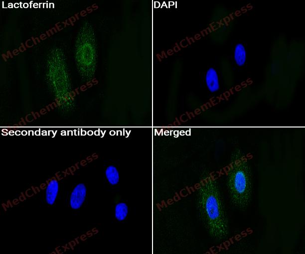 Immunocytochemistry analysis of HepG2 cells labeling Lactoferrin with Lactoferrin Antibody (HY-P82529) at 1/50 dilution. Cells were fixed in 4% paraformaldehyde for 15 minutes at room temperature, permeabilized with 0.1% Triton X-100 for 10 minutes at room temperature, then blocked with BSA for Immunol Staining for 10 min at room temperature. Cells were then incubated with Lactoferrin Antibody (HY-P82529) at 1/50 dilution in BSA for Immunol Staining at 4 ℃ overnight. AF488-conjugated AffiniPure Goat Anti-Rabbit IgG H&L(HY-P8002, Green) was used as the secondary antibody at 1/1,000 dilution. PBS instead of the primary antibody was used as the secondary antibody only control. The Nuclear counterstain was DAPI (Blue).
 -
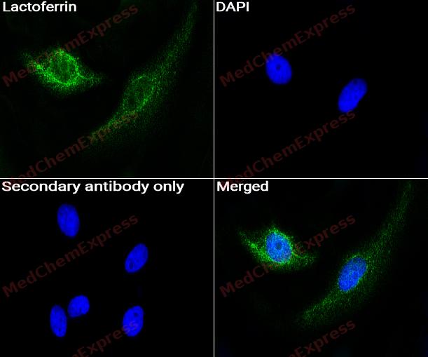 Immunocytochemistry analysis of HepG2 cells labeling Lactoferrin with Lactoferrin Antibody (HY-P82529) at 1/100 dilution. Cells were fixed in 4% paraformaldehyde for 15 minutes at room temperature, permeabilized with 0.1% Triton X-100 for 10 minutes at room temperature, then blocked with BSA for Immunol Staining for 10 min at room temperature. Cells were then incubated with Lactoferrin Antibody (HY-P82529) at 1/100 dilution in BSA for Immunol Staining at 4 ℃ overnight. AF488-conjugated AffiniPure Goat Anti-Rabbit IgG H&L(HY-P8002, Green) was used as the secondary antibody at 1/1,000 dilution. PBS instead of the primary antibody was used as the secondary antibody only control. The Nuclear counterstain was DAPI (Blue).


Background
-
Function
Lactoferrin is an iron-binding glycoprotein that supports innate immune defense, antimicrobial activity, and immune homeostasis[1][2]. Mechanistically, lactoferrin links iron sequestration with host-cell signaling, and studies describe effects on antigen-presenting cells, cytokine balance, and LPS-responsive inflammatory pathways[2][3][4]. In epithelial and macrophage inflammatory models, lactoferrin attenuates LPS-stimulated inflammatory responses and barrier impairment through NF-κB, MAPK, and Nrf2 pathway modulation[3][4]. In cancer models, bovine or recombinant human lactoferrin induces cell-cycle arrest, disrupts cytoskeletal organization, inhibits mTOR signaling, or promotes apoptosis in selected breast cancer cells[5][6]. Compared with related lactoferrin-derived peptides, intact lactoferrin provides the broader iron-binding and immune-modulatory scaffold, whereas lactoferricin and lactoferrampin are N-terminal antimicrobial peptides with antibacterial, antiviral, antifungal, and anti-inflammatory activities[7][8]. For experimental applications, lactoferrin-derived peptide chimeras show stronger membrane-perturbing antimicrobial activity than individual peptides, making them useful tools for structure-activity and membrane-interaction studies[8].
-
Subcellular Localization
Secreted; Cytoplasmic granule; Cytoplasm; Nucleus
-
Expression
Tissue_specificity:It is found in high concentrations in saliva and tears, moderate concentrations in serum and plasma, and low concentrations in urine. In the kidneys, this protein is detectable in the distal collecting ducts of the medulla, but not in the cortex or blood vessels. It is also detectable in peripheral blood neutrophils (protein level) . Isomers 1 and ΔLf are expressed in the mammary gland, prostate, spleen, pancreas, kidneys, small intestine, lungs, skeletal muscle, uterus, thymus, and fetal liver. Isomer 1 is expressed in the brain, testes, and peripheral blood leukocytes; isomer ΔLf is almost undetectable in these tissues. Isomer ΔLf is expressed in the placenta, liver, and ovary; isomer 1 is almost undetectable in these tissues. In the kidneys, isomer 1 is highly expressed in the medullary collecting ducts but very low in the cortex. -
Isoforms & Post-Translational Modification
P02788 has 2 isomers: P02788-1: 78182 Da (predicted); P02788-2: 73161 Da (predicted).
Phosphorylation at Ser-10 activates the transcriptional activity (PubMed:20404350). Phosphorylation at Ser-10 also promotes proteasomal degradation (PubMed:20404350). Alternatively can undergo O-GlcNAcylation at Ser-10 (PubMed:20404350);O-GlcNAcylation at Ser-10 inhibits DNA binding and negatively regulates the transcriptional activity (PubMed:20404350). Alternatively can undergo phosphorylation at Ser-10 (PubMed:20404350);Poly-N-acetyllactosaminic carbohydrate moiety seems to be needed for TLR4 activation -
Subunit
Monomer. Found in a complex with LTF, CLU, EPPIN and SEMG1. Found in a complex with MPO and LTF; interacts directly with CP, allows Fe(3+) incorporation into LTF and activation of CP ferroxidase activity (PubMed:23843990)
-
SwissProt ID
-
Synonyms
LTF; LF; Lactotransferrin; Lactoferrin; Talalactoferrin
-
Research Field
Cardiovascular


Documentation
-
Data Sheet (262 KB)
-
SDS (251 KB)
- English - EN (251 KB)
- Français - FR (251 KB)
- Deutsch - DE (251 KB)
- Norwegian - NO (251 KB)
- Español - ES (251 KB)
- Swedish - SV (251 KB)
- Italian - IT (251 KB)
- Korean - KR (251 KB)
- Portuguese - PT (251 KB)
-
User Guide for Antibodies (1077 KB)
References
[1]. Kell DB, et al. The Biology of Lactoferrin, an Iron-Binding Protein That Can Help Defend Against Viruses and Bacteria. Front Immunol. 2020 May 28;11:1221. [Content Brief]
[2]. Actor JK, et al. Lactoferrin as a natural immune modulator. Curr Pharm Des. 2009;15(17):1956-73. [Content Brief]
[3]. Hu P, et al. Lactoferrin attenuates lipopolysaccharide-stimulated inflammatory responses and barrier impairment through the modulation of NF-κB/MAPK/Nrf2 pathways in IPEC-J2 cells. Food Funct. 2020 Oct 21;11(10):8516-8526. [Content Brief]
[4]. Håversen L, et al. Lactoferrin down-regulates the LPS-induced cytokine production in monocytic cells via NF-kappa B. Cell Immunol. 2002 Dec;220(2):83-95. [Content Brief]
[5]. Itoh T, et al. A synthesized nostocionone derivative potentiates programmed cell death in human T-cell leukemia Jurkat cells through mitochondria via the release of endonuclease G. Nutr Cancer. 2014;66(8):1414-23. [Content Brief]
[6]. Iglesias-Figueroa BF, et al. Recombinant human lactoferrin induces apoptosis, disruption of F-actin structure and cell cycle arrest with selective cytotoxicity on human triple negative breast cancer cells. Apoptosis. 2019 Aug;24(7-8):562-577. [Content Brief]
[7]. Yin C, et al. Recent studies on the antimicrobial peptides lactoferricin and lactoferrampin. Curr Mol Med. 2014;14(9):1139-54. [Content Brief]
[8]. Haney EF, et al. Structural and biophysical characterization of an antimicrobial peptide chimera comprised of lactoferricin and lactoferrampin. Biochim Biophys Acta. 2012 Mar;1818(3):762-75. [Content Brief]

 Powered by Bioz
