



Lipin 1 Antibody (YA1909)
(Synonyms: Lipin-1; Lpin1; PAP1)
Based on 1 Customer Validation
Lipin 1 Antibody (YA1909) is a Rabbit-derived and non-conjugated IgG monoclonal antibody, targeting to Lipin 1.
-
Host:
Rabbit
-
Isotype:
IgG
-
Application:
WB, IHC-P, ICC/IF, IP, FC
-
Reactivity :
Human
-
Formulation:
Supplied in rabbit IgG in 50 mM Tris-Glycine, 0.15 M NaCl, 40% Glycerol, 0.01% sodium azide and 0.05% BSA, pH 7.4.
-
Conjugation:
Non-conjugated


To place orders, for customer services and technical support, please contact: MedChemExpress USA
Tel: 609-228-6898 E-mail: [email protected] [email protected]





Applications
| Application |
WB

WB: Western Blot
|
IHC-P
IHC-P: Immunohistochemistry-Paraffin
|
ICC/IF
ICC/IF: Immunocytochemistry/
Immunofluorescence |
IP
IP: Immunoprecipitation
|
FC
FC: Flow Cytometry
|
|---|---|---|---|---|---|
| Dilution Ratio | 1:500-1:1000 | 1:50-1:100 | 1:50-1:200 | 1:30 | 1:50-1:100 |


Product Details
Lipin 1 Antibody (YA1909) is a Rabbit-derived and non-conjugated IgG monoclonal antibody, targeting to Lipin 1.
-
Host Rabbit

-
Clonality Recombinant,Monoclonal

-
Species ReactivityHuman
-
Observed Molecular WeightObserved band size: 130 kDa
Note: Due to possible protein modifications or aggregation, the molecular weight should be confirmed by actual measurement, and the predicted value is for reference only.

-
Calculated Molecular Weight Predicted band size: 99 kDa

A synthesized peptide derived from human Lipin 1 aa150-200/890.
Endogenous
Affinity Chromatography
Non-conjugated
Unmodified
IgG


Product Properties
-
Appearance
Solution
-
Formulation
Supplied in rabbit IgG in 50 mM Tris-Glycine, 0.15 M NaCl, 40% Glycerol, 0.01% sodium azide and 0.05% BSA, pH 7.4.
-
Concentration
Batch-dependent, Please check the COA for the concentration of each lot. Check Lot Concentration
-
Storage & Stability
Stored at -20°C for 1 year. Avoid repeated freeze / thaw cycles.
-
Shipping
Shipping with blue ice.


Verification Images
-
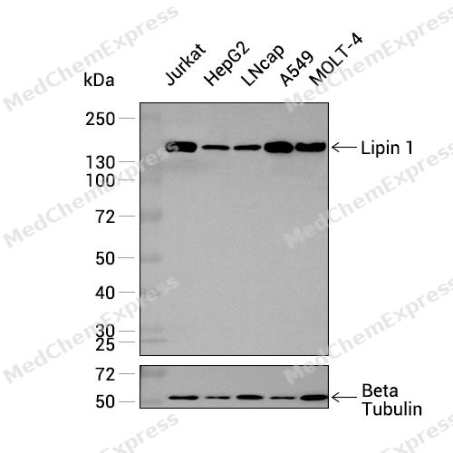 Western blot analysis was performed on extracts from Jurkat (lane 1, 15 μg), HepG2 (lane 2, 15 μg), LNcap (lane 3, 15 μg), A549 (lane 4, 15 μg), and MOLT-4 (lane 5, 15 μg) using Lipin 1 Rabbit mAb.Proteins were transferred to a PVDF membrane and blocked with 5% non-fat milk in TBST at 4°C overnight.The primary antibody (1:1000 dilution) and the loading control antibody (beta-Tubulin, HY-P80487, 1:60000 dilution) were incubated in 5% non-fat milk in TBST for 1 hour at 37°C.Goat Anti-Rabbit IgG-HRP Secondary Antibody (1:20000 dilution) was then applied for 40 minutes at 37°C.
 -
 Western blot analysis of extracts from K562 (lane 2(20μg), SH-SY-5Y(lane 3(20μg), HepG2 (lane 4(20μg) using Lipin 1 (HY-P82164) Rabbit mAb. Proteins were transferred to a PVDF membrane and blocked with 5% non-fat milk in TBST for 2 hour at room temperature. The primary antibody (1/1000) and Loading control antibody (Beta Actin, HY-P80438, 1/10000) was used in 5% non-fat milk in TBST at 4°C overnight. Goat Anti-Rabbit IgG-HRP Secondary Antibody (1/10000) was used for 1 hour at room temperature.
 -
 Immunohistochemical analysis of paraffin-embedded human lymphoma tissue using Lipin 1 antibody was performed. The section was pretreated using high-temperature mediated EDTA antigen retrieval buffer (pH 9.0), for 20 minutes. The tissues were incubated with primary antibody (HY-P82164, 1:200 dilution) at room temperature for 20 minutes. Detection was performed using an HRP conjugated compact polymer system. DAB was used as the chromogen. The tissues were counterstained with hematoxylin and mounted with neutral balsam mounting medium.
 -
 Immunohistochemical analysis of paraffin-embedded human breast cancer tissue using Lipin 1 antibody was performed. The section was pretreated using high-temperature mediated EDTA antigen retrieval buffer (pH 9.0), for 20 minutes. The tissues were incubated with primary antibody (HY-P82164, 1:200 dilution) at room temperature for 20 minutes. Detection was performed using an HRP conjugated compact polymer system. DAB was used as the chromogen. The tissues were counterstained with hematoxylin and mounted with neutral balsam mounting medium.
 -
 Immunohistochemical analysis of paraffin-embedded human esophagus tissue using Lipin 1 antibody was performed. The section was pretreated using high-temperature mediated EDTA antigen retrieval buffer (pH 9.0), for 20 minutes. The tissues were incubated with primary antibody (HY-P82164, 1:200 dilution) at room temperature for 20 minutes. Detection was performed using an HRP conjugated compact polymer system. DAB was used as the chromogen. The tissues were counterstained with hematoxylin and mounted with neutral balsam mounting medium.
 -
 Immunohistochemical analysis of paraffin-embedded human Endometrial carcinoma tissue using Lipin 1 antibody was performed. The section was pretreated using high-temperature mediated EDTA antigen retrieval buffer (pH 9.0), for 20 minutes. The tissues were incubated with primary antibody (HY-P82164, 1:200 dilution) at room temperature for 20 minutes. Detection was performed using an HRP conjugated compact polymer system. DAB was used as the chromogen. The tissues were counterstained with hematoxylin and mounted with neutral balsam mounting medium.
 -
 Immunohistochemical analysis of paraffin-embedded human gastric cancer tissue using Lipin 1 antibody was performed. The section was pretreated using high-temperature mediated EDTA antigen retrieval buffer (pH 9.0), for 20 minutes. The tissues were incubated with primary antibody (HY-P82164, 1:200 dilution) at room temperature for 20 minutes. Detection was performed using an HRP conjugated compact polymer system. DAB was used as the chromogen. The tissues were counterstained with hematoxylin and mounted with neutral balsam mounting medium.
 -
 Immunohistochemical analysis of paraffin-embedded human embryonal carcinoma tissue using Lipin 1 antibody was performed. The section was pretreated using high-temperature mediated EDTA antigen retrieval buffer (pH 9.0), for 20 minutes. The tissues were incubated with primary antibody (HY-P82164, 1:200 dilution) at room temperature for 20 minutes. Detection was performed using an HRP conjugated compact polymer system. DAB was used as the chromogen. The tissues were counterstained with hematoxylin and mounted with neutral balsam mounting medium.
 -
 Flow cytometric analysis of 1X10⁶ K562 cells labeling Lipin 1 Antibody (HY-P82164, red). Cells were fixed with 4% paraformaldehyde and permeabilised with 90% methanol. Then stained with the primary antibody at 1/50 dilution for an hour at 4℃. AF488-conjugated Goat Anti-Rabbit IgG H&L (HY-P8002) was used as the secondary antibody at 1/1,000 dilution for 30 minutes at 4℃. Rabbit IgG Isotype Control (HY-P80879, blue) was used as the isotype control, cells without incubation with primary antibody were used as the unlabeled control (black).
 -
 Immunocytochemistry analysis of HUVEC cells labeling Lipin 1 Antibody (HY-P82164) at 1/50 dilution. Cells were fixed in 4% paraformaldehyde for 15 minutes at room temperature, permeabilized with 0.1% Triton X-100 for 10 minutes at room temperature, then blocked with BSA for Immunol Staining for 10 min at room temperature. Cells were then incubated with Lipin 1 Antibody (HY-P82164) at 1/50 dilution in BSA for Immunol Staining at 4 ℃ Overnight. AF488-conjugated AffiniPure Goat Anti-Rabbit IgG H&L(HY-P8002, Green) was used as the secondary antibody at 1/1,000 dilution. PBS instead of the primary antibody was used as the secondary antibody only control. The Nuclear counterstain was DAPI (Blue).
 -
 Immunocytochemistry analysis of PC-3M cells labeling Lipin 1 Antibody (HY-P82164) at 1/50 dilution. Cells were fixed in 4% paraformaldehyde for 15 minutes at room temperature, permeabilized with 0.1% Triton X-100 for 10 minutes at room temperature, then blocked with BSA for Immunol Staining for 10 min at room temperature. Cells were then incubated with Lipin 1 Antibody (HY-P82164) at 1/50 dilution in BSA for Immunol Staining at 4 ℃ Overnight. AF488-conjugated AffiniPure Goat Anti-Rabbit IgG H&L(HY-P8002, Green) was used as the secondary antibody at 1/1,000 dilution. PBS instead of the primary antibody was used as the secondary antibody only control. The Nuclear counterstain was DAPI (Blue).


Background
-
Function
Lipin 1 acts as a magnesium-dependent phosphatidate phosphatase enzyme which catalyzes the conversion of phosphatidic acid to diacylglycerol during triglyceride, phosphatidylcholine and phosphatidylethanolamine biosynthesis and therefore controls the metabolism of fatty acids at different levels. Is involved in adipocyte differentiation. Recruited at the mitochondrion outer membrane and is involved in mitochondrial fission by converting phosphatidic acid to diacylglycerol. Acts also as nuclear transcriptional coactivator for PPARGC1A/PPARA regulatory pathway to modulate lipid metabolism gene expression[1][2][3][4].
-
Subcellular Localization
Cytoplasm, cytosol; Endoplasmic reticulum membrane; Nucleus membrane
-
Expression
Tissue_specificity:Specifically expressed in skeletal muscle. Also abundant in adipose tissue. Lower levels in some portions of the digestive tract -
Isoforms & Post-Translational Modification
Q14693 has 7 isomers: Q14693-1: 98664 Da (predicted); Q14693-2: 99367 Da (predicted); Q14693-3: 102313 Da (predicted); Q14693-4: 101636 Da (predicted); Q14693-5: 103016 Da (predicted); Q14693-6: 50528 Da (predicted); Q14693-7: 107861 Da (predicted).
Phosphorylated at multiple sites by mTOR in response to insulin, leading to its inactivation (PubMed:23426360). Phosphorylation does not affect the catalytic activity but regulates the localization (By similarity). Phosphorylation is decreased by epinephrine (By similarity). Dephosphorylated by the CTDNEP1-CNEP1R1 complex (By similarity). Dephosphorylation following mTOR inhibition promotes its activity (PubMed:31695197);Acetylation at Lys-425 and Lys-595 by KAT5 in response to fatty acids promotes translocation to the endoplasmic reticulum and synthesis of diacylglycerol;Sumoylated -
Subunit
Interacts (via LXXIL motif) with PPARA (By similarity). Interacts with PPARGC1A. Interaction with PPARA and PPARGC1A leads to the formation of a complex that modulates gene transcription (By similarity). Interacts with MEF2C (By similarity)
-
SwissProt ID
-
Synonyms
Lipin-1; Lpin1; PAP1
-
Research Field
Cardiovascular


Documentation
-
Data Sheet (234 KB)
-
SDS (251 KB)
- English - EN (251 KB)
- Français - FR (251 KB)
- Deutsch - DE (251 KB)
- Norwegian - NO (251 KB)
- Español - ES (251 KB)
- Swedish - SV (251 KB)
- Italian - IT (251 KB)
- Korean - KR (251 KB)
- Portuguese - PT (251 KB)
-
User Guide for Antibodies (1077 KB)

 Powered by Bioz
