



LKB1 Antibody (YA6032)
(Synonyms: STK11; LKB1; PJS; Serine/threonine-protein kinase STK11; Liver kinase B1; LKB1; hLKB1; Renal carcinoma antigen NY-REN-19)
Based on 1 Customer Validation
LKB1 Antibody (YA6032) is a Rabbit-derived and non-conjugated IgG monoclonal antibody, targeting to LKB1.
-
Host:
Rabbit
-
Isotype:
IgG
-
Application:
WB, ICC/IF, IP, ELISA
-
Reactivity :
Human, Mouse, Rat
-
Formulation:
Supplied in PBS, 50% glycerol, 0.05% Proclin 300, 0.05%BSA
-
Conjugation:
Non-conjugated


To place orders, for customer services and technical support, please contact: MedChemExpress USA
Tel: 609-228-6898 E-mail: [email protected] [email protected]





Applications
| Application |
WB

WB: Western Blot
|
ICC/IF
ICC/IF: Immunocytochemistry/
Immunofluorescence |
ELISA
ELISA: Enzyme Linked Immunosorbent Assay
|
IP
IP: Immunoprecipitation
|
|---|---|---|---|---|
| Dilution Ratio | 1:2000-1:10000 | 1:200-1:1000 | 1:5000-1:20000 | 1:50-1:200 |


Product Details
LKB1 Antibody (YA6032) is a Rabbit-derived and non-conjugated IgG monoclonal antibody, targeting to LKB1.
-
Host Rabbit

-
Clonality Monoclonal

-
Species ReactivityHuman, Mouse, Rat
-
Observed Molecular WeightObserved band size: 60 kDa
Note: Due to possible protein modifications or aggregation, the molecular weight should be confirmed by actual measurement, and the predicted value is for reference only.

-
Calculated Molecular Weight Predicted band size: 49 kDa

Protein A
Non-conjugated
Unmodified
IgG


Product Properties
-
Appearance
Liquid
-
Formulation
Supplied in PBS, 50% glycerol, 0.05% Proclin 300, 0.05%BSA
-
Concentration
Batch-dependent, Please check the COA for the concentration of each lot. Check Lot Concentration
-
Storage & Stability
Stored at -20°C for 1 year. Avoid repeated freeze / thaw cycles.
-
Shipping
Shipping with blue ice.


Verification Images
-
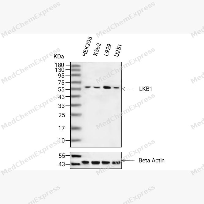 Western blot analysis of extracts from HEK293 (lane 2(20μg), K562 (lane 3(20μg), L929 (lane 4(20μg) and U251 (lane 5(20μg) using LKB1 Antibody (HY-P86340) Rabbit mAb. Proteins were transferred to a PVDF membrane and blocked with 5% non-fat milk in TBST for 2 hour at room temperature. The primary antibody (1/1000)and Loading control antibody (Beta Actin, HY-P80993, 1/10000) was used in 5% non-fat milk in TBST at 4°C overnight. Goat Anti-Rabbit IgG-HRP Secondary Antibody (1/10000) was used for 1 hour at room temperature.
 -
 Immunocytochemistry analysis of MCF-7 cells labeling LKB1 with LKB1 Antibody (HY-P86340) at 1/200 dilution. Cells were fixed in 4% paraformaldehyde for 15 minutes at room temperature, permeabilized with 0.1% Triton X-100 in PBS for 15 minutes at room temperature, then blocked with quick block buffer for 10 minutes at room temperature. Cells were then incubated with LKB1 Antibody (HY-P86340) at 1/200 dilution in quick block buffer overnight at 4 ℃. AF488-conjugated Goat Anti-Rabbit IgG H&L(HY-P8002, Green) was used as the secondary antibody at 1/1,000 dilution. PBS instead of the primary antibody was used as the secondary antibody only control. The Nuclear counterstain was DAPI (Blue).
 -
 Immunocytochemistry analysis of U2OS cells labeling LKB1 with LKB1 Antibody (HY-P86340) at 1/200 dilution. Cells were fixed in 4% paraformaldehyde for 15 minutes at room temperature, permeabilized with 0.1% Triton X-100 in PBS for 15 minutes at room temperature, then blocked with quick block buffer for 10 minutes at room temperature. Cells were then incubated with LKB1 Antibody (HY-P86340) at 1/200 dilution in quick block buffer overnight at 4 ℃. AF488-conjugated Goat Anti-Rabbit IgG H&L(HY-P8002, Green) was used as the secondary antibody at 1/1,000 dilution. PBS instead of the primary antibody was used as the secondary antibody only control. The Nuclear counterstain was DAPI (Blue).


Background
-
Function
LKB1 is a Tumor suppressor serine/threonine-protein kinase that controls the activity of AMP-activated protein kinase (AMPK) family members, thereby playing a role in various processes such as cell metabolism, cell polarity, apoptosis and DNA damage response. Acts by phosphorylating the T-loop of AMPK family proteins, thus promoting their activity: phosphorylates PRKAA1, PRKAA2, BRSK1, BRSK2, MARK1, MARK2, MARK3, MARK4, NUAK1, NUAK2, SIK1, SIK2, SIK3 and SNRK but not MELK. Also phosphorylates non-AMPK family proteins such as STRADA, PTEN and possibly p53/TP53. Acts as a key upstream regulator of AMPK by mediating phosphorylation and activation of AMPK catalytic subunits PRKAA1 and PRKAA2 and thereby regulates processes including: inhibition of signaling pathways that promote cell growth and proliferation when energy levels are low, glucose homeostasis in liver, activation of autophagy when cells undergo nutrient deprivation, and B-cell differentiation in the germinal center in response to DNA damage. Also acts as a regulator of cellular polarity by remodeling the actin cytoskeleton. Required for cortical neuron polarization by mediating phosphorylation and activation of BRSK1 and BRSK2, leading to axon initiation and specification. Involved in DNA damage response: interacts with p53/TP53 and recruited to the CDKN1A/WAF1 promoter to participate in transcription activation. Able to phosphorylate p53/TP53; the relevance of such result in vivo is however unclear and phosphorylation may be indirect and mediated by downstream STK11/LKB1 kinase NUAK1. Also acts as a mediator of p53/TP53-dependent apoptosis via interaction with p53/TP53: translocates to the mitochondrion during apoptosis and regulates p53/TP53-dependent apoptosis pathways. Regulates UV radiation-induced DNA damage response mediated by CDKN1A. In association with NUAK1, phosphorylates CDKN1A in response to UV radiation and contributes to its degradation which is necessary for optimal DNA repair; Has a role in spermiogenesis[1].
-
Subcellular Localization
Nucleus; Cytoplasm; Membrane; Mitochondrion; Nucleus; Cytoplasm
-
Expression
Tissue_specificity:Widely expressed. Strongest expression is observed in the testes and fetal liver. -
Isoforms & Post-Translational Modification
Q15831 has 2 isomers: Q15831-1: 48636 Da (predicted); Q15831-2: 45387 Da (predicted).
Phosphorylated by ATM at Thr-363 following ionizing radiation (IR). Phosphorylation at Ser-428 by RPS6KA1 and/or some PKA is required to inhibit cell growth. Phosphorylation at Ser-428 is also required during neuronal polarization to mediate phosphorylation of BRSK1 and BRSK2 (By similarity). Phosphorylation by PKC/PRKCZ at Ser-399 in isoform 2 promotes metformin (or peroxynitrite)-induced nuclear export of STK11 and activation of AMPK. UV radiation-induced phosphorylation at Thr-363 mediates CDKN1A degradation (By similarity);Acetylated. Deacetylation at Lys-48 enhances cytoplasmic localization and kinase activity in vitro -
Subunit
Catalytic component of a trimeric complex composed of STK11/LKB1, STRAD (STRADA or STRADB) and CAB39/MO25 (CAB39/MO25alpha or CAB39L/MO25beta): the complex tethers STK11/LKB1 in the cytoplasm and stimulates its catalytic activity. Found in a ternary complex composed of SMAD4, STK11/LKB1 and STK11IP. Interacts with p53/TP53, SMAD4, STK11IP and WDR6. Interacts with NR4A1. Interacts with NISCH; this interaction may increase STK11 activity. Interacts with PTEN; leading to PTEN phosphorylation. Interacts with SIRT1; the interaction deacetylates STK11. Interacts with CDKN1A
-
SwissProt ID
-
Synonyms
STK11; LKB1; PJS; Serine/threonine-protein kinase STK11; Liver kinase B1; LKB1; hLKB1; Renal carcinoma antigen NY-REN-19


Documentation
-
Data Sheet (235 KB)
-
SDS (252 KB)
- English - EN (252 KB)
- Français - FR (252 KB)
- Deutsch - DE (252 KB)
- Norwegian - NO (252 KB)
- Español - ES (252 KB)
- Swedish - SV (252 KB)
- Italian - IT (252 KB)
- Korean - KR (252 KB)
- Portuguese - PT (252 KB)
-
User Guide for Antibodies (1077 KB)

 Powered by Bioz
