



MDA5 Antibody (YA2399)
(Synonyms: Helicard; Hlcd; IDDM19; IFIH1; MDA-5; RH116; RIG I like receptor 2; RLR 2; RNA helicase DEAD box protein 116)
Based on 1 Customer Validation
MDA5 Antibody (YA2399) is a Rabbit-derived and non-conjugated IgG monoclonal antibody, targeting to MDA5.
-
Host:
Rabbit
-
Isotype:
IgG
-
Application:
WB, ICC/IF
-
Reactivity :
Human
-
Formulation:
Supplied in 50mM Tris-Glycine(pH 7.4), 0.15M NaCl, 40%Glycerol, 0.01% sodium azide and 0.05% BSA.
-
Conjugation:
Non-conjugated


To place orders, for customer services and technical support, please contact: MedChemExpress USA
Tel: 609-228-6898 E-mail: [email protected] [email protected]





Applications
| Application |
WB

WB: Western Blot
|
ICC/IF
ICC/IF: Immunocytochemistry/
Immunofluorescence |
|---|---|---|
| Dilution Ratio | 1:500-1:1000 | 1:50-1:200 |


Product Details
MDA5 Antibody (YA2399) is a Rabbit-derived and non-conjugated IgG monoclonal antibody, targeting to MDA5.
-
Host Rabbit

-
Clonality Recombinant,Monoclonal

-
Species ReactivityHuman
-
Observed Molecular WeightObserved band size: 117 kDa
Note: Due to possible protein modifications or aggregation, the molecular weight should be confirmed by actual measurement, and the predicted value is for reference only.

-
Calculated Molecular Weight Predicted band size: 117 kDa

A synthesized peptide derived from human MDA5 aa950-1025.
Endogenous
Affinity Chromatography
Non-conjugated
Unmodified
IgG


Product Properties
-
Appearance
Solution
-
Formulation
Supplied in 50mM Tris-Glycine(pH 7.4), 0.15M NaCl, 40%Glycerol, 0.01% sodium azide and 0.05% BSA.
-
Concentration
Batch-dependent, Please check the COA for the concentration of each lot. Check Lot Concentration
-
Storage & Stability
Stored at -20°C for 1 year. Avoid repeated freeze / thaw cycles.
-
Shipping
Shipping with blue ice.


Verification Images
-
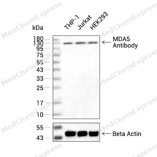 Western blot analysis of extracts from THP-1(lane 2(20ug) ,Jurkat(lane 3(20ug) and HEK293(lane 4(20ug) using MDA5 Antibody (HY-P82654) Rabbit mAb. Proteins were transferred to a PVDF membrane and blocked with 5% non-fat milk in TBST for 2 hour at room temperature. The primary antibody (1/1000) and Loading control antibody (Beta Actin, HY-P80438, 1/10000) was used in 5% non-fat milk in TBST at 4°C overnight. Goat Anti-Mouse/Rabbit IgG-HRP Secondary Antibody (1/10000) was used for 1 hour at room temperature.
 -
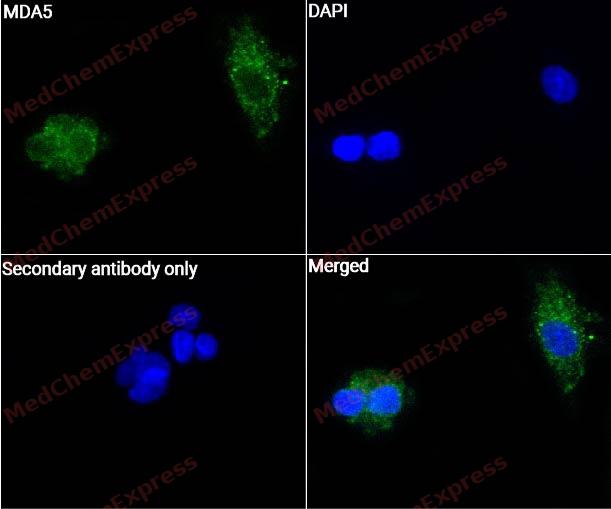 Immunocytochemistry analysis of HepG2 cells labeling MDA5 with MDA5 Antibody (HY-P82654) at 1/50 dilution. Cells were fixed in 4% paraformaldehyde for 15 minutes at room temperature, permeabilized with 0.1% Triton X-100 for 10 minutes at room temperature, then blocked with QuickBlock™ Blocking Buffer for Immunol Staining for 10 min at room temperature. Cells were then incubated with MDA5 Antibody (HY-P82654) at 1/50 dilution in QuickBlock™ Blocking Buffer for Immunol Staining at 4 ℃. Alexa Fluor® 488-conjugated AffiniPure Goat Anti-Rabbit IgG H&L(HY-P8002, Green) was used as the secondary antibody at 1/1,000 dilution. PBS instead of the primary antibody was used as the secondary antibody only control. The Nuclear counterstain was DAPI (Blue).
 -
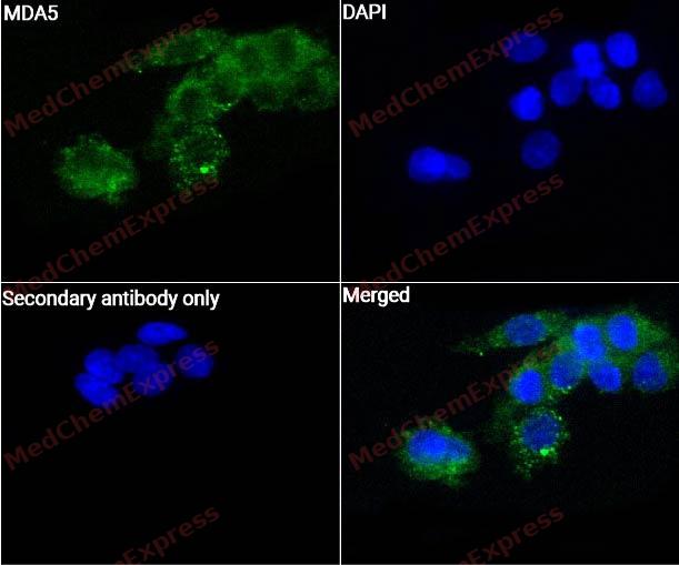 Immunocytochemistry analysis of HepG2 cells labeling MDA5 with MDA5 Antibody (HY-P82654) at 1/100 dilution. Cells were fixed in 4% paraformaldehyde for 15 minutes at room temperature, permeabilized with 0.1% Triton X-100 for 10 minutes at room temperature, then blocked with QuickBlock™ Blocking Buffer for Immunol Staining for 10 min at room temperature. Cells were then incubated with MDA5 Antibody (HY-P82654) at 1/100 dilution in QuickBlock™ Blocking Buffer for Immunol Staining at 4 ℃. Alexa Fluor® 488-conjugated AffiniPure Goat Anti-Rabbit IgG H&L(HY-P8002, Green) was used as the secondary antibody at 1/1,000 dilution. PBS instead of the primary antibody was used as the secondary antibody only control. The Nuclear counterstain was DAPI (Blue).


Background
-
Function
MDA5, encoded by IFIH1, functions as a cytoplasmic RNA sensor in innate immune responses[1]. Mechanistically, MDA5 recognizes long double-stranded RNA and assembles cooperative filaments that support antiviral signal activation[2][3]. ATP hydrolysis regulates filament dynamics, allowing MDA5 to discriminate viral dsRNA length during immune sensing[4]. RNA 2′-O-methylation also contributes to self-non-self RNA discrimination through an MDA5-dependent mechanism[5]. In disease models, MDA5 participates in antiviral defense against Paramyxoviridae infection in vivo[6]. Clinically, IFIH1/MDA5 is a dermatomyositis-specific autoantigen recognized by anti-CADM-140 antibody[1]. Compared with RIG-I, MDA5 preferentially detects longer dsRNA, whereas RIG-I responds more strongly to shorter dsRNA ligands[3]. This isoform distinction makes dsRNA length a practical design variable for experiments using poly (I:C) or viral RNA mimics[3].
-
Subcellular Localization
Cytoplasm; Nucleus; Mitochondrion
-
Expression
Tissue_specificity:Widely expressed, at a low level. Expression is detected at slightly highest levels in placenta, pancreas and spleen and at barely levels in detectable brain, testis and lung
Induction:By interferon (IFN) and TNF -
Isoforms & Post-Translational Modification
Q9BYX4 has 2 isomers: Q9BYX4-1: 116689 Da (predicted); Q9BYX4-2: 25129 Da (predicted).
Sumoylated. Sumoylation positively regulates its role in type I interferon induction and is enhanced by PIAS2-beta;Ubiquitinated by RNF125, leading to its degradation by the proteasome (PubMed:17460044). USP17/UPS17L2-dependent deubiquitination positively regulates the receptor (PubMed:20368735). Ubiquitinated by TRIM25 via 'Lys-63'-linked ubiquitination, promoting activation of IFIH1/MDA5 (PubMed:30193849). Ubiquitinated by TRIM40 via 'Lys-48'-linked ubiquitination; leading to proteasomal degradation (PubMed:29117565). Ubiquitinated by TRIM65 via 'Lys-63'-linked ubiquitination, promoting activation of IFIH1/MDA5 (PubMed:28594402);ISGylated by ISG15. ISGylation increases upon infection with dengue (DENV) or Zika (ZIKV) viruses. ISGylation at Lys-23 and Lys-43 is dependent of dephosphorylation at Ser-88, regulates mitochondrial translocation and oligomerization. Essential for IFIH1/MDA5-mediated cytokine responses and restriction of virus replication;Phosphorylated at Ser-88 (PubMed:23499489, PubMed:33727702). Dephosphorylated by phosphatases PPP11CA/PPP11CC; dephosphorylation precedes and is required for ISGylation (PubMed:23499489, PubMed:33727702);During apoptosis, processed into 3 cleavage products. The helicase-containing fragment, once liberated from the CARD domains, translocate from the cytoplasm to the nucleus. The processed protein significantly sensitizes cells to DNA degradation;(Microbial infection) Cleaved and inactivated by the protease 2A of coxsackievirus B3, poliovirus and enterovirus 71 allowing the virus to disrupt the host type I interferon production -
Subunit
Monomer in the absence of ligands and homodimerizes in the presence of dsRNA ligands. Can assemble into helical or linear polymeric filaments on long dsRNA (PubMed:33727702). Interacts with MAVS/IPS1. Interacts (via the CARD domains) with TKFC, the interaction is inhibited by viral infection (PubMed:17600090). Interacts with PCBP2. Interacts with NLRC5. Interacts with PIAS2-beta. Interacts with DDX60. Interacts with ANKRD17. Interacts with IKBKE (PubMed:17600090). Interacts with ATG5 and ATG12, either as ATG5 and ATG12 monomers or as ATG12-ATG5 conjugates (PubMed:17709747). Interacts with ZCCHC3; leading to activate IFIH1/MDA5 (PubMed:30193849). Interacts with RNF123 (PubMed:27312109). Interacts with DDX3X (PubMed:20127681). Interacts with NOD1; this interaction promotes transcription of antiviral genes and inhibition of viral replication (PubMed:32169843). Interacts with ECSIT; this interaction bridges IFIH1 to the MAVS complex at the mitochondrion (PubMed:25228397)
-
SwissProt ID
-
Synonyms
Helicard; Hlcd; IDDM19; IFIH1; MDA-5; RH116; RIG I like receptor 2; RLR 2; RNA helicase DEAD box protein 116
-
Research Field
Immunology


Documentation
-
Data Sheet (262 KB)
-
SDS (252 KB)
- English - EN (252 KB)
- Français - FR (252 KB)
- Deutsch - DE (252 KB)
- Norwegian - NO (252 KB)
- Español - ES (252 KB)
- Swedish - SV (252 KB)
- Italian - IT (252 KB)
- Korean - KR (252 KB)
- Portuguese - PT (252 KB)
-
User Guide for Antibodies (1077 KB)
References
[1]. Nakashima R, et al. The RIG-I-like receptor IFIH1/MDA5 is a dermatomyositis-specific autoantigen identified by the anti-CADM-140 antibody. Rheumatology (Oxford). 2010 Mar;49(3):433-40. [Content Brief]
[2]. Wu B, et al. Structural basis for dsRNA recognition, filament formation, and antiviral signal activation by MDA5. Cell. 2013 Jan 17;152(1-2):276-89. [Content Brief]
[3]. Kato H, et al. Length-dependent recognition of double-stranded ribonucleic acids by retinoic acid-inducible gene-I and melanoma differentiation-associated gene 5. J Exp Med. 2008 Jul 7;205(7):1601-10. [Content Brief]
[4]. Peisley A, et al. Cooperative assembly and dynamic disassembly of MDA5 filaments for viral dsRNA recognition. Proc Natl Acad Sci U S A. 2011 Dec 27;108(52):21010-5. [Content Brief]
[5]. Züst R, et al. Ribose 2'-O-methylation provides a molecular signature for the distinction of self and non-self mRNA dependent on the RNA sensor Mda5. Nat Immunol. 2011 Feb;12(2):137-43. [Content Brief]
[6]. Gitlin L, et al. Melanoma differentiation-associated gene 5 (MDA5) is involved in the innate immune response to Paramyxoviridae infection in vivo. PLoS Pathog. 2010 Jan 22;6(1):e1000734. [Content Brief]

 Powered by Bioz
