



Occludin/OCLN Antibody
(Synonyms: OCLN_HUMAN; Occludin; Ocl; BLCPMG; PPP1R115; PTORCH1; Tight junction protein occludin; )Based on 1 publication(s) in Google Scholar
Occludin/OCLN Antibody is a Rabbit-derived and non-conjugated IgG polyclonal antibody, targeting to Occludin/OCLN.
-
Host:
Rabbit
-
Isotype:
IgG
-
Application:
WB, IHC-P, ICC/IF
-
Reactivity :
Human, Mouse, Rat
-
Formulation:
Supplied in 0.01M TBS containing 0.03% Proclin300,50% Glycerol and 1% BSA, pH 7.4 or 50mM Tris-Glycine(pH 7.4), 0.15M NaCl, 40%Glycerol, 0.01% sodium azide and 0.05% BSA.
-
Conjugation:
Non-conjugated


To place orders, for customer services and technical support, please contact: MedChemExpress USA
Tel: 609-228-6898 E-mail: [email protected] [email protected]



-

HY-P83694
ZO-1/TJP1 Antibody (YA3430) -
HY-P8001
HRP-conjugated Goat Anti-Rabbit IgG H&L -
HY-P81222A
Claudin 1 Antibody (YA3551) -
HY-P80137
GAPDH Antibody (YA418) -
HY-P86250
ZO-1/TJP1 Antibody (YA5942) -
HY-P86518
Claudin 1 Antibody (YA6210) -
HY-P81051
Nrf2 Antibody(YA895) -
HY-P80704
HIF1 alpha Antibody -
HY-P81191
Phospho-eNOS (Ser1177) Antibody -
HY-P80927
VCAM1 Antibody (YA017)
Publications Citing Use of MedChemExpress (MCE) Occludin/OCLN Antibody
More

Applications
| Application |
WB

WB: Western Blot
|
IHC-P
IHC-P: Immunohistochemistry-Paraffin
|
ICC/IF
ICC/IF: Immunocytochemistry/
Immunofluorescence |
|---|---|---|---|
| Dilution Ratio | 1:500-1:1000 | 1:50-1:100 | 1:50-1:200 |


Product Details
Occludin/OCLN Antibody is a Rabbit-derived and non-conjugated IgG polyclonal antibody, targeting to Occludin/OCLN.
-
Host Rabbit

-
Clonality Polyclonal

-
Species ReactivityHuman, Mouse, Rat
-
Observed Molecular WeightObserved band size: 59 kDa
Note: Due to possible protein modifications or aggregation, the molecular weight should be confirmed by actual measurement, and the predicted value is for reference only.

-
Calculated Molecular Weight Predicted band size: 59 kDa

Entrez Gene: 100506658 Human ; 18260 Mouse ; 83497 Rat
SwissProt: Q16625 Human ; Q61146 Mouse ; Q6P6T5 Rat
OMIM: 251290 Human
A synthetic peptide of human Occludin KLH conjugated synthetic peptide derived from human Occludin: 151-250/522.
Endogenous
affinity purified
Non-conjugated
Unmodified
IgG


Product Properties
-
Appearance
Solution
-
Formulation
Supplied in 0.01M TBS containing 0.03% Proclin300,50% Glycerol and 1% BSA, pH 7.4 or 50mM Tris-Glycine(pH 7.4), 0.15M NaCl, 40%Glycerol, 0.01% sodium azide and 0.05% BSA.
-
Concentration
Batch-dependent, Please check the COA for the concentration of each lot. Check Lot Concentration
-
Storage & Stability
Stored at -20°C for 1 year. Avoid repeated freeze / thaw cycles.
-
Shipping
Shipping with blue ice.


Publications (1)
-
Journal Impact Factor
-
Most Recent


Verification Images
-
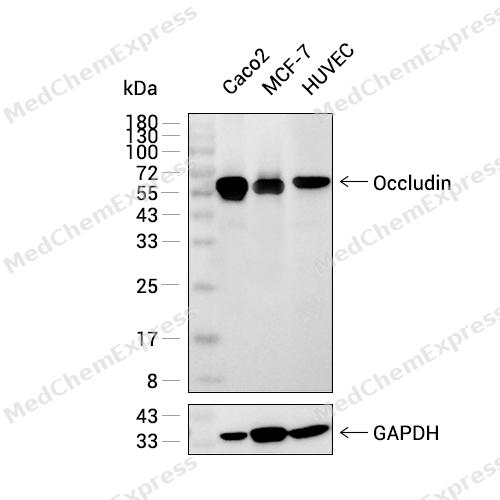 Western blot analysis was performed on extracts from Caco-2 (lane 1, 20 μg), MCF-7 (lane 2, 20 μg), and HUVEC (lane 3, 20 μg) using Occludin Rabbit pAb.Proteins were transferred to a PVDF membrane and blocked with 5% non - fat milk in TBST at 4°C overnight.The primary antibody (1:1000 dilution) and the loading control antibody (GAPDH, HY-P2804, 1:5000 dilution) were incubated in 5% non-fat milk in TBST for 1 hour at 37°C.Goat Anti - Rabbit IgG - HRP Secondary Antibody (1:20000 dilution) was then applied for 40 minutes at 37°C.
 -
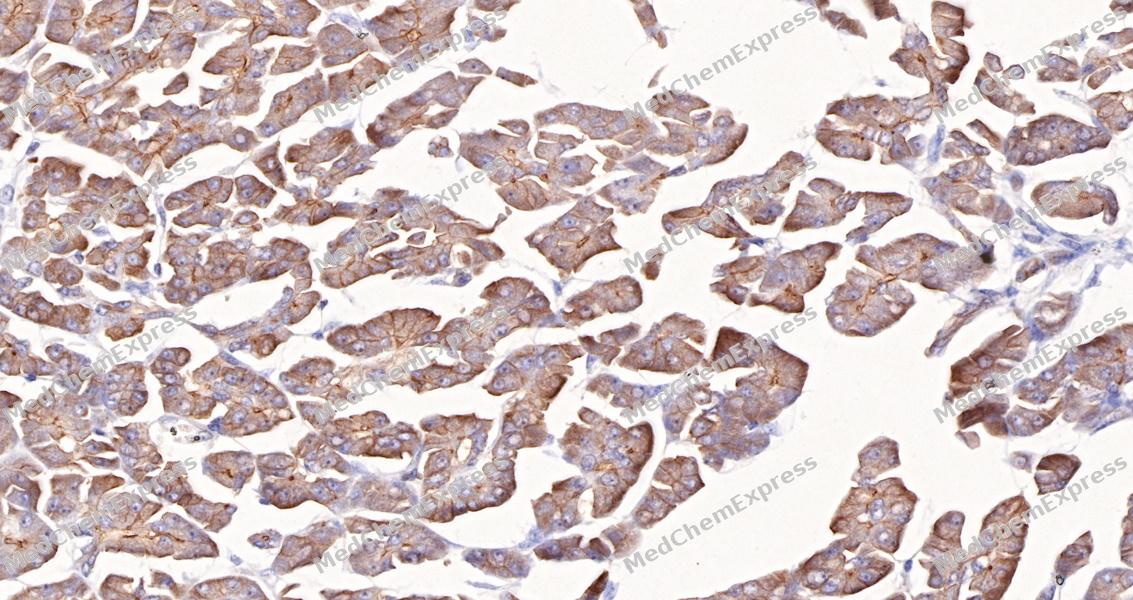 Immunohistochemical analysis of paraffin-embedded human pancreas tissue using Occludin Antibody (HY-P81155, 1/100). The section was pretreated using heat mediated antigen retrieval with sodium citrate buffer (pH 6.0) for 8 minutes. The tissues were blocked with quick block buffer for 0.5 hours at room temperature, washed with PBS and PBST, and then incubated with the primary antibody overnight at 4℃. The detection was performed using an HRP conjugated compact polymer system. DAB was used as the chromogen. Tissues were counterstained with hematoxylin and mounted with DPX.
 -
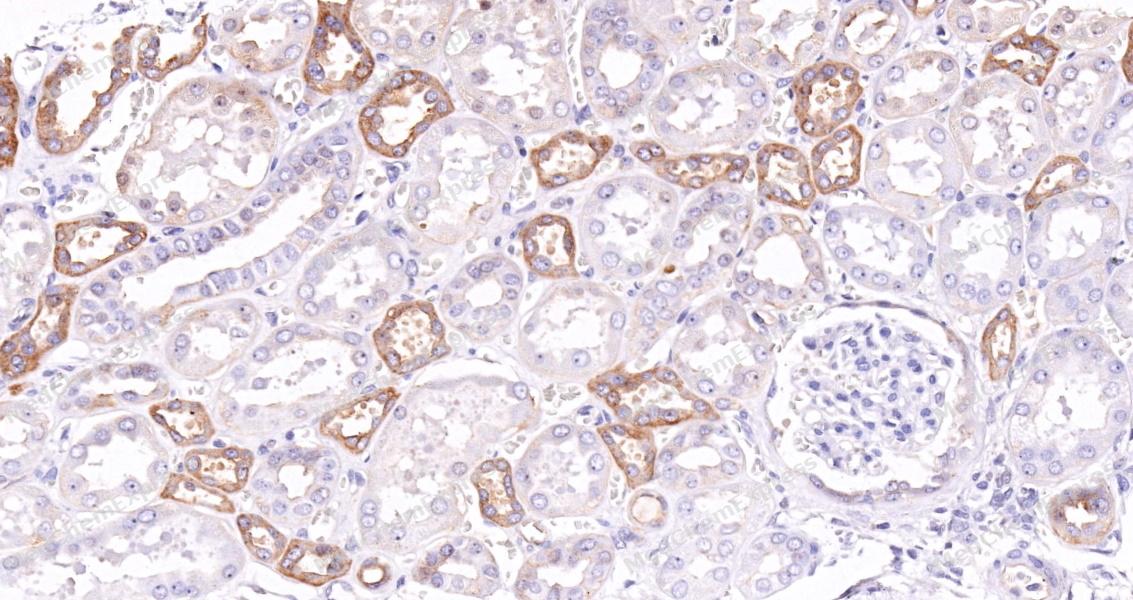 Immunohistochemical analysis of paraffin-embedded human kidney tissue using Occludin Antibody (HY-P81155, 1/100). The section was pretreated using heat mediated antigen retrieval with sodium citrate buffer (pH 6.0) for 8 minutes. The tissues were blocked with quick block buffer for 0.5 hours at room temperature, washed with PBS and PBST, and then incubated with the primary antibody overnight at 4℃. The detection was performed using an HRP conjugated compact polymer system. DAB was used as the chromogen. Tissues were counterstained with hematoxylin and mounted with DPX.
 -
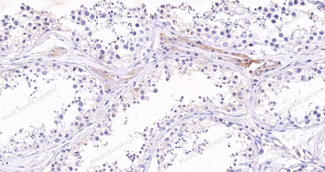 Immunohistochemical analysis of paraffin-embedded human testis tissue using Occludin Antibody (HY-P81155, 1/100). The section was pretreated using heat mediated antigen retrieval with sodium citrate buffer (pH 6.0) for 8 minutes. The tissues were blocked with quick block buffer for 0.5 hours at room temperature, washed with PBS and PBST, and then incubated with the primary antibody overnight at 4℃. The detection was performed using an HRP conjugated compact polymer system. DAB was used as the chromogen. Tissues were counterstained with hematoxylin and mounted with DPX.
 -
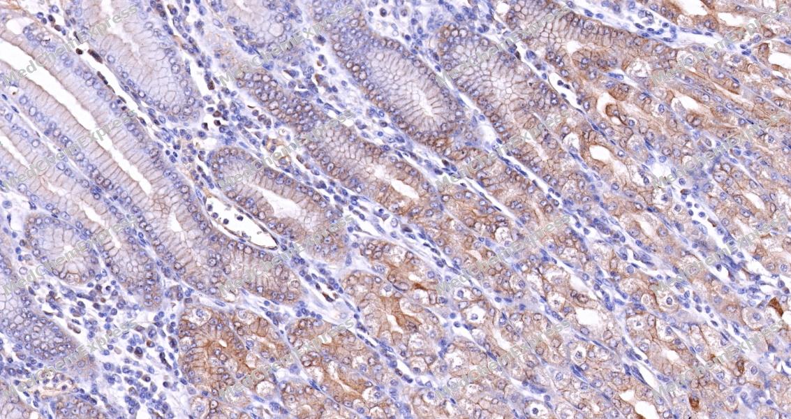 Immunohistochemical analysis of paraffin-embedded human stomach tissue using Occludin Antibody (HY-P81155, 1/100). The section was pretreated using heat mediated antigen retrieval with sodium citrate buffer (pH 6.0) for 8 minutes. The tissues were blocked with quick block buffer for 0.5 hours at room temperature, washed with PBS and PBST, and then incubated with the primary antibody overnight at 4℃. The detection was performed using an HRP conjugated compact polymer system. DAB was used as the chromogen. Tissues were counterstained with hematoxylin and mounted with DPX.
 -
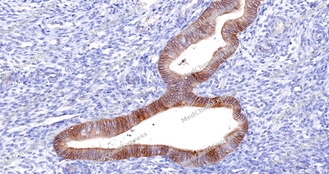 Immunohistochemical analysis of paraffin-embedded human endometrium tissue using Occludin Antibody (HY-P81155, 1/100). The section was pretreated using heat mediated antigen retrieval with sodium citrate buffer (pH 6.0) for 8 minutes. The tissues were blocked with quick block buffer for 0.5 hours at room temperature, washed with PBS and PBST, and then incubated with the primary antibody overnight at 4℃. The detection was performed using an HRP conjugated compact polymer system. DAB was used as the chromogen. Tissues were counterstained with hematoxylin and mounted with DPX.
 -
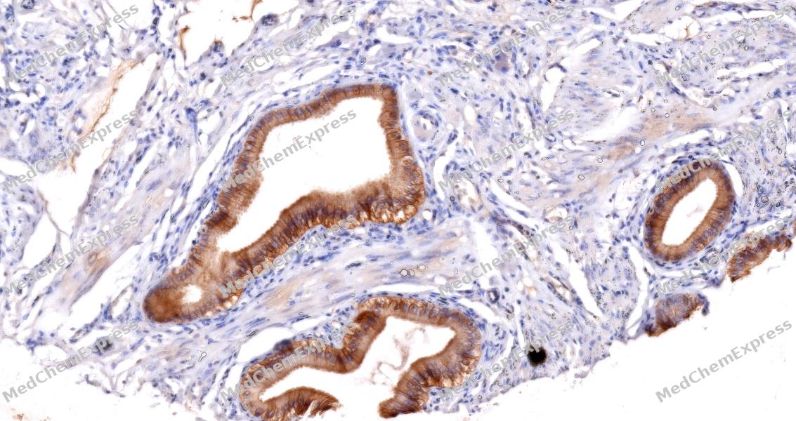 Immunohistochemical analysis of paraffin-embedded human gallbladder tissue using Occludin Antibody (HY-P81155, 1/100). The section was pretreated using heat mediated antigen retrieval with sodium citrate buffer (pH 6.0) for 8 minutes. The tissues were blocked with quick block buffer for 0.5 hours at room temperature, washed with PBS and PBST, and then incubated with the primary antibody overnight at 4℃. The detection was performed using an HRP conjugated compact polymer system. DAB was used as the chromogen. Tissues were counterstained with hematoxylin and mounted with DPX.
 -
 Immunocytochemistry analysis of A549 cells labeling Occludin/OCLN with Occludin/OCLN Antibody (HY-P81155) at 1/100 dilution. Cells were fixed in 4% paraformaldehyde for 15 minutes at room temperature, permeabilized with 0.1% Triton X-100 in PBS for 15 minutes at room temperature, then blocked with quick block buffer for 10 minutes at room temperature. Cells were then incubated with Occludin/OCLN Antibody (HY-P81155) at 1/100 dilution in quick block buffer overnight at 4 ℃. AF488-conjugated Goat Anti-Rabbit IgG H&L(HY-P8002, Green) was used as the secondary antibody at 1/1,000 dilution. PBS instead of the primary antibody was used as the secondary antibody only control. The Nuclear counterstain was DAPI (Blue).
 -
 Immunocytochemistry analysis of MCF-7 cells labeling Occludin/OCLN with Occludin/OCLN Antibody (HY-P81155) at 1/100 dilution. Cells were fixed in 4% paraformaldehyde for 15 minutes at room temperature, permeabilized with 0.1% Triton X-100 in PBS for 15 minutes at room temperature, then blocked with quick block buffer for 10 minutes at room temperature. Cells were then incubated with Occludin/OCLN Antibody (HY-P81155) at 1/100 dilution in quick block buffer overnight at 4 ℃. AF488-conjugated Goat Anti-Rabbit IgG H&L(HY-P8002, Green) was used as the secondary antibody at 1/1,000 dilution. PBS instead of the primary antibody was used as the secondary antibody only control. The Nuclear counterstain was DAPI (Blue).


Background
-
Function
Occludin/OCLN may play a role in the formation and regulation of the tight junction (TJ) paracellular permeability barrier. It is able to induce adhesion when expressed in cells lacking tight junctions; (Microbial infection) Acts as a coreceptor for hepatitis C virus (HCV) in hepatocytes
-
Subcellular Localization
Cell membrane; Multi-pass membrane protein; Cell junction, tight junction
-
Expression
Tissue_specificity:It is located at the tight junctions of epithelial and endothelial cells. It is highly expressed in the kidneys. It was not detected in the testes. -
Isoforms & Post-Translational Modification
Q16625 has 7 isomers: Q16625-1: 59144 Da (predicted); Q16625-2: 52706 Da (predicted); Q16625-3: 54124 Da (predicted); Q16625-4: 31602 Da (predicted); Q16625-5: 23324 Da (predicted); Q16625-6: 8033 Da (predicted); Q16625-7: 8175 Da (predicted).
Dephosphorylated by PTPRJ. The tyrosine phosphorylation on Tyr-398 and Tyr-402 reduces its ability to interact with TJP1. Phosphorylation at Ser-490 also attenuates the interaction with TJP1;(Microbial infection) Cleaved by S.pyogenes SpeB protease; leading to its degradation (PubMed:23532847). Degradation by SpeB promotes bacterial translocation across the host epithelial barrier (PubMed:23532847) -
Subunit
Interacts with TJP1/ZO1 (PubMed:19017651). Interacts with VAPA (PubMed:10523508). Interacts with CLDN1, CLDN6, CLDN9, CLDN11, CLDN12 and CLDN17 (PubMed:20375010). Interacts with PLSCR1 (PubMed:21806988). Interacts with LSR, ILDR1 and ILDR2 (PubMed:23239027). Interacts with TJP2/ZO2 (By similarity)
-
SwissProt ID
-
Synonyms
OCLN_HUMAN; Occludin; Ocl; BLCPMG; PPP1R115; PTORCH1; Tight junction protein occludin;
Documentation
-
Data Sheet (234 KB)
-
SDS (252 KB)
- English - EN (252 KB)
- Français - FR (252 KB)
- Deutsch - DE (252 KB)
- Norwegian - NO (252 KB)
- Español - ES (252 KB)
- Swedish - SV (252 KB)
- Italian - IT (252 KB)
- Korean - KR (252 KB)
- Portuguese - PT (252 KB)
-
User Guide for Antibodies (1077 KB)

 Powered by Bioz
