



Claudin 1 Antibody (YA6210)
(Synonyms: CLDN1; CLD1; SEMP1; Claudin-1; Senescence-associated epithelial membrane protein)
Based on 1 Customer Validation
Claudin 1 Antibody (YA6210) is a Rabbit-derived and non-conjugated IgG monoclonal antibody, targeting to Claudin 1.
-
Host:
Rabbit
-
Isotype:
IgG
-
Application:
WB, IHC-P, ICC/IF, IP, ELISA
-
Reactivity :
Human, Mouse, Rat
-
Formulation:
Supplied in PBS, 50% glycerol, 0.05% Proclin 300, 0.05%BSA
-
Conjugation:
Non-conjugated


To place orders, for customer services and technical support, please contact: MedChemExpress USA
Tel: 609-228-6898 E-mail: [email protected] [email protected]





Applications
| Application |
IHC-P

IHC-P: Immunohistochemistry-Paraffin
|
WB
WB: Western Blot
|
ICC/IF
ICC/IF: Immunocytochemistry/
Immunofluorescence |
ELISA
ELISA: Enzyme Linked Immunosorbent Assay
|
IP
IP: Immunoprecipitation
|
|---|---|---|---|---|---|
| Dilution Ratio | 1:200-1:1000 | 1:1000-1:5000 | 1:200-1:1000 | 1:5000-1:20000 | 1:50-1:200 |


Product Details
Claudin 1 Antibody (YA6210) is a Rabbit-derived and non-conjugated IgG monoclonal antibody, targeting to Claudin 1.
-
Host Rabbit

-
Clonality Monoclonal

-
Species ReactivityHuman, Mouse, Rat
-
Observed Molecular WeightObserved band size: 19 kDa
Note: Due to possible protein modifications or aggregation, the molecular weight should be confirmed by actual measurement, and the predicted value is for reference only.

-
Calculated Molecular Weight Predicted band size: 22 kDa

Protein A
Non-conjugated
Unmodified
IgG


Product Properties
-
Appearance
Liquid
-
Formulation
Supplied in PBS, 50% glycerol, 0.05% Proclin 300, 0.05%BSA
-
Concentration
Batch-dependent, Please check the COA for the concentration of each lot. Check Lot Concentration
-
Storage & Stability
Stored at -20°C for 1 year. Avoid repeated freeze / thaw cycles.
-
Shipping
Shipping with blue ice.


Verification Images
-
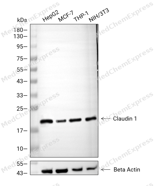 Western blot analysis of extracts from HepG2 (lane 2(20μg), MCF-7 (lane 3(20μg), THP-1 (lane 4(20μg), NIH/3T3 (lane 5(20μg) using Claudin 1 Antibody. Proteins were transferred to a PVDF membrane and blocked with 5% BSA in TBST for 2 hour at room temperature. The primary antibody (1/1000) and Loading control antibody (Beta Actin, HY-P80438, 1/3000) was used in 5% BSA in TBST at 4°C overnight. Goat Anti-Mouse/Rabbit IgG-HRP Secondary Antibody (HY-P8004/HY-P8001, 1/10,000) was used for 1 hour at room temperature.
 -
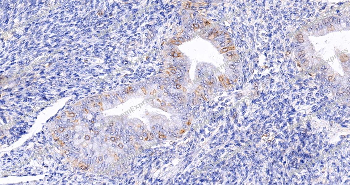 Immunohistochemical analysis of paraffin-embedded human endometrium tissue using Claudin 1 Antibody (HY-P86518, 1/1000). The section was pretreated using heat mediated antigen retrieval with sodium citrate buffer (pH 6.0) for 8 minutes. The tissues were blocked with quick block buffer for 0.5 hours at room temperature, washed with PBS and PBST, and then incubated with the primary antibody overnight at 4℃. The detection was performed using an HRP conjugated compact polymer system. DAB was used as the chromogen. Tissues were counterstained with hematoxylin and mounted with DPX.
 -
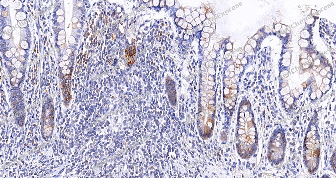 Immunohistochemical analysis of paraffin-embedded human small intestine tissue using Claudin 1 Antibody (HY-P86518, 1/1000). The section was pretreated using heat mediated antigen retrieval with sodium citrate buffer (pH 6.0) for 8 minutes. The tissues were blocked with quick block buffer for 0.5 hours at room temperature, washed with PBS and PBST, and then incubated with the primary antibody overnight at 4℃. The detection was performed using an HRP conjugated compact polymer system. DAB was used as the chromogen. Tissues were counterstained with hematoxylin and mounted with DPX.
 -
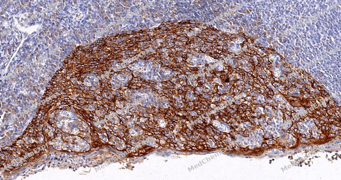 Immunohistochemical analysis of paraffin-embedded human tonsil tissue using Claudin 1 Antibody (HY-P86518, 1/1000). The section was pretreated using heat mediated antigen retrieval with sodium citrate buffer (pH 6.0) for 8 minutes. The tissues were blocked with quick block buffer for 0.5 hours at room temperature, washed with PBS and PBST, and then incubated with the primary antibody overnight at 4℃. The detection was performed using an HRP conjugated compact polymer system. DAB was used as the chromogen. Tissues were counterstained with hematoxylin and mounted with DPX.
 -
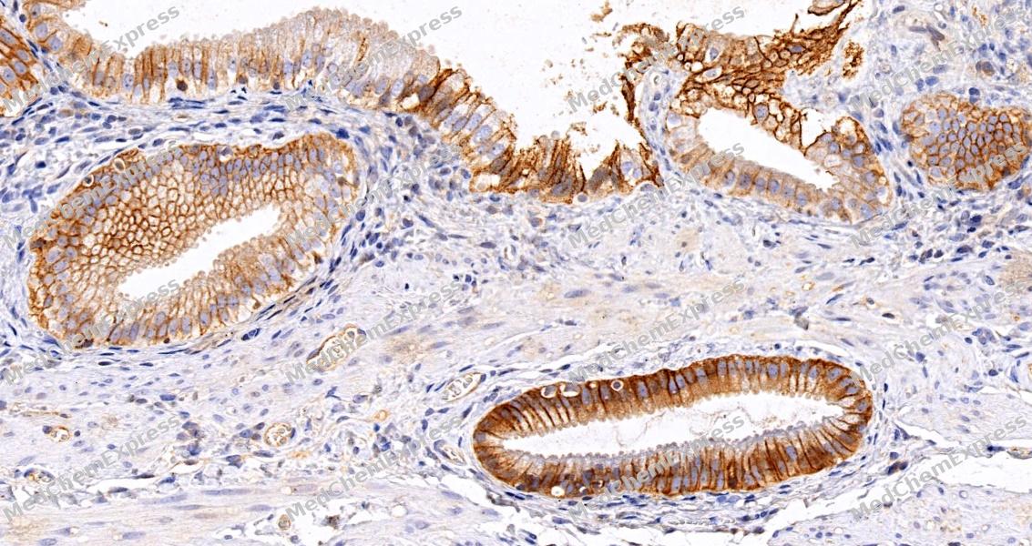 Immunohistochemical analysis of paraffin-embedded human gallbladder tissue using Claudin 1 Antibody (HY-P86518, 1/1000). The section was pretreated using heat mediated antigen retrieval with sodium citrate buffer (pH 6.0) for 8 minutes. The tissues were blocked with quick block buffer for 0.5 hours at room temperature, washed with PBS and PBST, and then incubated with the primary antibody overnight at 4℃. The detection was performed using an HRP conjugated compact polymer system. DAB was used as the chromogen. Tissues were counterstained with hematoxylin and mounted with DPX.
 -
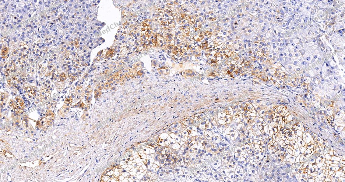 Immunohistochemical analysis of paraffin-embedded human adrenal gland tissue using Claudin 1 Antibody (HY-P86518, 1/1000). The section was pretreated using heat mediated antigen retrieval with sodium citrate buffer (pH 6.0) for 8 minutes. The tissues were blocked with quick block buffer for 0.5 hours at room temperature, washed with PBS and PBST, and then incubated with the primary antibody overnight at 4℃. The detection was performed using an HRP conjugated compact polymer system. DAB was used as the chromogen. Tissues were counterstained with hematoxylin and mounted with DPX.
 -
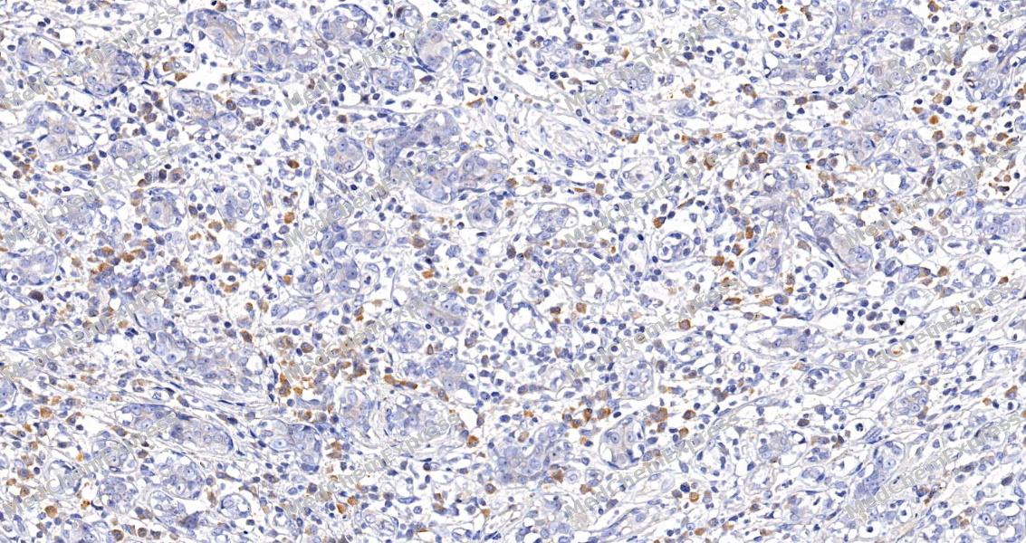 Immunohistochemical analysis of paraffin-embedded human breast tissue using Claudin 1 Antibody (HY-P86518, 1/1000). The section was pretreated using heat mediated antigen retrieval with sodium citrate buffer (pH 6.0) for 8 minutes. The tissues were blocked with quick block buffer for 0.5 hours at room temperature, washed with PBS and PBST, and then incubated with the primary antibody overnight at 4℃. The detection was performed using an HRP conjugated compact polymer system. DAB was used as the chromogen. Tissues were counterstained with hematoxylin and mounted with DPX.


Background
-
Function
Claudin 1 is a Claudins function as major constituents of the tight junction complexes that regulate the permeability of epithelia. While some claudin family members play essential roles in the formation of impermeable barriers, others mediate the permeability to ions and small molecules. Often, several claudin family members are coexpressed and interact with each other, and this determines the overall permeability. CLDN1 is required to prevent the paracellular diffusion of small molecules through tight junctions in the epidermis and is required for the normal barrier function of the skin. Required for normal water homeostasis and to prevent excessive water loss through the skin, probably via an indirect effect on the expression levels of other proteins, since CLDN1 itself seems to be dispensable for water barrier formation in keratinocyte tight junctions; (Microbial infection) Acts as a co-receptor for hepatitis C virus (HCV) in hepatocytes. Associates with CD81 and the CLDN1-CD81 receptor complex is essential for HCV entry into host cell. Acts as a receptor for dengue virus[1][2][3][4][5].
-
Subcellular Localization
Cell junction, tight junction; Cell membrane; Multi-pass membrane protein; Basolateral cell membrane
-
Expression
Tissue_specificity:It is highly expressed in the liver and kidneys. It is also expressed in the heart, brain, spleen, lungs, and testes. -
Subunit
Homopolymers interact with CLDN3, but not CLDN2, homopolymers. Can form homo- and heteropolymers with other claudin family members (PubMed:23704991).
-
SwissProt ID
-
Synonyms
CLDN1; CLD1; SEMP1; Claudin-1; Senescence-associated epithelial membrane protein


Documentation
-
Data Sheet (234 KB)
-
SDS (251 KB)
- English - EN (251 KB)
- Français - FR (251 KB)
- Deutsch - DE (251 KB)
- Norwegian - NO (251 KB)
- Español - ES (251 KB)
- Swedish - SV (251 KB)
- Italian - IT (251 KB)
- Korean - KR (251 KB)
- Portuguese - PT (251 KB)
-
User Guide for Antibodies (1077 KB)

 Powered by Bioz
