



PHAPI2/APRIL Antibody (YA1610)
(Synonyms: ANP32B; PAL31; PHAPI2; PHAPI2a; Ssp29)
Based on 1 Customer Validation
PHAPI2/APRIL Antibody (YA1610) is a Rabbit-derived and non-conjugated IgG monoclonal antibody, targeting to PHAPI2/APRIL.
-
Host:
Rabbit
-
Isotype:
IgG
-
Application:
WB, IHC-F, IHC-P, ICC/IF
-
Reactivity :
Human, Rat
-
Formulation:
Supplied in 50mM Tris-Glycine(pH 7.4), 0.15M NaCl, 40% Glycerol, 0.01% Sodium azide and 0.05% BSA
-
Conjugation:
Non-conjugated


To place orders, for customer services and technical support, please contact: MedChemExpress USA
Tel: 609-228-6898 E-mail: [email protected] [email protected]





Applications
| Application |
WB

WB: Western Blot
|
IHC-P
IHC-P: Immunohistochemistry-Paraffin
|
IHC-F
IHC-F: Immunohistochemistry-Frozen
|
ICC/IF
ICC/IF: Immunocytochemistry/
Immunofluorescence |
|---|---|---|---|---|
| Dilution Ratio | 1:500-1:1000 | 1:50-1:100 | 1:50-1:100 | 1:50-1:200 |


Product Details
PHAPI2/APRIL Antibody (YA1610) is a Rabbit-derived and non-conjugated IgG monoclonal antibody, targeting to PHAPI2/APRIL.
-
Host Rabbit

-
Clonality Recombinant,Monoclonal

-
Species ReactivityHuman, Rat
-
Observed Molecular WeightObserved band size: 29 kDa
Note: Due to possible protein modifications or aggregation, the molecular weight should be confirmed by actual measurement, and the predicted value is for reference only.

-
Calculated Molecular Weight Predicted band size: 29 kDa

Entrez Gene: 10541 Human ; 67628 Mouse ; 170724 Rat
SwissProt: Q92688 Human ; Q9EST5 Mouse ; Q9EST6 Rat
OMIM: 619823 Human
A synthetic peptide of human PHAPI2
Endogenous
Affinity Purified
Non-conjugated
Unmodified
IgG


Product Properties
-
Appearance
Solution
-
Formulation
Supplied in 50mM Tris-Glycine(pH 7.4), 0.15M NaCl, 40% Glycerol, 0.01% Sodium azide and 0.05% BSA
-
Concentration
Batch-dependent, Please check the COA for the concentration of each lot. Check Lot Concentration
-
Storage & Stability
Stored at -20°C for 1 year. Avoid repeated freeze / thaw cycles.
-
Shipping
Shipping with blue ice.


Verification Images
-
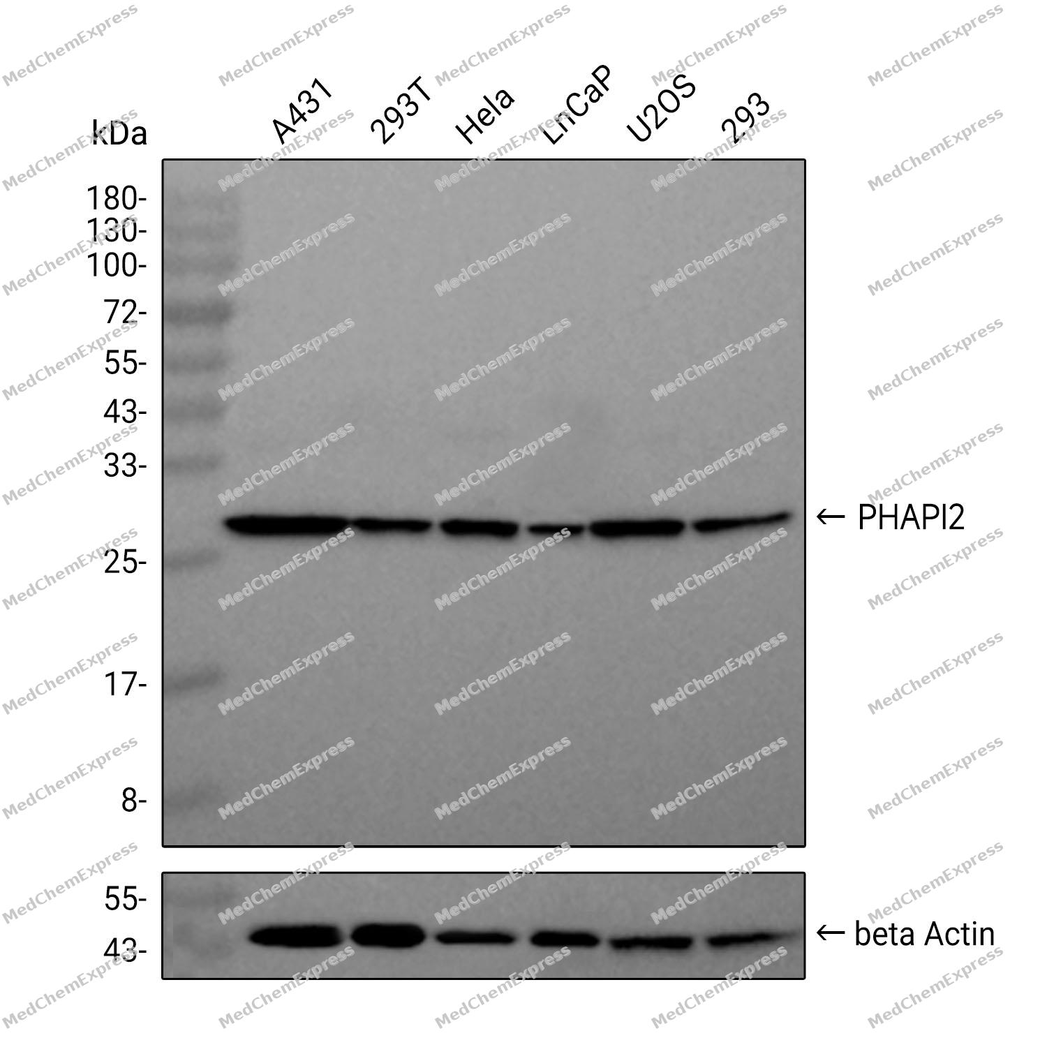
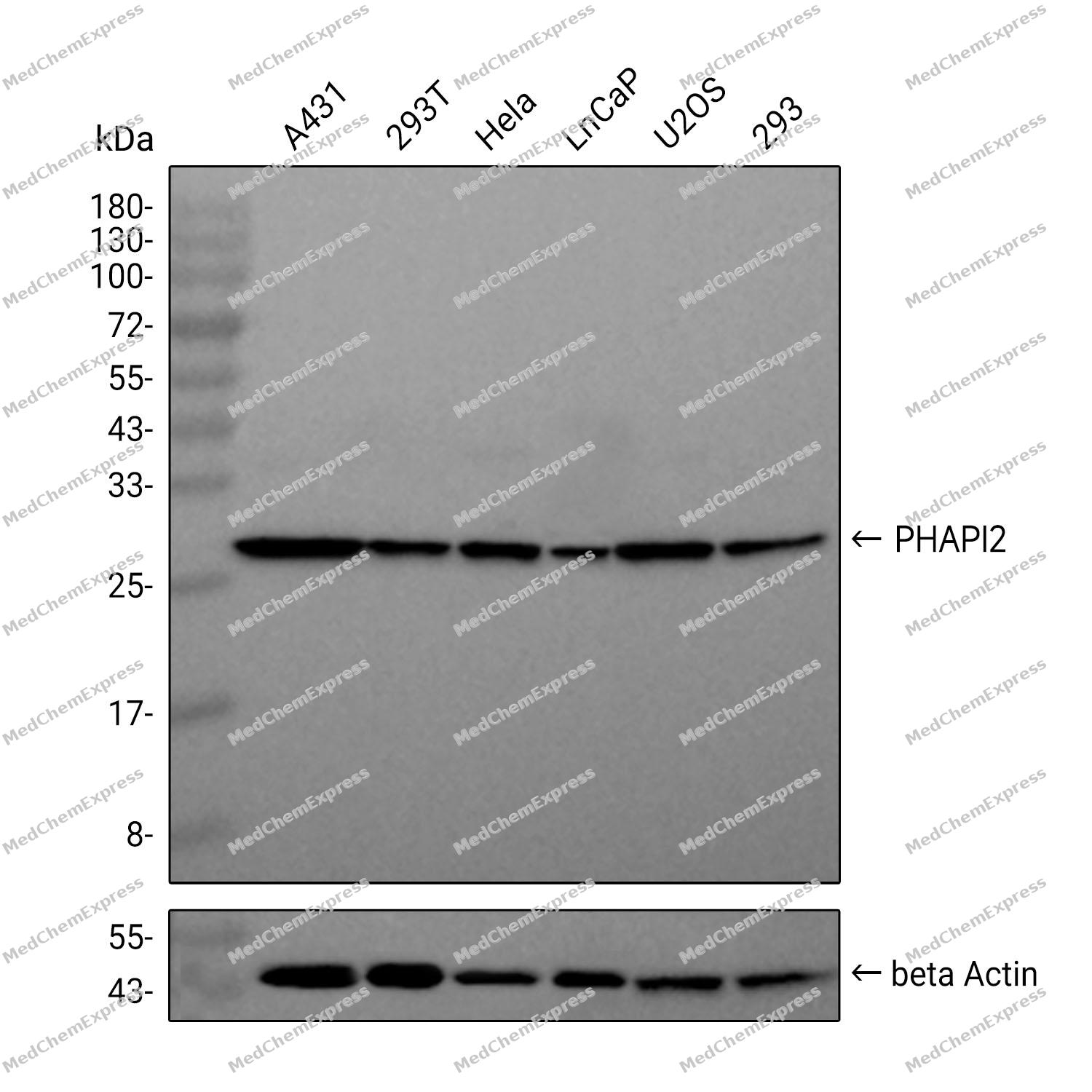 Western blot analysis was performed on extracts from A431 (lane 1, 15 μg), 293T (lane 2, 15 μg), Hela (lane 3, 15 μg), LnCaP (lane 4, 15 μg), U2OS (lane 5, 15 μg), and 293 (lane 6, 15 μg) using PHAPI2 Rabbit mAb.Proteins were transferred to a PVDF membrane and blocked with 5% non-fat milk in TBST at 4°C overnight.The primary antibody (1:1000 dilution) and the loading control antibody (beta-Actin, HY-P80438, 1:5000 dilution) was incubated in 5% non-fat milk in TBST for 1 hour at 37°C.Goat Anti-Rabbit IgG-HRP Secondary Antibody (1:20000 dilution) was then applied for 40 minutes at 37°C.
 -
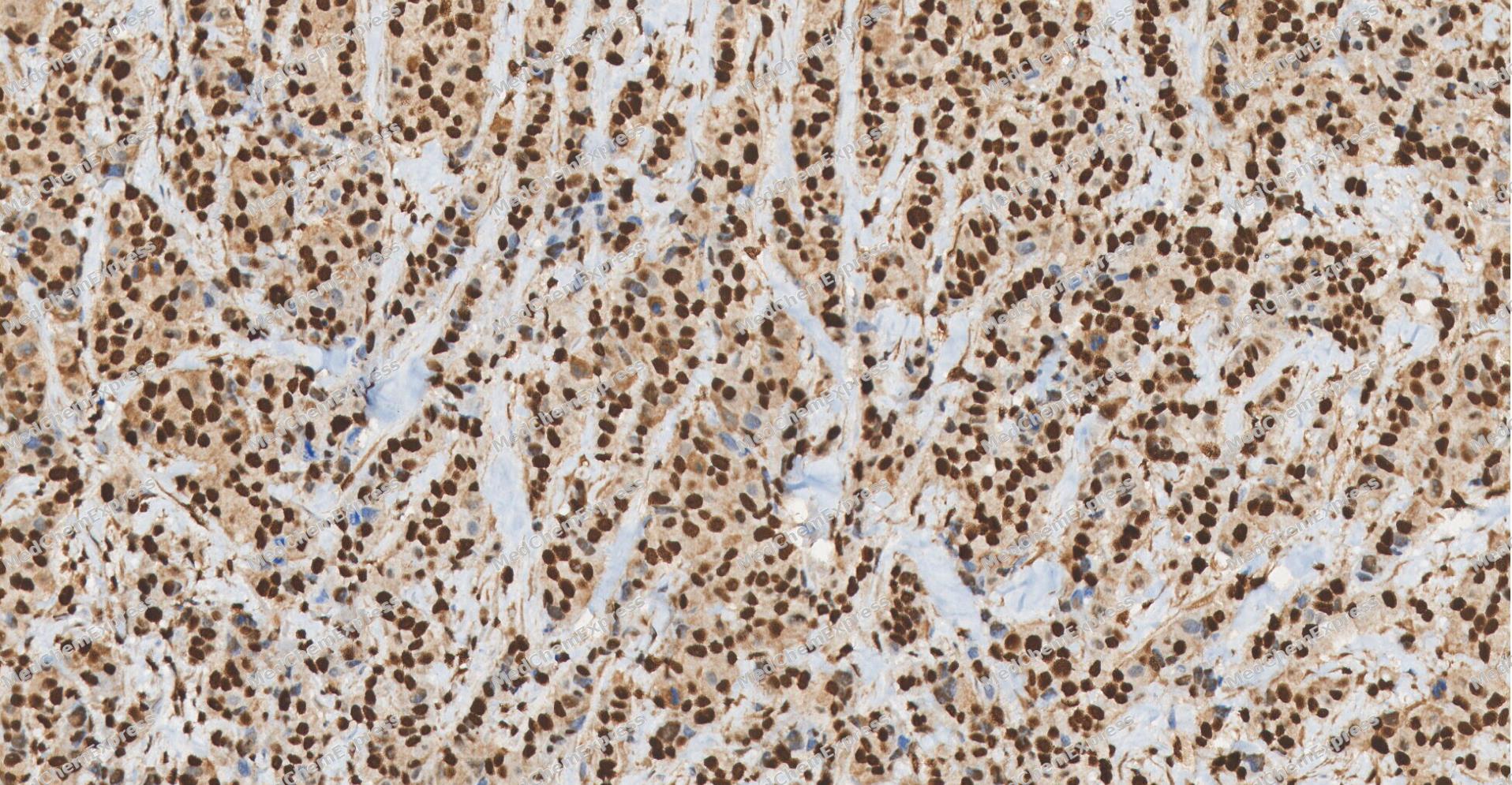 Immunohistochemical analysis of paraffin-embedded human Breast Cancer tissue using PHAPI2/APRIL antibody was performed. The section was pretreated using high-temperature and high-pressure mediated EDTA antigen retrieval buffer (pH 9.0), for 5 minutes. The tissues were incubated with primary antibody (HY-P81865, 1:100 dilution) at room temperature for 60 minutes. Detection was performed using an HRP conjugated compact polymer system. DAB was used as the chromogen. The tissues were counterstained with hematoxylin and mounted with neutral balsam mounting medium.
 -
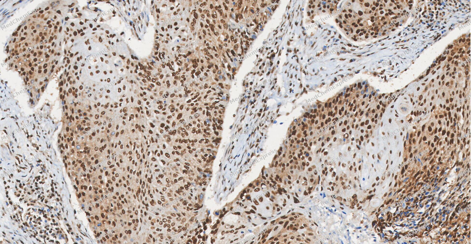 Immunohistochemical analysis of paraffin-embedded human Esophageal Carcinoma tissue using PHAPI2/APRIL antibody was performed. The section was pretreated using high-temperature and high-pressure mediated EDTA antigen retrieval buffer (pH 9.0), for 5 minutes. The tissues were incubated with primary antibody (HY-P81865, 1:100 dilution) at room temperature for 60 minutes. Detection was performed using an HRP conjugated compact polymer system. DAB was used as the chromogen. The tissues were counterstained with hematoxylin and mounted with neutral balsam mounting medium.
 -
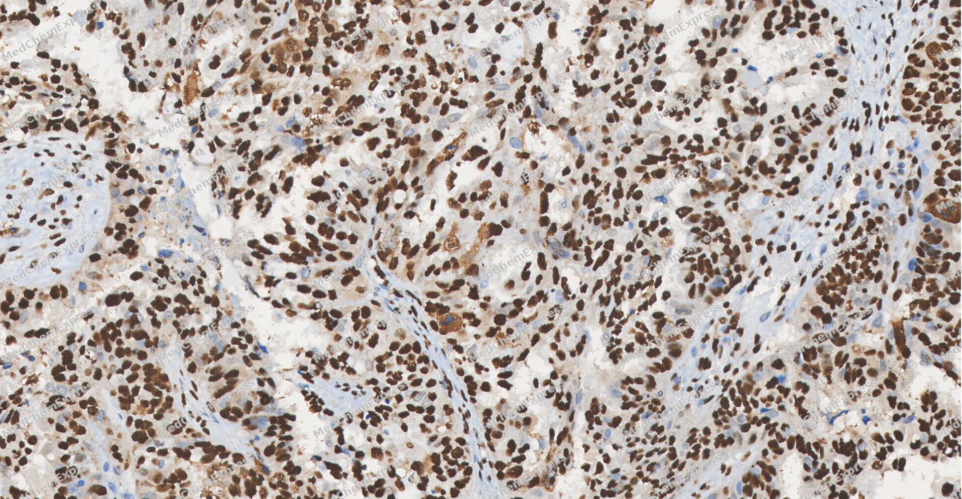 Immunohistochemical analysis of paraffin-embedded human ovarian carcinoma tissue using PHAPI2/APRIL antibody was performed. The section was pretreated using high-temperature and high-pressure mediated EDTA antigen retrieval buffer (pH 9.0), for 5 minutes. The tissues were incubated with primary antibody (HY-P81865, 1:100 dilution) at room temperature for 60 minutes. Detection was performed using an HRP conjugated compact polymer system. DAB was used as the chromogen. The tissues were counterstained with hematoxylin and mounted with neutral balsam mounting medium.
 -
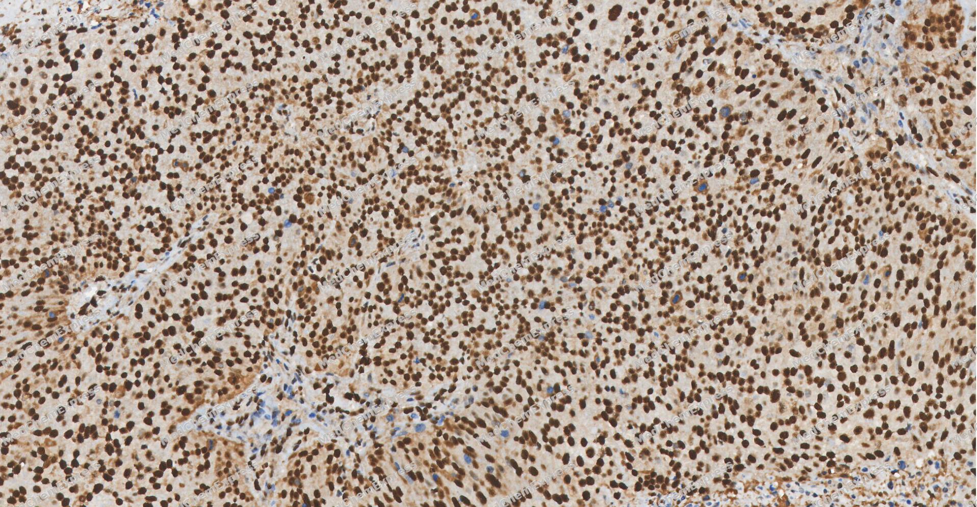 Immunohistochemical analysis of paraffin-embedded human Cervical cancer tissue using PHAPI2/APRIL antibody was performed. The section was pretreated using high-temperature and high-pressure mediated EDTA antigen retrieval buffer (pH 9.0), for 5 minutes. The tissues were incubated with primary antibody (HY-P81865, 1:100 dilution) at room temperature for 60 minutes. Detection was performed using an HRP conjugated compact polymer system. DAB was used as the chromogen. The tissues were counterstained with hematoxylin and mounted with neutral balsam mounting medium.
 -
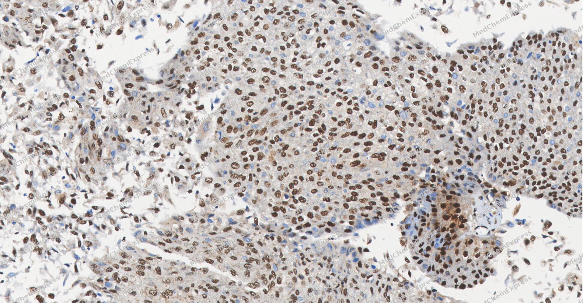 Immunohistochemical analysis of paraffin-embedded human bladder cancer tissue using PHAPI2/APRIL antibody was performed. The section was pretreated using high-temperature and high-pressure mediated EDTA antigen retrieval buffer (pH 9.0), for 5 minutes. The tissues were incubated with primary antibody (HY-P81865, 1:100 dilution) at room temperature for 60 minutes. Detection was performed using an HRP conjugated compact polymer system. DAB was used as the chromogen. The tissues were counterstained with hematoxylin and mounted with neutral balsam mounting medium.
 -
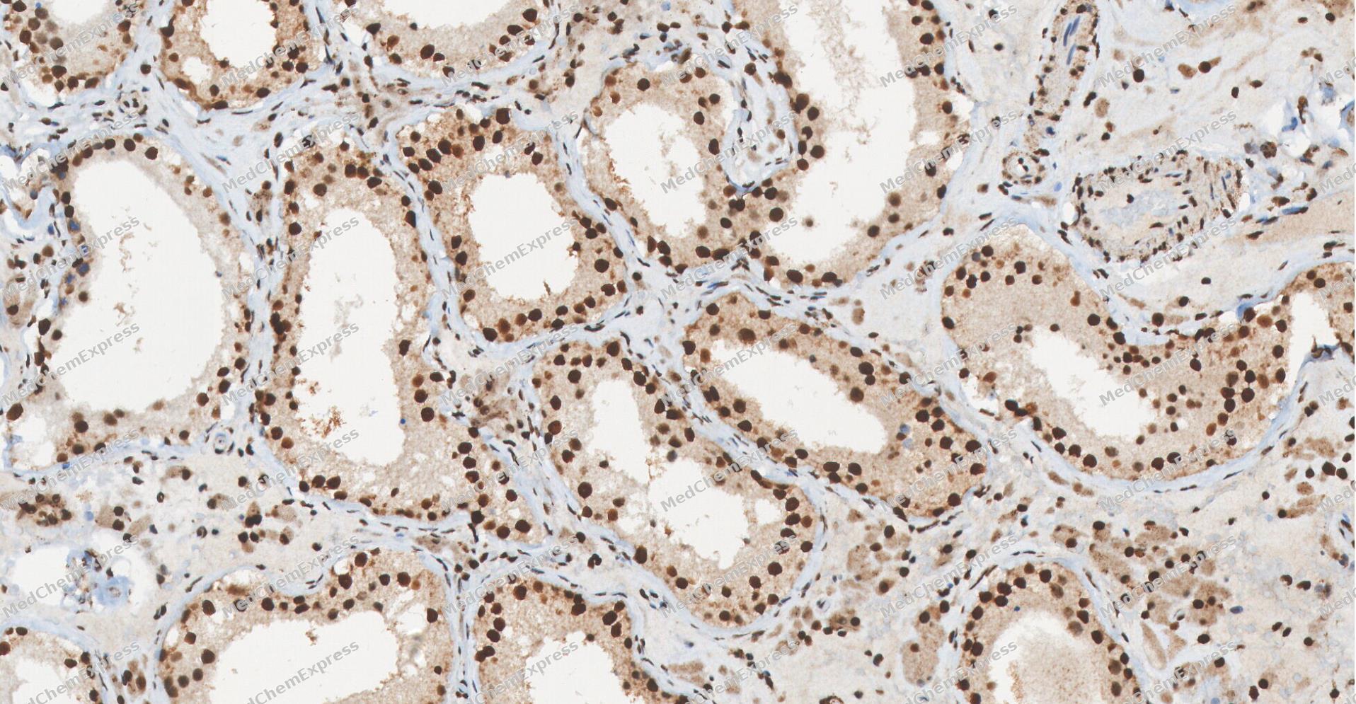 Immunohistochemical analysis of paraffin-embedded human Testis tissue using PHAPI2/APRIL antibody was performed. The section was pretreated using high-temperature and high-pressure mediated EDTA antigen retrieval buffer (pH 9.0), for 5 minutes. The tissues were incubated with primary antibody (HY-P81865, 1:100 dilution) at room temperature for 60 minutes. Detection was performed using an HRP conjugated compact polymer system. DAB was used as the chromogen. The tissues were counterstained with hematoxylin and mounted with neutral balsam mounting medium.
 -
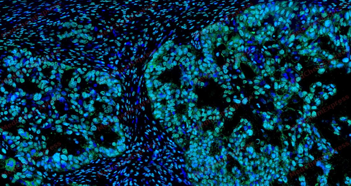 Tyramide signaling amplification based immunofluorescence was performed on paraffin-embedded human ovarian carcinoma tissue using PHAPI2/APRIL antibody. Antigen retrieval was performed in EDTA buffer pH 9.0 (95 °C, 20 min) followed by cooling to RT. Then incubated with primary antibody (HY-P81865, 1:300 dilution) at room temperature for 60 minutes and HRP conjugated secondary antibody for 10 minutes. Fluorescence was then developed with Vari Fluor 532 TSA (200×)(HY-D1832). The tissues were counterstained with DAPI and mounted with Anti-fade mounting medium.
 -
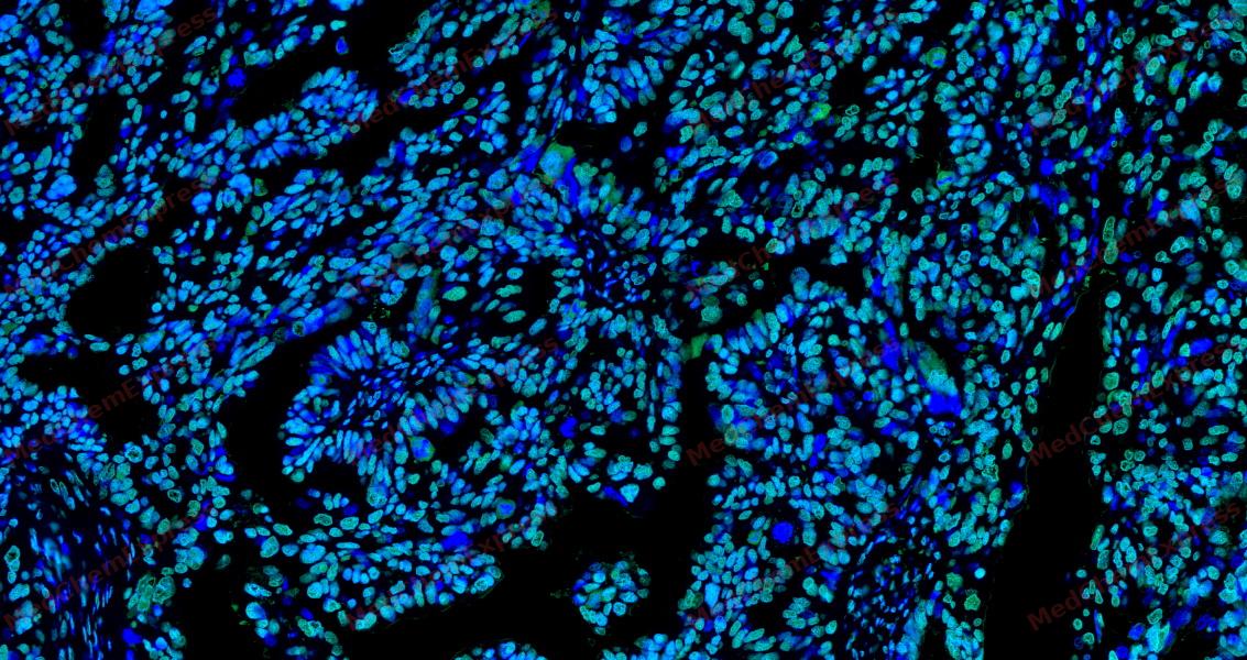 Tyramide signaling amplification based immunofluorescence was performed on paraffin-embedded human ovarian carcinoma tissue using PHAPI2/APRIL antibody. Antigen retrieval was performed in EDTA buffer pH 9.0 (95 °C, 20 min) followed by cooling to RT. Then incubated with primary antibody (HY-P81865, 1:300 dilution) at room temperature for 60 minutes and HRP conjugated secondary antibody for 10 minutes. Fluorescence was then developed with Vari Fluor 532 TSA (200×)(HY-D1832). The tissues were counterstained with DAPI and mounted with Anti-fade mounting medium.
 -
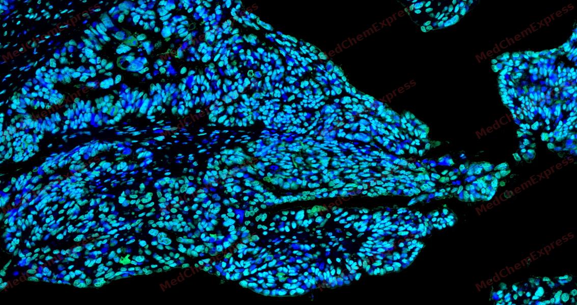 Tyramide signaling amplification based immunofluorescence was performed on paraffin-embedded human ovarian carcinoma tissue using PHAPI2/APRIL antibody. Antigen retrieval was performed in EDTA buffer pH 9.0 (95 °C, 20 min) followed by cooling to RT. Then incubated with primary antibody (HY-P81865, 1:300 dilution) at room temperature for 60 minutes and HRP conjugated secondary antibody for 10 minutes. Fluorescence was then developed with Vari Fluor 532 TSA (200×)(HY-D1832). The tissues were counterstained with DAPI and mounted with Anti-fade mounting medium.
 -
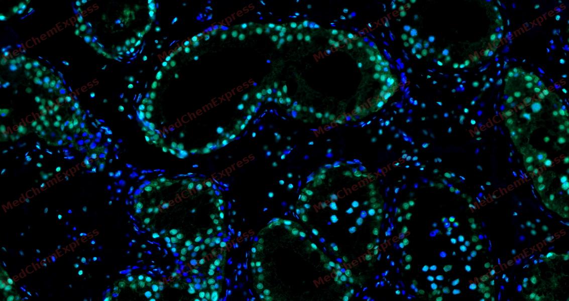 Tyramide signaling amplification based immunofluorescence was performed on paraffin-embedded human Testis tissue using PHAPI2/APRIL antibody. Antigen retrieval was performed in EDTA buffer pH 9.0 (95 °C, 20 min) followed by cooling to RT. Then incubated with primary antibody (HY-P81865, 1:300 dilution) at room temperature for 60 minutes and HRP conjugated secondary antibody for 10 minutes. Fluorescence was then developed with Vari Fluor 532 TSA (200×)(HY-D1832). The tissues were counterstained with DAPI and mounted with Anti-fade mounting medium.
 -
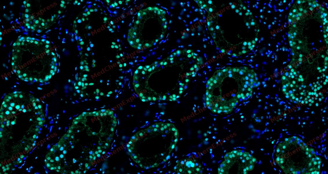 Tyramide signaling amplification based immunofluorescence was performed on paraffin-embedded human Testis tissue using PHAPI2/APRIL antibody. Antigen retrieval was performed in EDTA buffer pH 9.0 (95 °C, 20 min) followed by cooling to RT. Then incubated with primary antibody (HY-P81865, 1:300 dilution) at room temperature for 60 minutes and HRP conjugated secondary antibody for 10 minutes. Fluorescence was then developed with Vari Fluor 532 TSA (200×)(HY-D1832). The tissues were counterstained with DAPI and mounted with Anti-fade mounting medium.
 -
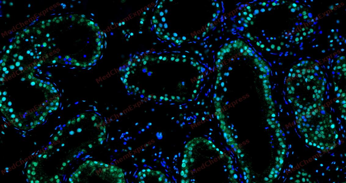 Tyramide signaling amplification based immunofluorescence was performed on paraffin-embedded human Testis tissue using PHAPI2/APRIL antibody. Antigen retrieval was performed in EDTA buffer pH 9.0 (95 °C, 20 min) followed by cooling to RT. Then incubated with primary antibody (HY-P81865, 1:300 dilution) at room temperature for 60 minutes and HRP conjugated secondary antibody for 10 minutes. Fluorescence was then developed with Vari Fluor 532 TSA (200×)(HY-D1832). The tissues were counterstained with DAPI and mounted with Anti-fade mounting medium.
 -
 Immunocytochemistry analysis of Hela cells labeling PHAPI2/APRIL with PHAPI2/APRIL Antibody (HY-P81865) at 1/100 dilution. Cells were fixed in 4% paraformaldehyde for 15 minutes at room temperature, permeabilized with 0.1% Triton X-100 in PBS for 15 minutes at room temperature, then blocked with quick block buffer for 10 minutes at room temperature. Cells were then incubated with PHAPI2/APRIL Antibody (HY-P81865) at 1/100 dilution in quick block buffer overnight at 4 ℃. AF488-conjugated Goat Anti-Rabbit IgG H&L(HY-P8002, Green) was used as the secondary antibody at 1/1,000 dilution. PBS instead of the primary antibody was used as the secondary antibody only control. The Nuclear counterstain was DAPI (Blue).
 -
 Immunocytochemistry analysis of MCF-7 cells labeling PHAPI2/APRIL with PHAPI2/APRIL Antibody (HY-P81865) at 1/100 dilution. Cells were fixed in 4% paraformaldehyde for 15 minutes at room temperature, permeabilized with 0.1% Triton X-100 in PBS for 15 minutes at room temperature, then blocked with quick block buffer for 10 minutes at room temperature. Cells were then incubated with PHAPI2/APRIL Antibody (HY-P81865) at 1/100 dilution in quick block buffer overnight at 4 ℃. AF488-conjugated Goat Anti-Rabbit IgG H&L(HY-P8002, Green) was used as the secondary antibody at 1/1,000 dilution. PBS instead of the primary antibody was used as the secondary antibody only control. The Nuclear counterstain was DAPI (Blue).


Background
-
Function
PHAPI2/APRIL multifunctional protein that is involved in the regulation of many processes including cell proliferation, apoptosis, cell cycle progression or transcription. Regulates the proliferation of neuronal stem cells, differentiation of leukemic cells and progression from G1 to S phase of the cell cycle. As negative regulator of caspase-3-dependent apoptosis, may act as an antagonist of ANP32A in regulating tissue homeostasis. Exhibits histone chaperone properties, able to recruit histones to certain promoters, thus regulating the transcription of specific genes. Also plays an essential role in the nucleocytoplasmic transport of specific mRNAs via the uncommon nuclear mRNA export receptor XPO1/CRM1. Participates in the regulation of adequate adaptive immune responses by acting on mRNA expression and cell proliferation; (Microbial infection) Plays an essential role in influenza A and B viral genome replication. Also plays a role in foamy virus mRNA export from the nucleus to the cytoplasm[1][2][3][4][5][6][7].
-
Subcellular Localization
Nucleus; Cytoplasm; Cytoplasm
-
Expression
Tissue_specificity:Expressed in heart, lung, pancreas, prostate and in spleen, thymus and placenta -
Isoforms & Post-Translational Modification
Q92688 has 2 isomers: Q92688-1: 28788 Da (predicted); Q92688-2: 22277 Da (predicted).
Some glutamate residues are glycylated by TTLL8. This modification occurs exclusively on glutamate residues and results in a glycine chain on the gamma-carboxyl group (By similarity);Directly cleaved by caspase-3/CASP3 -
Subunit
Interacts with histones H3 and H4 (PubMed:20538007). Interacts with KLF5; this interaction induces promoter region-specific histone incorporation and inhibition of histone acetylation by ANP32B (PubMed:18039846)
-
SwissProt ID
-
Synonyms
ANP32B; PAL31; PHAPI2; PHAPI2a; Ssp29
-
Research Field
Cell Biology


Documentation
-
Data Sheet (261 KB)
-
SDS (251 KB)
- English - EN (251 KB)
- Français - FR (251 KB)
- Deutsch - DE (251 KB)
- Norwegian - NO (251 KB)
- Español - ES (251 KB)
- Swedish - SV (251 KB)
- Italian - IT (251 KB)
- Korean - KR (251 KB)
- Portuguese - PT (251 KB)
-
User Guide for Antibodies (1077 KB)
References
[1]. Munemasa Y, et al. Promoter region-specific histone incorporation by the novel histone chaperone ANP32B and DNA-binding factor KLF5. Mol Cell Biol. 2008 Feb;28(3):1171-81. [Content Brief]
[2]. Shen SM, et al. Downregulation of ANP32B, a novel substrate of caspase-3, enhances caspase-3 activation and apoptosis induction in myeloid leukemic cells. Carcinogenesis. 2010 Mar;31(3):419-26. [Content Brief]
[3]. Tochio N, et al. Solution structure of histone chaperone ANP32B: interaction with core histones H3-H4 through its acidic concave domain. J Mol Biol. 2010 Aug 6;401(1):97-114. [Content Brief]
[4]. Fries B, et al. Analysis of nucleocytoplasmic trafficking of the HuR ligand APRIL and its influence on CD83 expression. J Biol Chem. 2007 Feb 16;282(7):4504-4515. [Content Brief]
[5]. Staller E, et al. ANP32 Proteins Are Essential for Influenza Virus Replication in Human Cells. J Virol. 2019 Sep 1;93(17):. [Content Brief]
[6]. Zhang Z, et al. Selective usage of ANP32 proteins by influenza B virus polymerase: Implications in determination of host range. PLoS Pathog. 2020 Oct;16(10):e1008989. [Content Brief]
[7]. Bodem J, et al. Foamy virus nuclear RNA export is distinct from that of other retroviruses. J Virol. 2011 Mar;85(5):2333-41. [Content Brief]

 Powered by Bioz
