



Phospho-Nrf2 (Ser40) Antibody (YA169)
(Synonyms: NFE2L2; NRF2; Nuclear factor erythroid 2-related factor 2; NF-E2-related factor 2; NFE2-related factor 2; HEBP1; Nuclear factor; erythroid derived 2; like 2)
Based on 1 Customer Validation
Phospho-Nrf2 (Ser40) Antibody (YA169) is a Rabbit-derived and non-conjugated IgG monoclonal antibody, targeting to Phospho-Nrf2 (Ser40).
-
Host:
Rabbit
-
Isotype:
IgG
-
Application:
WB, IHC-P
-
Reactivity :
Human
-
Formulation:
Supplied in 50 mM Tris-Glycine (pH 7.4), 0.15 M NaCl, 40% Glycerol and 0.05% BSA. Preservative: 0.01% Sodium azide
-
Conjugation:
Non-conjugated


To place orders, for customer services and technical support, please contact: MedChemExpress USA
Tel: 609-228-6898 E-mail: [email protected] [email protected]





Applications
| Application |
WB

WB: Western Blot
|
IHC-P
IHC-P: Immunohistochemistry-Paraffin
|
|---|---|---|
| Dilution Ratio | 1:500-1:1000 | 1:50-1:100 |


Product Details
Phospho-Nrf2 (Ser40) Antibody (YA169) is a Rabbit-derived and non-conjugated IgG monoclonal antibody, targeting to Phospho-Nrf2 (Ser40).
-
Host Rabbit

-
Clonality Recombinant,Monoclonal

-
Species ReactivityHuman
-
Observed Molecular WeightObserved band size: 100 kDa
Note: Due to possible protein modifications or aggregation, the molecular weight should be confirmed by actual measurement, and the predicted value is for reference only.

-
Calculated Molecular Weight Predicted band size: 68 kDa

Synthetic phosphopeptide corresponding to residues surrounding Ser40 of Human Nrf2.The exact sequence is proprietary to MCE.
Endogenous
affinity purified
Non-conjugated
Phosphorylated
IgG


Product Properties
-
Appearance
Solution
-
Formulation
Supplied in 50 mM Tris-Glycine (pH 7.4), 0.15 M NaCl, 40% Glycerol and 0.05% BSA. Preservative: 0.01% Sodium azide
-
Concentration
Batch-dependent, Please check the COA for the concentration of each lot. Check Lot Concentration
-
Storage & Stability
Stored at -20°C for 1 year. Avoid repeated freeze / thaw cycles.
-
Shipping
Shipping with blue ice.


Verification Images
-
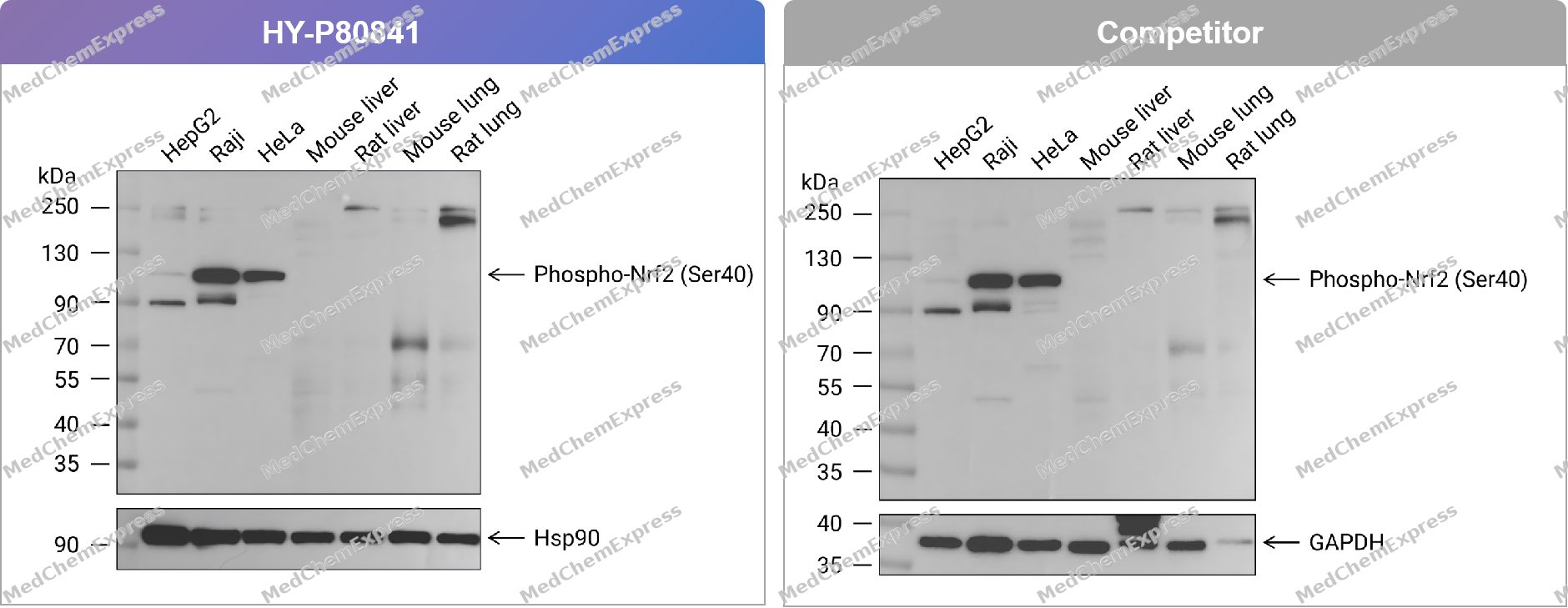 Western blot analysis of extracts from HepG2 (lane 1(20μg)) 、Raji (lane 2(20μg)) 、HeLa (lane 3(20μg)) 、Mouse liver (lane 4(20μg)) 、Rat liver (lane 5(20μg)) 、Mouse lung (lane 6(20μg)) and Rat lung (lane 7(20μg)) using Phospho-Nrf2 (Ser40) Antibody. Proteins were transferred to a PVDF membrane and blocked with 5% BSA in TBST for 1.5 hour at room temperature. The primary antibody (HY-P80481, 1/1000) , competitor's antibody (1/1000) and Loading control antibody (Hsp90, 1/10000; GAPDH, 1/10000) was used in 5% BSA in TBST at 4℃ overnight. Goat Anti-Rabbit IgG-HRP Secondary Antibody (1/10,000) was used for 1 hour at room temperature.
 -
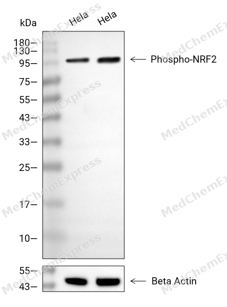 Western blot analysis of extracts from Hela (lane 2(20μg), Hela (lane 3(40μg), using Phospho-Nrf2 Antibody. Proteins were transferred to a PVDF membrane and blocked with 5% BSA in TBST for 2 hour at room temperature. The primary antibody and Loading control antibody (Beta Actin, HY-P80438, 1/3000) was used in 5% BSA in TBST at 4°C overnight. Goat Anti-Mouse/Rabbit IgG-HRP Secondary Antibody (HY-P8004/HY-P8001, 1/10,000) was used for 1 hour at room temperature.
 -
 Western blot analysis of extracts from HepG2 (lane 1(20μg)) 、Raji (lane 2(20μg)) 、HeLa (lane 3(20μg)) 、Mouse liver (lane 4(20μg)) 、Rat liver (lane 5(20μg)) 、Mouse lung (lane 6(20μg)) and Rat lung (lane 7(20μg)) using Phospho-Nrf2 (Ser40) Antibody (HY-P80841) . Proteins were transferred to a PVDF membrane and blocked with 5% BSA in TBST for 1.5 hour at room temperature. The primary antibody (1/1000) and Loading control antibody (Hsp90, 1/10000) was used in 5% BSA in TBST at 4℃ overnight. Goat Anti-Rabbit IgG-HRP Secondary Antibody (1/10,000) was used for 1 hour at room temperature.
 -
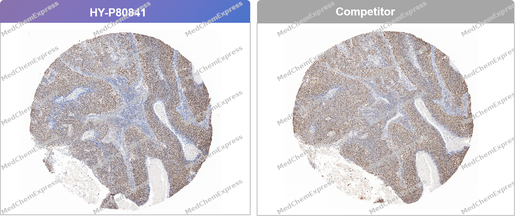 Immunohistochemical analysis of paraffin-embedded human cervical cancer tissue using Phospho-Nrf2 (Ser40) Antibody. The section was pre-treated using heat mediated antigen retrieval with Tris/EDTA buffer (pH 9.0) for 20 minutes. The tissues were probed with the primary antibody (HY-P80841, 1/100) , competitor's antibody (1/100) overnight at 4℃. The detection was performed using an HRP conjugated compact polymer system. DAB was used as the chromogen. Tissues were counterstained with hematoxylin and mounted with neutral balsam.
 -
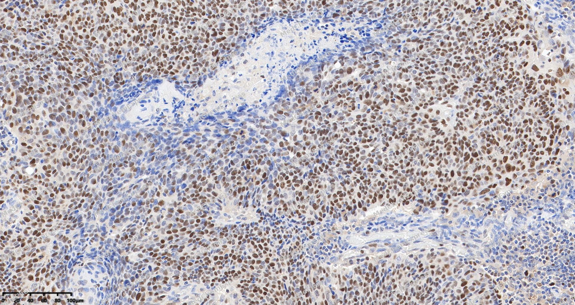 Immunohistochemical analysis of paraffin-embedded human cervical cancer tissue using Phospho-Nrf2 (Ser40) Antibody. The section was pre-treated using heat mediated antigen retrieval with Tris/EDTA buffer (pH 9.0) for 20 minutes. The tissues were probed with the primary antibody (HY-P80841, 1/100) overnight at 4℃. The detection was performed using an HRP conjugated compact polymer system. DAB was used as the chromogen. Tissues were counterstained with hematoxylin and mounted with neutral balsam.
 -
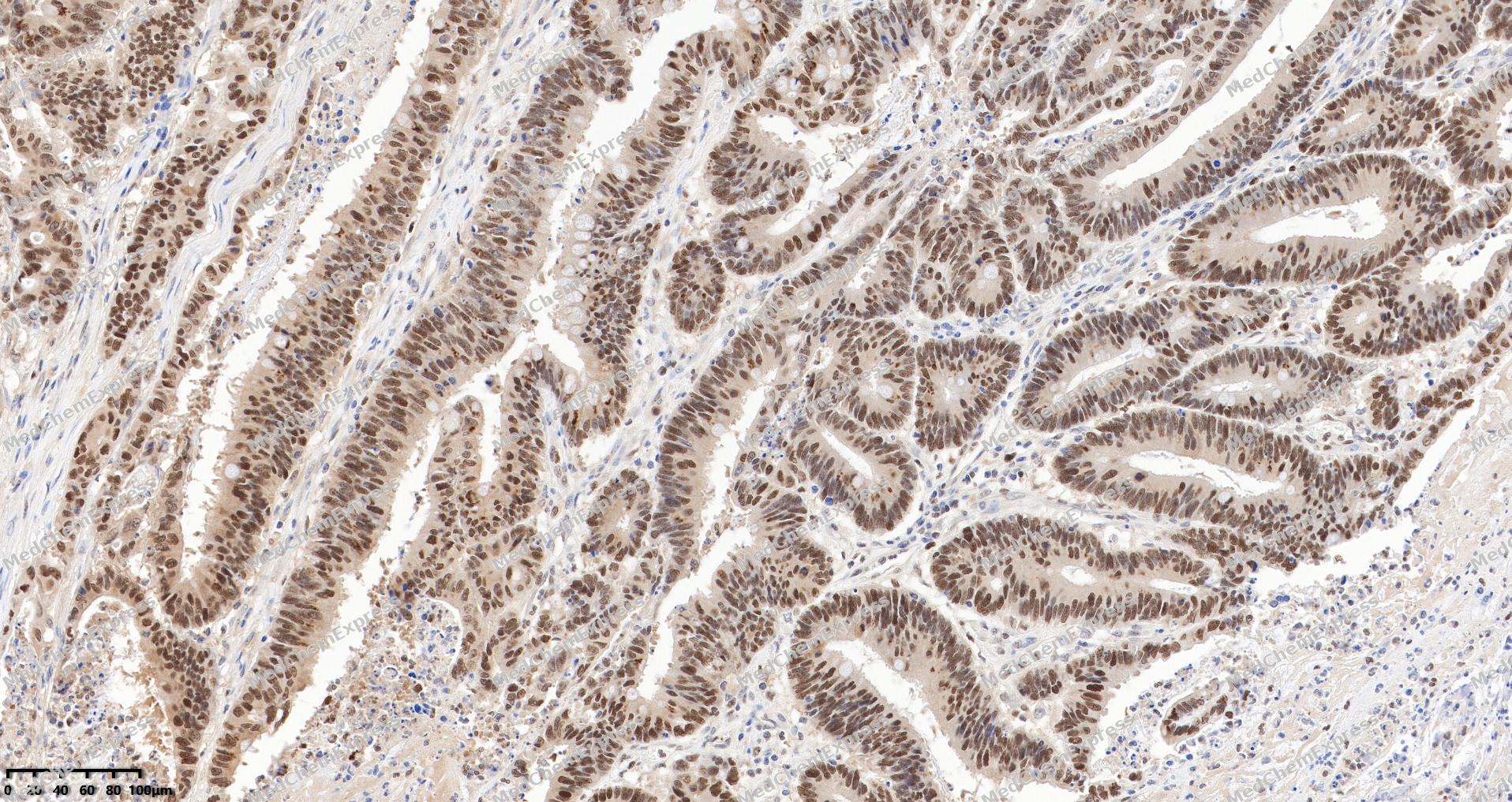 Immunohistochemical analysis of paraffin-embedded human colon cancer tissue using Phospho-Nrf2 (Ser40) Antibody. The section was pre-treated using heat mediated antigen retrieval with Tris/EDTA buffer (pH 9.0) for 20 minutes. The tissues were probed with the primary antibody (HY-P80841, 1/100) overnight at 4℃. The detection was performed using an HRP conjugated compact polymer system. DAB was used as the chromogen. Tissues were counterstained with hematoxylin and mounted with neutral balsam.
 -
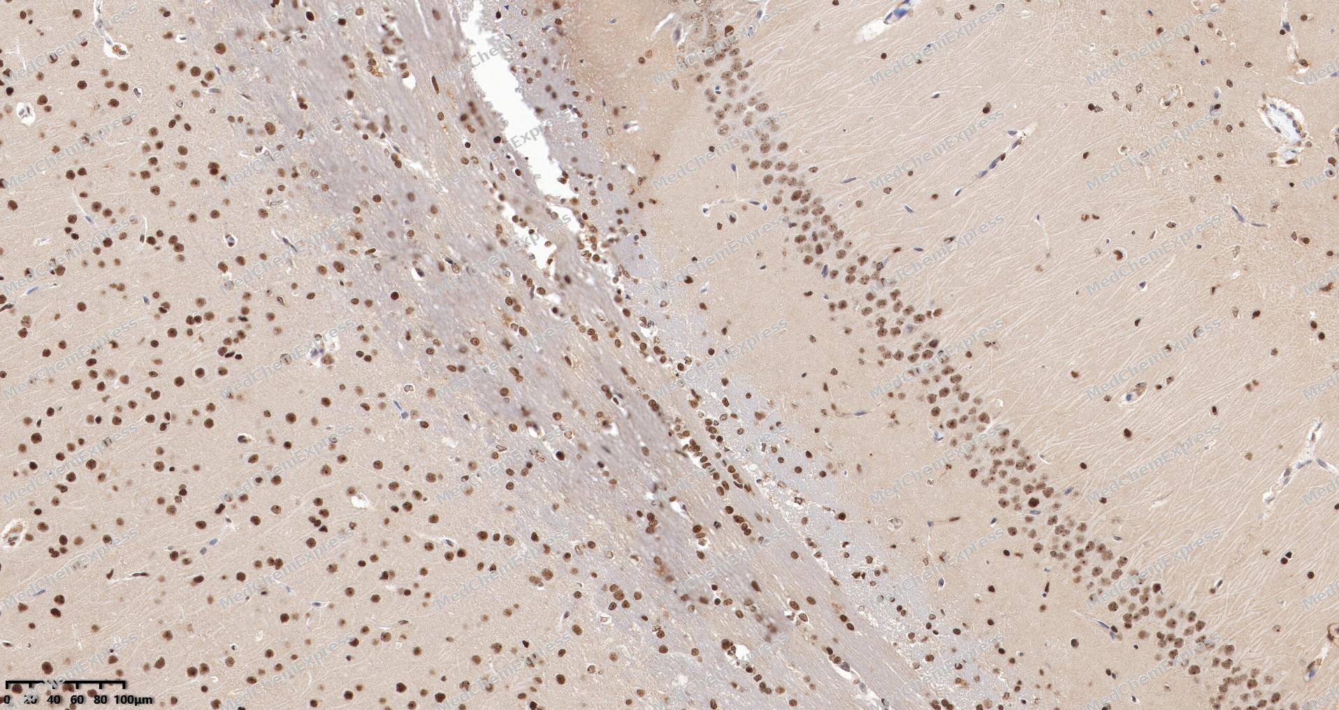 Immunohistochemical analysis of paraffin-embedded rat brain tissue using Phospho-Nrf2 (Ser40) Antibody. The section was pre-treated using heat mediated antigen retrieval with Tris/EDTA buffer (pH 9.0) for 20 minutes. The tissues were probed with the primary antibody (HY-P80841, 1/100) overnight at 4℃. The detection was performed using an HRP conjugated compact polymer system. DAB was used as the chromogen. Tissues were counterstained with hematoxylin and mounted with neutral balsam.
 -
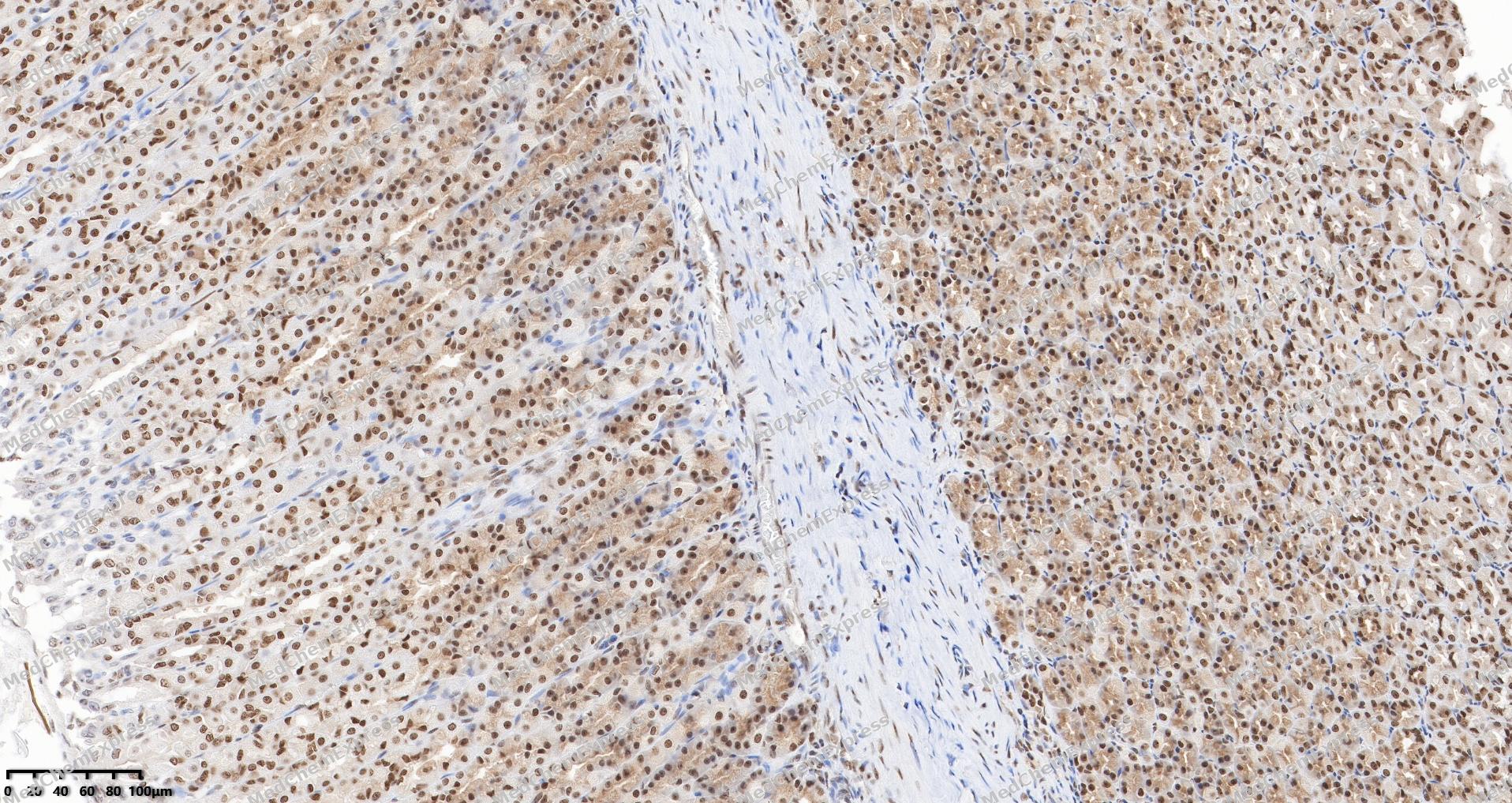 Immunohistochemical analysis of paraffin-embedded rat stomach tissue using Phospho-Nrf2 (Ser40) Antibody. The section was pre-treated using heat mediated antigen retrieval with Tris/EDTA buffer (pH 9.0) for 20 minutes. The tissues were probed with the primary antibody (HY-P80841, 1/100) overnight at 4℃. The detection was performed using an HRP conjugated compact polymer system. DAB was used as the chromogen. Tissues were counterstained with hematoxylin and mounted with neutral balsam.
 -
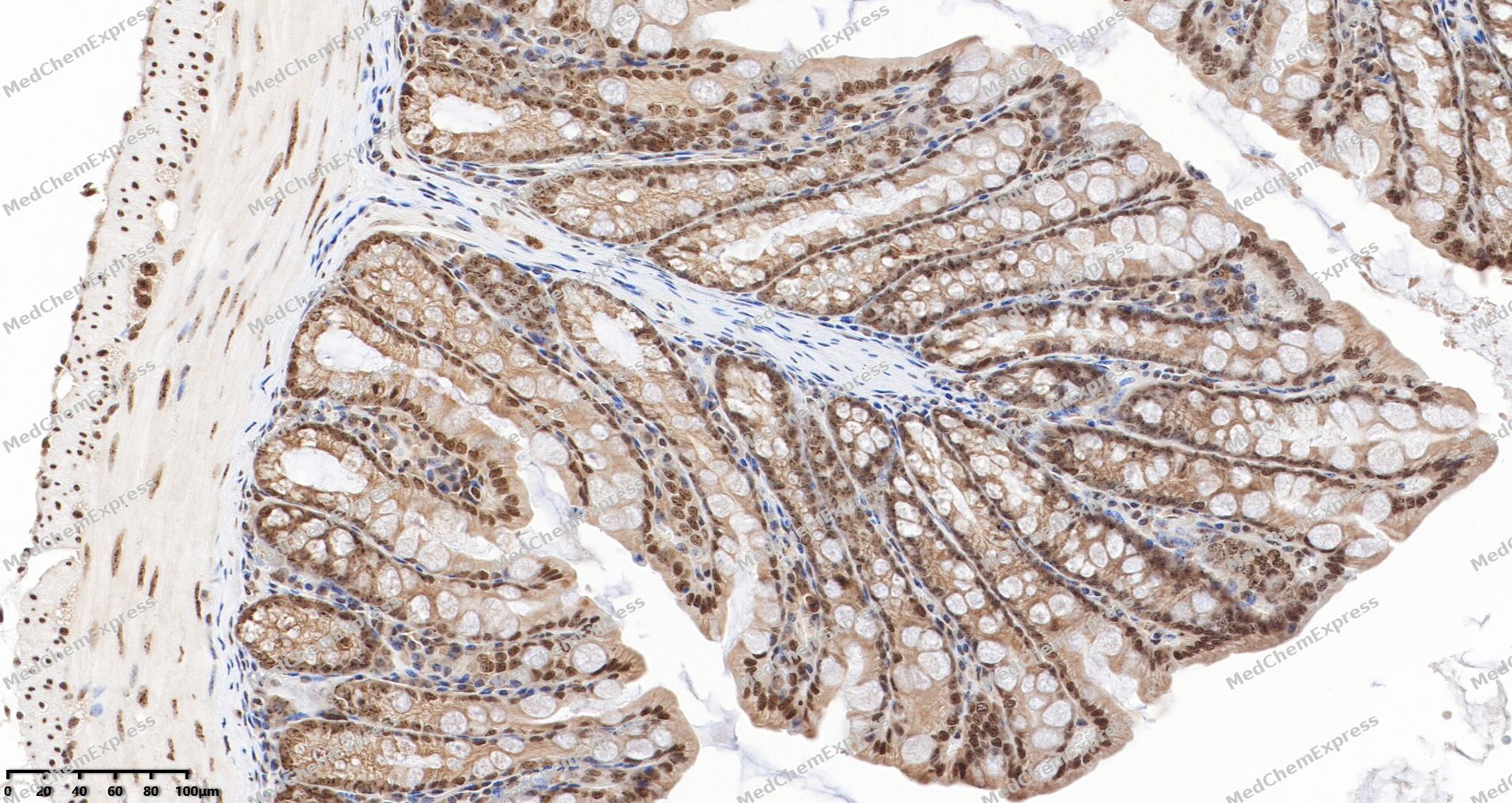 Immunohistochemical analysis of paraffin-embedded mouse colon tissue using Phospho-Nrf2 (Ser40) Antibody. The section was pre-treated using heat mediated antigen retrieval with Tris/EDTA buffer (pH 9.0) for 20 minutes. The tissues were probed with the primary antibody (HY-P80841, 1/100) overnight at 4℃. The detection was performed using an HRP conjugated compact polymer system. DAB was used as the chromogen. Tissues were counterstained with hematoxylin and mounted with neutral balsam.


Background
-
Function
Nrf2 transcription factor that plays a key role in the response to oxidative stress: binds to antioxidant response (ARE) elements present in the promoter region of many cytoprotective genes, such as phase 2 detoxifying enzymes, and promotes their expression, thereby neutralizing reactive electrophiles. In normal conditions, ubiquitinated and degraded in the cytoplasm by the BCR(KEAP1) complex. In response to oxidative stress, electrophile metabolites inhibit activity of the BCR(KEAP1) complex, promoting nuclear accumulation of NFE2L2/NRF2, heterodimerization with one of the small Maf proteins and binding to ARE elements of cytoprotective target genes. The NFE2L2/NRF2 pathway is also activated in response to selective autophagy: autophagy promotes interaction between KEAP1 and SQSTM1/p62 and subsequent inactivation of the BCR(KEAP1) complex, leading to NFE2L2/NRF2 nuclear accumulation and expression of cytoprotective genes. The NFE2L2/NRF2 pathway is also activated during the unfolded protein response (UPR), contributing to redox homeostasis and cell survival following endoplasmic reticulum stress. May also be involved in the transcriptional activation of genes of the beta-globin cluster by mediating enhancer activity of hypersensitive site 2 of the beta-globin locus control region. Also plays an important role in the regulation of the innate immune response and antiviral cytosolic DNA sensing. It is a critical regulator of the innate immune response and survival during sepsis by maintaining redox homeostasis and restraint of the dysregulation of pro-inflammatory signaling pathways like MyD88-dependent and -independent and TNF-alpha signaling. Suppresses macrophage inflammatory response by blocking pro-inflammatory cytokine transcription and the induction of IL6. Binds to the proximity of pro-inflammatory genes in macrophages and inhibits RNA Pol II recruitment. The inhibition is independent of the NRF2-binding motif and reactive oxygen species level. Represses antiviral cytosolic DNA sensing by suppressing the expression of the adapter protein STING1 and decreasing responsiveness to STING1 agonists while increasing susceptibility to infection with DNA viruses. Once activated, limits the release of pro-inflammatory cytokines in response to human coronavirus SARS-CoV-2 infection and to virus-derived ligands through a mechanism that involves inhibition of IRF3 dimerization. Also inhibits both SARS-CoV-2 replication, as well as the replication of several other pathogenic viruses including Herpes Simplex Virus-1 and-2, Vaccinia virus, and Zika virus through a type I interferon (IFN)-independent mechanism[1][2][3][4][5][6][7][8][9][10].
-
Subcellular Localization
Cytoplasm, cytosol; Nucleus
-
Expression
Tissue_specificity:Widely expressed. Highest expression levels were observed in human muscle, kidneys, lungs, liver, and fetal muscle.
Induction:Down-regulated by ENC1 via a proteasomal ubiquitin-independent protein catabolic process -
Isoforms & Post-Translational Modification
Q16236 has 3 isomers: Q16236-1: 67827 Da (predicted); Q16236-2: 66076 Da (predicted); Q16236-3: 65356 Da (predicted).
Ubiquitinated in the cytoplasm by the BCR(KEAP1) E3 ubiquitin ligase complex leading to its degradation (PubMed:15601839, PubMed:15983046, PubMed:19489739). In response to oxidative stress, electrophile metabolites, such as sulforaphane, modify KEAP1, leading to inhibit activity of the BCR(KEAP1) complex, promoting NFE2L2/NRF2 nuclear accumulation and activity (PubMed:19489739, PubMed:29590092). In response to autophagy, the BCR(KEAP1) complex is inactivated (By similarity);Phosphorylated by EIF2AK3/PERK following unfolded protein response (UPR), promoting dissociation from its cytoplasmic inhibitor KEAP1, followed by its translocation into the nucleus (By similarity). Phosphorylation of Ser-40 by PKC in response to oxidative stress dissociates NFE2L2 from its cytoplasmic inhibitor KEAP1, promoting its translocation into the nucleus (By similarity);Acetylation at Lys-596 and Lys-599 increases nuclear localization whereas deacetylation by SIRT1 enhances cytoplasmic presence;Glycation impairs transcription factor activity by preventing heterodimerization with small Maf proteins (PubMed:31398338). Deglycation by FN3K restores activity (PubMed:31398338) -
Subunit
Interacts with KEAP1; the interaction is direct and promotes ubiquitination by the BCR(KEAP1) E3 ubiquitin ligase complex (PubMed:15601839, PubMed:16888629).
-
SwissProt ID
-
Synonyms
NFE2L2; NRF2; Nuclear factor erythroid 2-related factor 2; NF-E2-related factor 2; NFE2-related factor 2; HEBP1; Nuclear factor; erythroid derived 2; like 2
-
Research Field
Epigenetics and Nuclear Signaling


Documentation
-
Data Sheet (262 KB)
-
SDS (252 KB)
- English - EN (252 KB)
- Français - FR (252 KB)
- Deutsch - DE (252 KB)
- Norwegian - NO (252 KB)
- Español - ES (252 KB)
- Swedish - SV (252 KB)
- Italian - IT (252 KB)
- Korean - KR (252 KB)
- Portuguese - PT (252 KB)
-
User Guide for Antibodies (1077 KB)
References
[1]. Huang HC, et al. Regulation of the antioxidant response element by protein kinase C-mediated phosphorylation of NF-E2-related factor 2. Proc Natl Acad Sci U S A. 2000 Nov 7;97(23):12475-80. [Content Brief]
[2]. Eggler AL, et al. Cul3-mediated Nrf2 ubiquitination and antioxidant response element (ARE) activation are dependent on the partial molar volume at position 151 of Keap1. Biochem J. 2009 Jul 29;422(1):171-80. [Content Brief]
[3]. Huppke P, et al. Activating de novo mutations in NFE2L2 encoding NRF2 cause a multisystem disorder. Nat Commun. 2017 Oct 10;8(1):818. [Content Brief]
[4]. Sanghvi VR, et al. The Oncogenic Action of NRF2 Depends on De-glycation by Fructosamine-3-Kinase. Cell. 2019 Aug 8;178(4):807-819.e21. [Content Brief]
[5]. Furukawa M, et al. BTB protein Keap1 targets antioxidant transcription factor Nrf2 for ubiquitination by the Cullin 3-Roc1 ligase. Mol Cell Biol. 2005 Jan;25(1):162-71. [Content Brief]
[6]. Mills EL, et al. Itaconate is an anti-inflammatory metabolite that activates Nrf2 via alkylation of KEAP1. Nature. 2018 Apr 5;556(7699):113-117. [Content Brief]
[7]. Jain A, et al. p62/SQSTM1 is a target gene for transcription factor NRF2 and creates a positive feedback loop by inducing antioxidant response element-driven gene transcription. J Biol Chem. 2010 Jul 16;285(29):22576-91. [Content Brief]
[8]. Moi P, et al. Isolation of NF-E2-related factor 2 (Nrf2), a NF-E2-like basic leucine zipper transcriptional activator that binds to the tandem NF-E2/AP1 repeat of the beta-globin locus control region. Proc Natl Acad Sci U S A. 1994 Oct 11;91(21):9926-30. [Content Brief]
[9]. Olagnier D, et al. Nrf2 negatively regulates STING indicating a link between antiviral sensing and metabolic reprogramming. Nat Commun. 2018 Aug 29;9(1):3506. [Content Brief]
[10]. Olagnier D, et al. SARS-CoV2-mediated suppression of NRF2-signaling reveals potent antiviral and anti-inflammatory activity of 4-octyl-itaconate and dimethyl fumarate. Nat Commun. 2020 Oct 2;11(1):4938. [Content Brief]

 Powered by Bioz
