



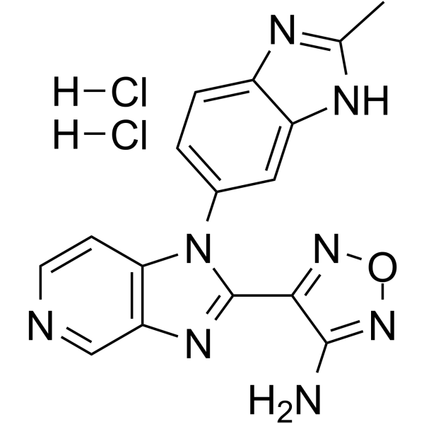
AS2863619 GMP is AS2863619 (HY-126675) produced by using GMP guidelines. GMP small molecules works appropriately as an auxiliary reagent for cell therapy manufacture. AS2863619 is an orally active CDK8/19 inhibitor that also inhibits BMP2, MDA5 and RIG-I receptors. AS2863619 targets Stat5a-CDK8/19 to promote the differentiation of CD4+ T cells into regulatory T cells and induce FOXP3 expression, thereby restoring immune homeostasis and establishing transplant immune tolerance. AS2863619 also enhances the BMP2/SMAD signaling pathway to promote osteogenic differentiation and inhibit adipogenic differentiation. AS2863619 exerts osteoprotective effects by alleviating inflammation-induced impairment of osteogenic function and inducing neutrophil apoptosis (apoptosis). AS2863619 can be applied to research in related fields such as periodontitis-induced bone defects.
For research use only. We do not sell to patients.
- CAS No.: 2241300-51-4
- Formula: C16H14Cl2N8O
- Molecular Weight:405.24
-
Storage:
Please store the product under the recommended conditions in the Certificate of Analysis.


To place orders, for customer services and technical support, please contact: MedChemExpress USA
Tel: 609-228-6898 E-mail: [email protected] [email protected]


-
 Biological Activity
-
Chemical Information
- Protocols
- Purity & Documentation
- References
-
 Help & FAQs




Biological Activity
Description
In Vitro
AS2863619 GMP, combination with IL-2 and TGF-β, enhances the differentiation of primary CD4+ T cells isolated from the spleens of DBA/1 mice into Treg, and its efficacy is superior to that of the regimens using IL-2 alone combined with either TGF-β or AS2863619[1].
AS2863619 GMP (125 nM-2 μM; 7-10 d) enhances osteogenic differentiation of human periodontal ligament stem cells, with concentrations of 250 nM and 500 nM increasing ALP activity, upregulating key osteogenic markers, and accelerating mineralized nodule formation on day 10[2].
AS2863619 GMP (250 nM-500 nM; 7-21 d) inhibits adipogenic differentiation of human periodontal ligament stem cells by reducing lipid droplet formation and downregulating key adipogenic genes[2].
AS2863619 GMP (250 nM-500 nM; 7-10 d) alleviates rhTNF-α-induced osteogenic differentiation impairment of human periodontal ligament stem cells by inhibiting the RIG-I/MDA5 receptor-mediated RLR signaling pathway, and the 500 nM concentration reverses the inflammatory impairment blocked by the RLR agonist Poly (I:C)[2].
Combination of AS2863619 GMP (1 mM/mL; 5 d) with IL-2 and TGF-β1 induces transdifferentiation of allogeneic antigen-specific Balb/c CD4+ T cells into ag-Treg, while upregulating the expression of Treg-specific genes and enriching pathways associated with T cell tolerance induction[3].
AS2863619 GMP (0.01-10 μM; 2 h) significantly increases the apoptosis level of human polymorphonuclear neutrophils (PMNs) stimulated by IC, with a value comparable to that of the negative control group, while it does not significantly reduce cell viability[4].
MedChemExpress (MCE) has not independently confirmed the accuracy of these methods. They are for reference only.
-
Cell Line:human periodontal ligament stem cells (PDLSCs)
-
Concentration:125 nM-2 μM (ALP activity and gene expression); 250 nM-500 nM (ARS staining)
-
Incubation Time:7 days (ALP activity and gene expression); 10 days (ARS staining)
-
Result:Enhanced ALP activity at 125 nM, with stronger effects observed at 500 nM to 2 μM.
Significantly augmented ALP activity, upregulated mRNA and protein expression of osteogenic markers ALP, osteopontin (OPN), and osterix (SP7), and induced mineralized nodule formation within 10 days at 250 nM and 500 nM.
-
Cell Line:human periodontal ligament stem cells (PDLSCs)
-
Concentration:250 nM-500 nM
-
Incubation Time:7 days (gene expression); 21 days (ORO staining)
-
Result:Substantially inhibited lipid droplet formation at 250 nM and 500 nM.
Downregulated mRNA expression of adipogenic markers peroxisome proliferator-activated receptor-gamma (PPAR-γ), lipoprotein lipase (LPL), and fatty acid binding protein 4 (FABP4) at 250 nM and 500 nM.
In Vivo
MedChemExpress (MCE) has not independently confirmed the accuracy of these methods. They are for reference only.
- Phagocytosis Functional Assay
- RNA extraction experimental
- Detection of Apoptosis
- TUNEL staining for apoptotic DNA fragmentation
- Annexin V plus membrane-impermeant dye apoptosis staining
- Mesenchymal stromal/stem cell osteogenic differentiation
- Apoptosis Solutions
- Research Protocol for Inflammation-related Diseases


Chemical Information
-
CAS No. 2241300-51-4
-
Molecular Weight 405.24
-
Formula C16H14Cl2N8O
-
SMILES
NC1=NON=C1C(N2C3=CC=C4N=C(C)NC4=C3)=NC5=C2C=CN=C5.[H]Cl.[H]Cl
-
Shipping
Room temperature in continental US; may vary elsewhere.
-
Storage
Please store the product under the recommended conditions in the Certificate of Analysis.


Purity & Documentation
References
[1]. Fang K, et al. Progress and Future Perspectives of Treg Cell Therapy. Front Biosci (Landmark Ed). 2025;30(11):41250. [Content Brief]
[2]. He H, et al. AS2863619 boosts osteogenesis in periodontal ligament stem cells and mitigates inflammatory impairment. Int Immunopharmacol. 2025;161:115101. [Content Brief]
[3]. Bulygin A, et al. Assessment and Characterization of Induced Alloantigen-Specific Regulatory T Cells Obtained by the Inhibition of CDK8/19 with the AS2863619 Compound. Int J Mol Sci. 2025;26(22):10957. Published 2025 Nov 12. [Content Brief]
[5]. Akamatsu M, et al. Conversion of antigen-specific effector/memory T cells into Foxp3-expressing Treg cells by inhibition of CDK8/19. Sci Immunol. 2019 Oct 25;4(40). pii: eaaw2707. [Content Brief]

Calculators
Concentration (start) × Volume (start) = Concentration (final) × Volume (final)


AS2863619 (GMP) Related Classifications
HY-126675G Related Classifications
 Powered by Bioz