



Reozalimab

Based on 1 Customer Validation
Reozalimab (LY3434172; IBI-318) is an IgG1 monoclonal antibody targeting PD-1/PD-L1, with an IC50 of 2.27 nM and an EC50 of 0.080 nM against human PD-1, as well as an IC50 of 0.75 nM and an EC50 of 0.455 nM against human PD-L1. Reozalimab blocks the binding of PD-1 to PD-L1/PD-L2, as well as the binding of PD-L1 to PD-1/CD80, thereby inhibiting the inhibitory signal transduction of the PD-1 pathway. Reozalimab enhances the functional activation of T cells and induces anti-tumor activity in mouse tumor models reconstituted with human immune cells. Reozalimab can be used in research related to cancers such as non-small cell lung cancer and ovarian cancer.
Nur für Forschungszwecke. Wir verkaufen nicht an Patienten.
- Reinheit : 99.90%
- CAS. Nr.: 2445259-99-2
- Molecular Weight:143.55 kDa
-
Speicherung:
Please store the product under the recommended conditions in the Certificate of Analysis.


To place orders, for customer services and technical support, please contact: MedChemExpress USA
Tel: 609-228-6898 E-mail: [email protected] [email protected]


-
 Biologische Aktivität
-
Chemical Information
- Protokoll
- Reinheit & Dokumentation
- Verweise
-
 Help & FAQs


-
HY-P9902
PembrolizumabPD-1 Inhibitor -

HY-P9904
AtezolizumabAnti-PD-L1 Antibody -
HY-P9971
CamrelizumabPD-1 Neutralizing Antibody -
HY-P9903
NivolumabPD-1 Inhibitor -

HY-P9919
DurvalumabPD-L1 Blocking Antibody -

HY-P99052
TislelizumabPD-1 Inhibitor -

HY-P9978
ToripalimabPD-1 Inhibitor -

HY-108730
AvelumabAnti-PD-L1 Antibody -
HY-P99048
SintilimabPD-1/PD-L1 Inhibitor -
HY-P9919A
Durvalumab (anti-PD-L1)PD-1/PD-L1 Inhibitor -

HY-P99203
CemiplimabPD-1 Inhibitor -
HY-P99118
SerplulimabAnti-PD-1 Antibody


Biologische Aktivität
Beschreibung
Isotype
Ig(G1-kappa_G1-lambda2)
Recommend Isotype Controls
Species Reactivity
Human
IC50 & Target
PDCD1 & CD274
In Vitro
Reozalimab (1 h) blocks the interactions of PD-1/PD-L2 and PD-L1/CD80, with corresponding IC50 values of 2.27 nM and 0.75 nM, respectively, and also inhibits the binding of PD-1/PD-L1[2].
Reozalimab binds to human and cynomolgus monkey PD-1 and PD-L1 with high affinity, but does not bind to mouse or rat PD-1 or PD-L1[2].
Reozalimab (1 h) specifically binds to CHO-K1 cells expressing PD-1 and PD-L1, with corresponding EC50 values of 2.39 nM and 0.589 nM[2].
Reozalimab (6 h) enhances T cell activation in the co-culture system of CHO-K1 cells expressing PD-L1 or co-expressing PD-L1+PD-L2 with NFAT-luc2/PD1 Jurkat cells, with EC50 values of 0.12 nM and 0.18 nM, respectively[2].
Reozalimab (66 h) enhances the activation of naive human CD4 T cells in allogeneic MLR, with an EC50 of 0.01 nM[2].
Reozalimab enhances primary human pan-T cell-mediated killing of HEL 92.1.7 tumor cells, with an EC50 of 0.005 nM[2].
MedChemExpress (MCE) has not independently confirmed the accuracy of these methods. They are for reference only. Further protocols information, click here.
Parmacokinetics
| Species | Dose | Route | CL | T1/2 | Vss |
|---|---|---|---|---|---|
| Mice[2] | 10 mg/kg | i.v. | 0.15 | 293 | 68.1 mL/kg |
In Vivo
Reozalimab (0.0025-0.25 mg/kg; i.p.; once weekly) exhibits significant anti-tumor activity in the prophylactic NCI-H292 NSCLC humanized mouse model[2].
MedChemExpress (MCE) has not independently confirmed the accuracy of these methods. They are for reference only.
-
Animal Model:NOD/SCID Gamma (NSG) (female, 7 weeks of age)[2]
-
Dosage:2 mg/kg; 0.2 mg/kg; 0.02 mg/kg
-
Administration:i.p.; once weekly
-
Result:Achieved a %T/C of 3.9 at 2 mg/kg/wk.
Achieved a %T/C of 19.4 at 0.2 mg/kg/wk.
Achieved a %T/C of 50.3 at 0.02 mg/kg/wk.
Caused no significant body weight changes across all doses.
-
Animal Model:NOD/SCID Gamma (NSG) (female, 7 weeks of age)[2]
-
Dosage:0.25 mg/kg; 0.025 mg/kg; 0.0025 mg/kg
-
Administration:i.p.; once weekly
-
Result:Achieved a %T/C of 6 with 5/8 complete responders (CRs) at 0.25 mg/kg/wk.
Induced 1/8 CRs at 0.025 mg/kg/wk with %T/C not specified.
Induced 0/8 CRs at 0.0025 mg/kg/wk with %T/C not specified.
Was significantly more efficacious than anti-PD-L1 monotherapy and the combination of anti-PD-1 and anti-PD-L1.
Caused no significant body weight changes across all doses.
Clinical Trial
| NCT Number | Sponsor | Condition | Start Date |
Phase

|
|---|---|---|---|---|
| NCT01329991 | Plexxikon| | 2011-05 | PHASE1 |
Gene ID
Accession
Conjugated
Unconjugated
Reconsititution
The product can be reconstituted/diluted with sterile PBS or saline.
Format
-
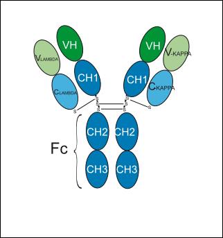
Ig(G1-kappa_G1-lambda2)
Anwendung
ELISA, FACS, Functional assay


Chemical Information
-
CAS. Nr. 2445259-99-2
-
Appearance Liquid
-
Molecular Weight 143.55 kDa
-
Color Colorless to light yellow
-
SMILES
[Reozalimab]
-
Synonyms
LY3434172; IBI-318
-
Versand
Shipping with dry ice.
-
Formulation
Please refer to the lot-specific COA for specific buffer information.
-
Speicherung
Please store the product under the recommended conditions in the Certificate of Analysis.

Protokoll
-
Cell Cytotoxicity Assay
Cytotoxicity assays are usually based on the assessment of cell membrane damage, which can also be indirectly detected by measuring cell viability. Detection methods include MTT assay, CKK-8 assay, LDH assay and ATP assay, etc.


Reinheit & Dokumentation
-
Data Sheet (262 KB)
-
SDS (251 KB)
- English - EN (251 KB)
- Français - FR (251 KB)
- Deutsch - DE (251 KB)
- Norwegian - NO (251 KB)
- Español - ES (251 KB)
- Swedish - SV (251 KB)
- Italian - IT (251 KB)
- Korean - KR (251 KB)
- Portuguese - PT (251 KB)
-
Inhibitory Antibodies User Guide (603 KB)
Verweise

Calculators
Konzentration (Stammlösung) × Volumen (Stammlösung) = Konzentration (Ziellösung) × Volumen (Ziellösung)


Reozalimab Related Classifications
HY-P9996 Related Classifications
 Powered by Bioz