



OAB-14

Based on 1 Customer Validation
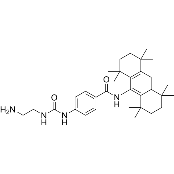
OAB-14, is a Bexarotene (HY-14171) derivative, improves Alzheimer's disease-related pathologies and cognitive impairments by increasing β-amyloid clearance in APP/PS1 mice. OAB-14 effectively ameliorates the dysfunction of the endosomal-autophagic-lysosomal pathway in APP/PS1 transgenic mice.
For research use only. We do not sell to patients.
- Purity : 99.21%
- CAS No.: 2140911-49-3
- Formula: C32H46N4O2
- Molecular Weight:518.73
-
Storage:Powder -20°C, 3 years , 4°C, 2 years ; In solvent -80°C, 6 months , -20°C, 1 month

To place orders, for customer services and technical support, please contact: MedChemExpress USA
Tel: 609-228-6898 E-mail: [email protected] [email protected]


-
 Biological Activity
-
Chemical Information
-
Solvent & Solubility
- Protocols
- Purity & Documentation
- References
-
 Help & FAQs


-
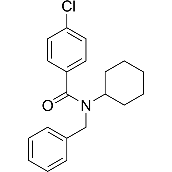
HY-19370
FPS-ZM1Amyloid-β Inhibitor -
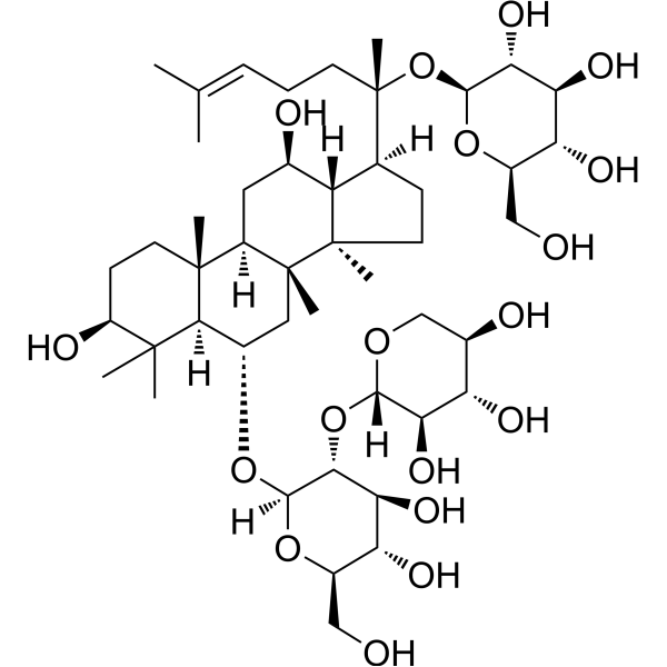
HY-N0615
Notoginsenoside R1Apoptosis Inhibitor -
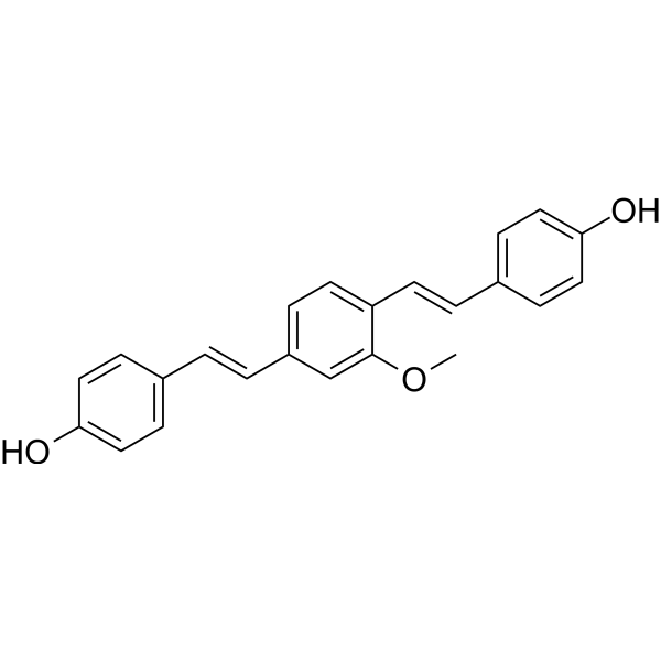
HY-103240
Methoxy-X04Fluorescent Dye -
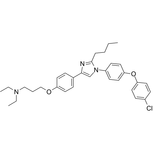
HY-50682
AzeliragonRAGE Inhibitor -
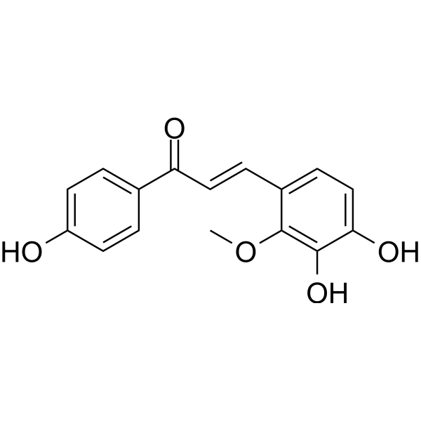
HY-N0373
Licochalcone BApoptosis Inducer -
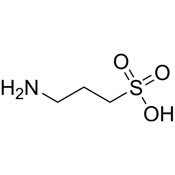
HY-14602
TramiprosateNeuroprotection Agent -
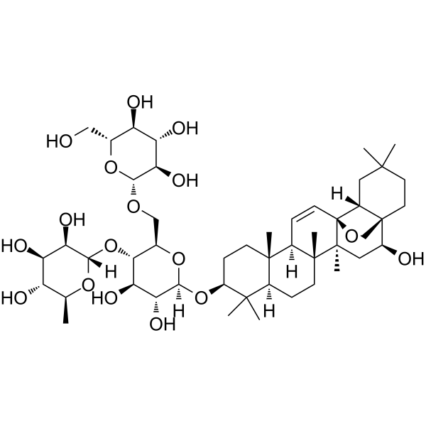
HY-N0249
Saikosaponin CMMP-2 Inducer -
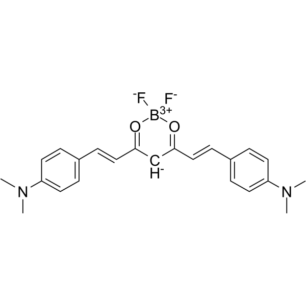
HY-103242
CRANAD-2Fluorescent Probe -
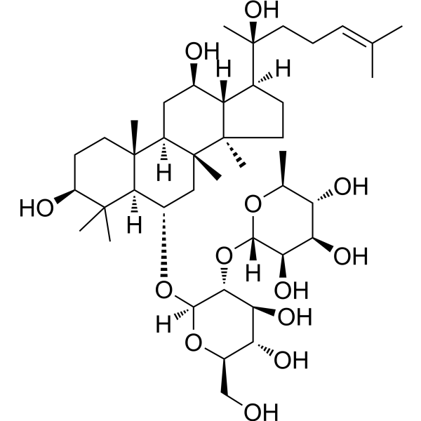
HY-N0602
Ginsenoside Rg2NF-κB Inhibitor -
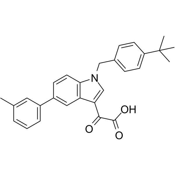
HY-14759
AleplasininPAI-1 Inhibitor -
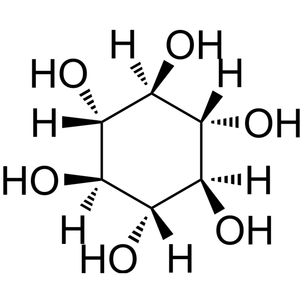
HY-W010041
Scyllo-Inositolα-synuclein/Amyloid-β Aggregation Inhibitor -
HY-18292
ARN2966APP Translation Modulator


Biological Activity
Description
In Vivo
OAB-14 facilitates receptor-mediated endocytosis and restores autophagy flux via the AMPK/mTOR pathway. OAB-14 enhances the lysosomal activity, and reduced Aβ accumulation in lysosomes is observed in OAB-14-treated AD mice[2].
MedChemExpress (MCE) has not independently confirmed the accuracy of these methods. They are for reference only.


Chemical Information
-
CAS No. 2140911-49-3
-
Appearance Solid
-
Molecular Weight 518.73
-
Formula C32H46N4O2
-
Color White to off-white
-
SMILES
O=C(NCCN)NC1=CC=C(C(NC2=C(C(C)(CCC3(C)C)C)C3=CC4=C2C(C)(C)CCC4(C)C)=O)C=C1
-
Shipping
Room temperature in continental US; may vary elsewhere.
-
Storage
Powder -20°C 3 years 4°C 2 years In solvent -80°C 6 months -20°C 1 month


Solvent & Solubility
In Vitro:
DMSO : 20 mg/mL (38.56 mM; ultrasonic and warming and adjust pH to 3 with HCl and heat to 60°C; Hygroscopic DMSO has a significant impact on the solubility of product, please use newly opened DMSO)
Please refer to the solubility information to select the appropriate solvent. Once prepared, please aliquot and store the solution to prevent product inactivation from repeated freeze-thaw cycles.
Storage method and period of stock solution: -80°C, 6 months; -20°C, 1 month. When stored at -80°C, please use it within 6 months. When stored at -20°C, please use it within 1 month.
Please refer to the solubility information to select the appropriate solvent. Once prepared, please aliquot and store the solution to prevent product inactivation from repeated freeze-thaw cycles.
Storage method and period of stock solution: -80°C, 6 months; -20°C, 1 month. When stored at -80°C, please use it within 6 months. When stored at -20°C, please use it within 1 month.
Concentration (start) × Volume (start) = Concentration (final) × Volume (final)

Protocols
-
Protocol for Pharmacokinetic Study
Pharmacokinetic studies quantify how an organism handles a drug over time through absorption, distribution, metabolism, and excretion, and the core experimental readout is the concentration-time profile of parent drug and, when relevant, metabolites in biological matrices such as plasma, whole blood, urine, bile, or tissue. Pharmacokinetic analysis links dose, route, exposure, clearance, half-life, distribution, bioavailability, and systemic exposure to drug efficacy and toxicity hypotheses rather than measuring a signaling pathway directly. The literature links pharmacokinetics to drug-development phenotypes by showing that drug metabolism and pharmacokinetics influence compound progression, exposure-response interpretation, safety margins, dosing strategy, and failure risk during discovery and development. DMPK science contributes to compound optimization by integrating physicochemical properties, in vitro metabolism, transporter behavior, in vivo exposure, and pharmacodynamic contex
-
Autophagy
Autophagy is a process in which eukaryotic cells use lysosomes to degrade their own cytoplasmic proteins and damaged organelles under the regulation of autophagy related gene (Atg). Microtubule-associated proteins light chain 3 (LC3) is recognized as autophagy marker, which transfers from cytoplasmic LC3 (LC3-I) to membrane type (LC3-II). LC3-II/I ratio could be detected by Western Blot and fluorescence microscopy.
-
Lysosome and acidic-vesicle live-cell staining
Lysosome and acidic-vesicle live-cell staining detects acidic intracellular compartments by using membrane-permeant acidotropic probes that accumulate in low-pH vesicles, including lysosomes, late endosomes, autolysosomes, and acidic phagosomes. LysoTracker staining is commonly used as an intensity-based readout of acidic lysosomal compartment abundance or enlargement, while acridine orange produces green fluorescence in less concentrated compartments and red fluorescence after concentration-dependent accumulation in acidic vesicular organelles. Loss or reduction of acridine-orange red signal can be used as a readout of lysosomal membrane permeabilization or reduced acidic-vesicle integrity. This protocol is designed for live cultured cells and can be adapted for fluorescence microscopy, high-content imaging, plate-reader readout, or flow cytometry when the selected literature supports the readout. Because these dyes report acidotropic accumulation rather than lysosome identity alone,
-
Amyloid: Congo Red Amyloid Staining
Congo red amyloid staining is a histochemical method used to detect extracellular amyloid deposits in tissue sections based on the affinity of Congo red dye for β-pleated sheet-rich protein aggregates. When bound to amyloid, Congo red produces characteristic apple-green birefringence under polarized light microscopy, which is widely regarded as a diagnostic feature of amyloid deposition in histopathology. The diagnostic principle relies on the combination of dye binding (congophilia) and optical anisotropy under polarized illumination, which distinguishes amyloid from most non-amyloid eosinophilic extracellular deposits in routine histological evaluation. Amyloid identification by Congo red staining remains a cornerstone in diagnostic pathology despite the availability of adjunct methods such as immunohistochemistry and mass spectrometry, particularly because of its ability to localize deposits directly within tissue architecture. The specificity of Congo red-positive deposits is incre
-
Alzheimer’s Disease Modeling
Alzheimer’s Disease (AD) is a neurodegenerative disorder characterized by a progressive decline in cognitive functions and loss of specific types of neurons and synapses. Alzheimer's symptoms can be simulated in mice by injecting drugs (such as Aβ) or genetically modified.


Purity & Documentation
-
Data Sheet (271 KB)
-
SDS (251 KB)
- English - EN (251 KB)
- Français - FR (251 KB)
- Deutsch - DE (251 KB)
- Norwegian - NO (251 KB)
- Español - ES (251 KB)
- Swedish - SV (251 KB)
- Italian - IT (251 KB)
- Korean - KR (251 KB)
- Portuguese - PT (251 KB)
-
Handling Instructions (2659 KB)
References
[1]. Guo X, et al. OAB-14 Effectively Ameliorates the Dysfunction of the Endosomal-Autophagic-Lysosomal Pathway in APP/PS1 Transgenic Mice. ACS Chem Neurosci. 2021;12(21):3985-3993. [Content Brief]
[2]. Yuan C, et al. OAB-14, a bexarotene derivative, improves Alzheimer's disease-related pathologies and cognitive impairments by increasing β-amyloid clearance in APP/PS1 mice. Biochim Biophys Acta Mol Basis Dis. 2019;1865(1):161-180. [Content Brief]


Complete Stock Solution Preparation Table
Please refer to the solubility information to select the appropriate solvent. Once prepared, please aliquot and store the solution to prevent product inactivation from repeated freeze-thaw cycles.
Storage method and period of stock solution: -80°C, 6 months; -20°C, 1 month. When stored at -80°C, please use it within 6 months. When stored at -20°C, please use it within 1 month.
| Optional Solvent | Concentration Solvent Mass | 1 mg | 5 mg | 10 mg | 25 mg |
|---|---|---|---|---|---|
| DMSO | 1 mM | 1.9278 mL | 9.6389 mL | 19.2779 mL | 48.1946 mL |
| 5 mM | 0.3856 mL | 1.9278 mL | 3.8556 mL | 9.6389 mL | |
| 10 mM | 0.1928 mL | 0.9639 mL | 1.9278 mL | 4.8195 mL | |
| 15 mM | 0.1285 mL | 0.6426 mL | 1.2852 mL | 3.2130 mL | |
| 20 mM | 0.0964 mL | 0.4819 mL | 0.9639 mL | 2.4097 mL | |
| 25 mM | 0.0771 mL | 0.3856 mL | 0.7711 mL | 1.9278 mL | |
| 30 mM | 0.0643 mL | 0.3213 mL | 0.6426 mL | 1.6065 mL |


OAB-14 Related Classifications
HY-139973 Related Classifications
 Powered by Bioz