



Curdione
Based on 2 publication(s) in Google Scholar
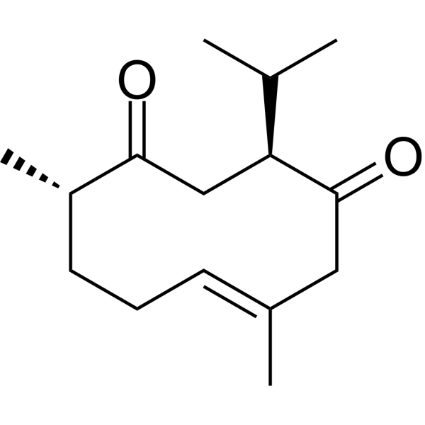
Curdione ((+)-Curdione) is an orally active sesquiterpenoid. Curdione inhibits platelet aggregation. Curdione induces ferroptosis in colorectal cancer via m6A methylation mediated by METTL14 and YTHDF2. Curdione inhibits ferroptosis in Isoproterenol (HY-B0468)-induced myocardial infarction by regulating the Keap1/Trx1/GPX4 signaling pathway, suppressing oxidative stress (ROS) and apoptosis. Curdione ameliorates Doxorubicin (HY-15142)-induced cardiotoxicity by inhibiting oxidative stress (ROS) and activating the Nrf2/HO-1 pathway. Curdione ameliorates sepsis-induced lung injury by inhibiting platelet-mediated neutrophil extracellular trap formation. Curdione ameliorates Bleomycin (HY-17565A)-induced pulmonary fibrosis by inhibiting TGF-β-induced fibroblast-to-myofibroblast differentiation. Curdione exhibits neuroprotective effects against focal cerebral ischemia-reperfusion injury in rats. Curdione exerts antiproliferative effects against human uterine leiomyosarcoma by targeting IDO1. Curdione protects vascular endothelial cells and atherosclerosis by regulating DNMT1-mediated ERBB4 promoter methylation. Curdione inhibits inducible prostaglandin E2 production (IC50 = 1.1 μM) and cyclooxygenase 2 expression.
For research use only. We do not sell to patients.
- Purity: 99.80%
- CAS No.: 13657-68-6
- Formula: C15H24O2
- Molecular Weight:236.35
-
Storage:Powder -20°C, 3 years , 4°C, 2 years ; In solvent -80°C, 2 years , -20°C, 1 year

To place orders, for customer services and technical support, please contact: MedChemExpress USA
Tel: 609-228-6898 E-mail: [email protected] [email protected]


-
 Biological Activity
-
Chemical Information
-
Solvent & Solubility
- Purity & Documentation
- References
-
 Help & FAQs
-

Apoptosis Compound Library
HY-L003
-
Immunology/Inflammation Compound Library
HY-L007
-
Metabolism/Protease Compound Library
HY-L012
-
NF-κB Signaling Compound Library
HY-L014
-
Stem Cell Signaling Compound Library
HY-L017
-
TGF-beta/Smad Compound Library
HY-L018
-
Natural Product Library
HY-L021
-
Anti-Cancer Compound Library
HY-L025
-
Autophagy Compound Library
HY-L029
-
Small Molecule Immuno-Oncology Compound Library
HY-L031
-
Anti-Aging Compound Library
HY-L034
-
Covalent Screening Library
HY-L036
-
Antioxidant Compound Library
HY-L037
-
Oxygen Sensing Compound Library
HY-L045
-
Anti-Cardiovascular Disease Compound Library
HY-L046
-
Ferroptosis Compound Library
HY-L051
-
Medicine Food Homology Compound Library
HY-L055
-
Terpenoids Library
HY-L056
-
Orally Active Compound Library
HY-L061
-
Traditional Chinese Medicine Active Compound Library
HY-L065
-
Neuroprotective Compound Library
HY-L070
-
Anti-Pancreatic Cancer Compound Library
HY-L077
-
Anti-Cancer Metabolism Compound Library
HY-L083
-
Anti-Parkinson's Disease Compound Library
HY-L085
-
Neurodegenerative Disease-related Compound Library
HY-L086
-
Angiogenesis-Related Compound Library
HY-L088
-
Transcription Factor-Targeted Library
HY-L090
-
Food-Sourced Compound Library
HY-L094
-
Anti-Colorectal Cancer Compound Library
HY-L103
-
Anti-Cancer Natural Product Library
HY-L107
-
Anti-inflammatory Traditional Chinese Medicine Active Compound Library
HY-L114
-
Plant-Sourced Natural Product Library
HY-L115
-
Human Metabolite Library
HY-L123
-
Anti-Pulmonary Fibrosis Compound Library
HY-L125
-
Osteogenesis Compound Library
HY-L131
-
Coagulation and Anti-coagulation Compound Library
HY-L136
-
Metabolic Enzyme Compound Library
HY-L146
-
Membrane Protein-targeted Compound Library
HY-L149
-
Membrane Receptor-targeted Compound Library
HY-L150
-
Cysteine Targeted Covalent Library
HY-L153
-
Cell Death Library
HY-L162
-
Multi-Target Compound Library
HY-L176
-
Radioprotector Library
HY-L178
-
Bioactive Compound Library Max
HY-L181
-
MCE Bioactive Compound Library
HY-L001V
-
Covalent Screening Library Plus
HY-L036P
-
Natural Product Library Plus
HY-L021P
-
Natural Product and Natural Product-Like Compound Library
HY-L021M
-
Bioactive Compound Library
HY-L001
-
Miao Ethnicity Medicine Compound Library
HY-L190
-
PBCRBS Traditional Chinese Medicine Compound Library
HY-L193
-
Heat-Clearing and Detoxification Traditional Chinese Medicine Compound Library
HY-L194
-
Non-Alcoholic Fatty Liver Disease (NAFLD) Compound Library
HY-L199
-
RO5 Drug-like Natural Product Library
HY-L200
-
High-Throughput Bioactive Compound Library
HY-L205
-
High-Throughput Natural Product Library
HY-L206
-
Ancient Chinese Classical Formulas Traditional Chinese Medicine Active Compound Library
HY-L209
-
Mass Spectrometry Human Metabolite Library
HY-L215
-
Diarrhea-Related Traditional Chinese Medicine Active Compound Library
HY-L224
-
Mongolian Medicine Compound Library
HY-L238
-
Natural Product Diversity Scaffold Library
HY-L245
-
Lactylation Compound Library
HY-L249
-
Mass Spectrometry Natural Product Library
HY-L262


Publications Citing Use of MedChemExpress (MCE) Curdione
More

Biological Activity
|
GPX4 |
Curdione (12.5-50 μM, 48 h) decreases the cell viability, promotes intracellular ROS production, increases the expression levels of METTL14 and YTHDF2, and decreases the expression levels of SLC7A11, SLC3A2, HOXA13, and GPX4 in CT26 cells[1].
Curdione (12.5-50 μM, 48 h) decreases GSH concentration, increases MDA, m6A, ferrous iron and LPO levels and increases the mRNA levels of SLC7A11 and HOXA13 in CT26 and SW480 cells[1].
Curdione (50 μM, 48 h) reduces cellular ROS concentration, Fe2+ and MDA levels, and increases GSH activity, induces ferroptosis via m6A modification of the XC system and the methylation transferase METTL14 in shRNA-METTL14 CT26 and SW480 cells[1].
Curdione (12.5-50 μM, 48 h) induces cell apoptosis, and its apoptosis inhibitors (Z-VAD-FMK (HY-16658B)) have no effect on ferroptosis in SW480 cells[1].
Curdione (25-100 μM, 24 h) improves the survival rate in Isoproterenol (ISO) (HY-B0468)-induced H9c2 cells, decreases the cell injury in Erastin (HY-15763)-induced H9c2 cells[2].
Curdione (25-100 μM, 24 h) bounds to Keap1, regulates the Keap1/Trx1/GPX4 signaling pathway in H9c2 cells[2].
Curdione (100-200 μM) inhibits neutrophil extracellular trap (NET) formation in neutrophils isolated from mouse bone marrow stimulated with Phorbol 12-myristate 13-acetate (PMA) (HY-18739) or pyogenic platelets[3].
Curdione (160-500 μM, 48 h) does not affect cell viability, inhibits the differentiation of fibroblasts into myofibroblasts, inhibits TGF-β/Smad3 signaling in HPFs[5].
Curdione (0-500 μM, 12-72 h) reduces cell viability, inhibits the proliferation mediated by IDO1 against SK-UT-1 and SK-LMS-1 cells, with IC50s of 327 and 309.9 μM[7].
Curdione (0-100 μM, 24 h) induces G2/M phase arrest, apoptosis and autophagy mediated by IDO1 in SK-UT-1 and SK-LMS-1 cells[7].
Curdione (0-200 μg/mL, 72 h) induces apoptosis, impaires mitochondrial membrane potential occurred in MCF-7 cells (IC50 = 125.632 μg/mL)[9].
MedChemExpress (MCE) has not independently confirmed the accuracy of these methods. They are for reference only.
-
Cell Line:CT26 cells
-
Concentration:12.5 μM, 25 μM, 50 μM
-
Incubation Time:48 h
-
Result:Increased the expression levels of METTL14 and YTHDF2, and decreased the expression levels of SLC7A11, SLC3A2, HOXA13, and GPX4.
-
Cell Line:shRNA-METTL14 CT26 and SW480 cells
-
Concentration:50 μM
-
Incubation Time:48 h
-
Result:Increased expression of SLC7A11, HOXA13, SLC3A2 and GPX4.
-
Cell Line:SW480 cells
-
Concentration:12.5 μM, 25 μM, 50 μM
-
Incubation Time:48 h
-
Result:Inducted apoptosis, promoted the development of ferroptosis.
Had no statistically significant difference of ferroptosis when combined with Z-VAD-FMK (HY-16658B) (10 μM).
-
Cell Line:H9c2 cells
-
Concentration:100 μmol/L
-
Incubation Time:1 h
-
Result:Increased the stability of Keap1.
-
Cell Line:H9c2 cells/ Flag-Keap1, HA-GPX4, and Myc-Trx1 plasmids and tranfected to 293T cells
-
Concentration:25 μM, 50 μM, 100 μM
-
Incubation Time:24 h
-
Result:Inhibited Keap1 expression and increased ISO-induced Trx1 expression, reduced interaction between Keap1 and Trx1 and increased complex between Trx1 and GPX4.
-
Cell Line:HPFs
-
Concentration:160 μM, 300 μM
-
Incubation Time:48 h
-
Result:Reduced fibronectin, collagen 1, and α-SMA levels.
Attenuated TGF-β1-induced p-Smad3 activation, but not p-Smad2.
-
Cell Line:HPFs
-
Concentration:160 μM, 300 μM
-
Incubation Time:48 h
-
Result:Reduced fibronectin, collagen 1, and α-SMA levels, increased Smad7 inhibits p-Smad3
-
Cell Line:HPFs
-
Concentration:160 μM, 300 μM
-
Incubation Time:48 h
-
Result:Reduced fibronectin, collagen 1, and α-SMA levels.
-
Cell Line:SK-UT-1 and SK-LMS-1 cells
-
Concentration:0 μM, 25 μM, 50 μM, 100 μM
-
Incubation Time:24 h
-
Result:Reduced the expression of EdU and Ki67.
-
Cell Line:SK-UT-1 and SK-LMS-1 cells
-
Concentration:0 μM, 25 μM, 50 μM, 100 μM
-
Incubation Time:24 h
-
Result:Induced G2/M phase arrest.
-
Cell Line:SK-UT-1 and SK-LMS-1 cells
-
Concentration:0 μM, 25 μM, 50 μM, 100 μM
-
Incubation Time:24 h
-
Result:Up-regulated the cell cycle checkpoint proteins P21 and CyclinB1 and down-regulated Cdc2.
Increased cleavage of caspase 3, 6, and 9 without affecting caspase 8.
Up-regulateed LC3 and Beclin-1, and down-regulated P62, IDO1.
-
Cell Line:SK-UT-1 and SK-LMS-1 cells
-
Concentration:0 μM, 25 μM, 50 μM, 100 μM
-
Incubation Time:24 h
-
Result:Increased the percentage of early and late apoptotic cells and induced cell death.
-
Cell Line:MCF-7 cells
-
Concentration:0, 25, 50, 100, 150, 200 μg/mL
-
Incubation Time:72 h
-
Result:Induced apoptosis, increased the expressions of Bax, cleaved caspase-3 and caspase-9, decreased Bcl-2 expression.
Curdione (25-100 mg/kg, i.g., once a day, 7 days) attenuates myocardial injury, alleviates ferroptosis via regulating Keap1/Trx1/GPX4 signaling pathway in ISO-induced myocardial infarction male (MI) mice model[2].
Curdione (50-100 mg/kg, i.p., once) improves lung injury, reduce inflammation and oxidative stress levels, inhibit the activation of MAPK kinase and NF-κB P65, lung neutrophil infiltration and NET formation, regulate platelet activation, and reduce the interaction between neutrophils and platelets in the lungin cecal ligation and puncture (CLP) sepsis mice model[3].
Curdione (100 mg/kg, i.p., every 2 days, 21 days) reduces pulmonary fibrosis, expression of fibrosis-specific markers, and inhibits differentiation of fibroblasts into myofibroblasts Bleomycin (BLM) (HY-17565A)-induced Idiopathic pulmonary fibrosis (IPF) mouse model[5].
Curdione (100 mg/kg, i.g., once a day, 7 days) exerts neuroprotective effects against cerebral ischemia/reperfusion-induced brain injury through antioxidant and anti-apoptotic effects middle cerebral artery occlusion (MCAO) SD rats model[6].
Curdione (100-200 mg/kg, i.p., once a day, 21 days) suppresses the growth of uLMS by targeting IDO1 and activating apoptosis and autophagy in SK-UT-1 xenograft model[7].
Curdione (50-150 mg/kg, every two days, 16 days) inhibits tumor growth in MCE-7 xenograft BALB/c nude mice model[9]
MedChemExpress (MCE) has not independently confirmed the accuracy of these methods. They are for reference only.
-
Animal Model:CRC (0.5 × 10⁷, CT26 cells) xenograft nude mice[1]
-
Dosage:50 mg/kg, 100 mg/kg, 200 mg/kg
-
Administration:i.v., once a day, 22 days
-
Result:Suppressed tumor volume and mass, exhibited pronounced ruffling and nuclear chromatin boundary setting, accompanied by necrosis and fibrosis, and decreased cell density.
Increased iron, MDA, and LPO levels, and decreased GSH levels.
Increased protein expression of METTL14, YTHDF2, SLC7A11, and decreased protein expression levels of SLC3A2, HOXA13, and GPX4.
Increased mRNA expression of YTHDF2, SLC7A11, SLC3A2, HOXA13, and PTGS2 and decreased mRNA expression of GPX4.
-
Animal Model:ISO (100 mg/kg)-induced MI C57BL/6 mice (21-25 g, 6-8 weeks) model[2]
-
Dosage:25 mg/kg, 50 mg/kg, 100 mg/kg
-
Administration:i.g., once a day, 7 days
-
Result:Improved ejection fraction (EF), reduced ISO-increased CK-MB levels, restored normal muscle fiber structure, and reduced the degree of inflammatory cell infiltration in cardiac tissue.
Lowered MDA and iron levels and increaseed GSH, GPX4, FTH1 levels.
Inhibited Keap1 expression and increased ISO-induced Trx1 expression.
-
Animal Model:CLP sepsis mice (8-10 weeks old, Male) model[3]
-
Dosage:50 mg/kg, 100 mg/kg
-
Administration:i.p., once
-
Result:Reduced lung congestion, alveolar wall thickening, and inflammatory cell infiltration in lung sections, reduced lung injury scores, pulmonary edema, and reduced exudate protein levels in BALF and lung W/D ratio, as well as LDH activity in BALF.
Reduced CLP-induced increases in DHE and MDA, increased superoxide dismutase (SOD) levels, and decreased IL-1β, IL-6, and TNF-α levels in BALF.
Inhibited the phosphorylation of MAPK kinase and NF-κB P65, and reduced the activation of NF-κB P65.
Reduced CXCL4 and CXCL7 in BALF and reduced platelet activation in lung tissue.
Decreased MPO-positive neutrophils, total cell counts in BALF, and the number of infiltrating neutrophils in BALF.
Reduced fluorescence intensity of platelet activation markers CD42d/GP5, neutrophil marker Ly6G and the formation of NETs in the lungs.
-
Animal Model:BLM-induced IPF mouse (8 weeks old, male) model[5]
-
Dosage:100 mg/kg
-
Administration:i.p., every 2 days, 21 days
-
Result:Reduced lung injury, lung fibrosis, and inflammatory cell (lymphocyte and macrophage) infiltration levels, reduced hydroxyproline content.
Reduced protein and mRNA expression levels of fibronectin, collagen 1, and α-SMA.
Reduced α-SMA positive cells and reduced fibroblast differentiation.
-
Animal Model:MCAO SD rats (240-270 g, adult male) model[6]
-
Dosage:100 mg/kg
-
Administration:i.g., once a day, 7 days
-
Result:Reduced infarct size and neurological deficits and promoted motor function and cognitive function recovery.
Reversed the obvious pathological abnormalities of the MCAO group, including loose arrangement of neurons, nuclear consolidation, loss of color staining or dark color.
Increased the activities of SOD, CAT and GSH-PX, suppressed the increase in the MDA content caused by the injury with cerebral ischemia/reperfusion.
Down-regulated Bax expression and up-regulated Bcl-2 expression, thereby increasing the Bcl-2/Bax ratio, reduced Cyt-C, c-caspase-3, and c-caspase-9 protein levels.
-
Animal Model:SK-UT-1 (1 × 10⁷) xenograft BALB/c nude mice (6-7 weeks, female, 18 g) model[7]
-
Dosage:100 mg/kg, 200 mg/kg
-
Administration:i.p., once a day, 21 days
-
Result:Exhibited anti-uLMS growth efficacy with minimal systemic toxicity, down-regulated IDO1, ki67, and p62, and up-regulated the cleaved caspase-3, Beclin1 and LC3 in tumor tissues.
-
Animal Model:MCE-7 (1 × 10⁷) xenograft BALB/c nude mice (5-6 weeks, female, 20 g) model[9]
-
Dosage:50 mg/kg, 100 mg/kg, 150 mg/kg
-
Administration:every two days, 16 days
-
Result:Inhibited tumor growth.


Chemical Information
-
CAS No. 13657-68-6
-
Appearance Solid
-
Molecular Weight 236.35
-
Formula C15H24O2
-
Color White to off-white
-
SMILES
O=C1C[C@@H](C(C)C)C(C/C(C)=C/CC[C@@H]1C)=O
-
Synonyms
(+)-Curdione
-
Structure Classification
-
Initial Source
-
Shipping
Room temperature in continental US; may vary elsewhere.
-
Storage
Powder -20°C 3 years 4°C 2 years In solvent -80°C 2 years -20°C 1 year


Publications (2)
-
Journal Impact Factor
-
Most Recent
-
Front Cell Dev Biol
Curdione and Schisandrin C Synergistically Reverse Hepatic Fibrosis via Modulating the TGF-β Pathway and Inhibiting Oxidative Stress. [Abstract]2021 Nov 10;9:763864. PMID: 34858986 -
Tohoku J Exp Med
2025 Nov 13. PMID: 41224330


Solvent & Solubility
DMSO : 100 mg/mL (423.10 mM; Need ultrasonic; Hygroscopic DMSO has a significant impact on the solubility of product, please use newly opened DMSO)
Please refer to the solubility information to select the appropriate solvent. Once prepared, please aliquot and store the solution to prevent product inactivation from repeated freeze-thaw cycles.
Storage method and period of stock solution: -80°C, 2 years; -20°C, 1 year. When stored at -80°C, please use it within 2 years. When stored at -20°C, please use it within 1 year.
Please refer to the solubility information to select the appropriate solvent. Once prepared, please aliquot and store the solution to prevent product inactivation from repeated freeze-thaw cycles.
Storage method and period of stock solution: -80°C, 2 years; -20°C, 1 year. When stored at -80°C, please use it within 2 years. When stored at -20°C, please use it within 1 year.
Concentration (start) × Volume (start) = Concentration (final) × Volume (final)
Select the appropriate dissolution method based on your experimental animal and administration route.
- For the following dissolution methods, please ensure to first prepare a clear stock solution using an In Vitro approach and then sequentially add co-solvents:
- To ensure reliable experimental results, the clarified stock solution can be appropriately stored based on storage conditions. As for the working solution for In Vivo experiments, it is recommended to prepare freshly and use it on the same day.
- The percentages shown for the solvents indicate their volumetric ratio in the final prepared solution. If precipitation or phase separation occurs during preparation, heat and/or sonication can be used to aid dissolution.
Add each solvent one by one: 10% DMSO 40% PEG300 5% Tween-80 45% Saline
Solubility: ≥ 2.75 mg/mL (11.64 mM); Clear solution
This protocol yields a clear solution of ≥ 2.75 mg/mL (saturation unknown).
Taking 1 mL working solution as an example, add 100 μL DMSO stock solution (27.5 mg/mL) to 400 μL PEG300, and mix evenly; then add 50 μL Tween-80 and mix evenly; then add 450 μL Saline to adjust the volume to 1 mL.
Preparation of Saline: Dissolve 0.9 g sodium chloride in ddH₂O and dilute to 100 mL to obtain a clear Saline solution.
Add each solvent one by one: 10% DMSO 90% (20% SBE-β-CD in Saline)
Solubility: ≥ 2.75 mg/mL (11.64 mM); Clear solution
This protocol yields a clear solution of ≥ 2.75 mg/mL (saturation unknown).
Taking 1 mL working solution as an example, add 100 μL DMSO stock solution (27.5 mg/mL) to 900 μL 20% SBE-β-CD in Saline, and mix evenly.
Preparation of 20% SBE-β-CD in Saline (4°C, storage for one week): 2 g SBE-β-CD powder is dissolved in 10 mL Saline, completely dissolve until clear.
Please enter the basic information of animal experiments:
-
-
-
-
Recommended: Prepare an additional quantity of animals to account for potential losses during experiments.
Please enter your animal formula composition:
-
%DMSO +
Recommended: Keep the proportion of DMSO in working solution below 2% if your animal is weak.
-
%+
-
+%Tween-80 + +
-
%Saline +
The co-solvents required include: DMSO, . All of co-solvents are available by MedChemExpress (MCE). , Tween 80. All of co-solvents are available by MedChemExpress (MCE).
Working solution concentration: 0.22 mg/mL
Method for preparing stock solution: mg drug dissolved in μL DMSO. Stock solution concentration: mg/mL.
1. Take μL DMSO stock solution;
2. Add μL .
μL , mix evenly;
3. Then add μL Tween 80, mix evenly;
4. Then add μL
Please ensure that the stock solution in the first step is dissolved to a clear state, and add co-solvents in sequence. You can use ultrasonic heating (ultrasonic cleaner, recommended frequency 20-40 kHz), vortexing, etc. to assist dissolution.


Purity & Documentation
-
Data Sheet (303 KB)
-
SDS (396 KB)
- English - EN (396 KB)
- Français - FR (396 KB)
- Deutsch - DE (396 KB)
- Norwegian - NO (396 KB)
- Español - ES (396 KB)
- Swedish - SV (396 KB)
- Italian - IT (396 KB)
- Korean - KR (396 KB)
- Portuguese - PT (396 KB)
-
Handling Instructions (2659 KB)
References
[1]. Wang F, et al. Curdione induces ferroptosis mediated by m6A methylation via METTL14 and YTHDF2 in colorectal cancer. Chin Med. 2023 Sep 21;18(1):122. [Content Brief]
[2]. Wang H, et al. Curdione inhibits ferroptosis in isoprenaline-induced myocardial infarction via regulating Keap1/Trx1/GPX4 signaling pathway. Phytother Res. 2023 Nov;37(11):5328-5340. [Content Brief]
[3]. Yang K, et al. Curdione ameliorates sepsis-induced lung injury by inhibiting platelet-mediated neutrophil extracellular trap formation. Int Immunopharmacol. 2023 May;118:110082. [Content Brief]
[4]. Ma Y, et al. Curdione Relieved Isoproterenol-Induced Myocardial Damage through Inhibiting Oxidative Stress and Apoptosis. Am J Chin Med. 2023;51(1):73-89. [Content Brief]
[5]. Liu P, et al. Curdione ameliorates bleomycin-induced pulmonary fibrosis by repressing TGF-β-induced fibroblast to myofibroblast differentiation. Respir Res. 2020 Feb 19;21(1):58. [Content Brief]
[6]. Li XJ, et al.Neuroprotective effects of curdione against focal cerebral ischemia reperfusion injury in rats. Neuropsychiatr Dis Treat. 2017 Jun 30;13:1733-1740. [Content Brief]
[7]. Wei C, et al. Curdione Induces Antiproliferation Effect on Human Uterine Leiomyosarcoma via Targeting IDO1. Front Oncol. 2021 Feb 26;11:637024. [Content Brief]
[8]. Wu Z, Zai W, Chen W, Han Y, Jin X, Liu H. Curdione Ameliorated Doxorubicin-Induced Cardiotoxicity Through Suppressing Oxidative Stress and Activating Nrf2/HO-1 Pathway. J Cardiovasc Pharmacol. 2019 Aug;74(2):118-127. [Content Brief]
[9]. Li J, et al. Curdione inhibits proliferation of MCF-7 cells by inducing apoptosis. Asian Pac J Cancer Prev. 2014;15(22):9997-10001. [Content Brief]
[10]. Xia Q, Wang X, Xu DJ, Chen XH, Chen FH. Inhibition of platelet aggregation by curdione from Curcuma wenyujin essential Oil. Thromb Res. 2012 Sep;130(3):409-14. [Content Brief]
[11]. Wu Y, et al. Curdione protects vascular endothelial cells and atherosclerosis via the regulation of DNMT1-mediated ERBB4 promoter methylation. Open Med (Wars). 2025 Jul 14;20(1):20251223. [Content Brief]
[12]. Oh OJ, et al. Inhibition of inducible prostaglandin E2 production and cyclooxygenase-2 expression by curdione from Curcuma zedoaria. Arch Pharm Res. 2007 Oct;30(10):1236-9. [Content Brief]


Complete Stock Solution Preparation Table
Please refer to the solubility information to select the appropriate solvent. Once prepared, please aliquot and store the solution to prevent product inactivation from repeated freeze-thaw cycles.
Storage method and period of stock solution: -80°C, 2 years; -20°C, 1 year. When stored at -80°C, please use it within 2 years. When stored at -20°C, please use it within 1 year.
| Optional Solvent | Concentration Solvent Mass | 1 mg | 5 mg | 10 mg | 25 mg |
|---|---|---|---|---|---|
| DMSO | 1 mM | 4.2310 mL | 21.1551 mL | 42.3101 mL | 105.7753 mL |
| 5 mM | 0.8462 mL | 4.2310 mL | 8.4620 mL | 21.1551 mL | |
| 10 mM | 0.4231 mL | 2.1155 mL | 4.2310 mL | 10.5775 mL | |
| 15 mM | 0.2821 mL | 1.4103 mL | 2.8207 mL | 7.0517 mL | |
| 20 mM | 0.2116 mL | 1.0578 mL | 2.1155 mL | 5.2888 mL | |
| 25 mM | 0.1692 mL | 0.8462 mL | 1.6924 mL | 4.2310 mL | |
| 30 mM | 0.1410 mL | 0.7052 mL | 1.4103 mL | 3.5258 mL | |
| 40 mM | 0.1058 mL | 0.5289 mL | 1.0578 mL | 2.6444 mL | |
| 50 mM | 0.0846 mL | 0.4231 mL | 0.8462 mL | 2.1155 mL | |
| 60 mM | 0.0705 mL | 0.3526 mL | 0.7052 mL | 1.7629 mL | |
| 80 mM | 0.0529 mL | 0.2644 mL | 0.5289 mL | 1.3222 mL | |
| 100 mM | 0.0423 mL | 0.2116 mL | 0.4231 mL | 1.0578 mL |


Curdione Related Classifications
HY-N0353 Related Classifications
 Powered by Bioz
- Curdione
- 13657-68-6
- (+)-Curdione
- Ferroptosis
- Apoptosis
- Reactive Oxygen Species (ROS)
- Autophagy
- Glutathione Peroxidase
- Keap1-Nrf2
- Heme Oxygenase (HO)
- TGF-β Receptor
- Indoleamine 2,3-Dioxygenase (IDO)
- apoptosis
- MCF-7 cells
- CT26 cells
- SW480 cells
- H9c2 cells
- HPFs
- SK-UT-1 cells
- SK-LMS-1 cells
- MI
- IPF
- MCAO
- Inhibitor
- inhibitor
- inhibit