



Copper tripeptide
Based on 1 publication(s) in Google Scholar
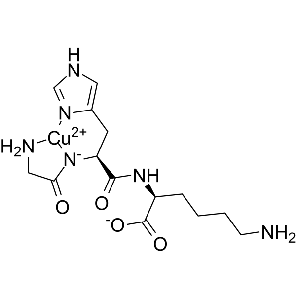
Copper tripeptide (GHK-Cu) is a tripeptide. During wound healing, Copper tripeptide may be freed from existing extracellular proteins via proteolysis and serves as a chemoattractant for inflammatory and endothelial cells. Copper tripeptide has been shown to increase messenger RNA production for collagen, elastin, proteoglycans, and glycosaminoglycans in fibroblasts. Copper tripeptide is a natural modulator of multiple cllular pathways in skin regeneration.
For research use only. We do not sell to patients.
- Purity : 99.94%
- CAS No.: 89030-95-5
- Formula: C14H22CuN6O4
- Molecular Weight:401.91
-
Storage:
Sealed storage, away from moisture and light.
Powder -80°C, 2 years , -20°C, 1 year* In solvent : -80°C, 6 months; -20°C, 1 month (sealed storage, away from moisture and light)

To place orders, for customer services and technical support, please contact: MedChemExpress USA
Tel: 609-228-6898 E-mail: [email protected] [email protected]


-
 Biological Activity
-
Chemical Information
-
Solvent & Solubility
- Protocols
- Purity & Documentation
- References
-
 Help & FAQs


-
HY-105174
BPC 157Gastric Pentadecapeptide -
HY-P0170
TB500Thymosin β 4 Synthetic Molecule -
HY-W013494
L-CarnosineFree Radical Scavenger -

HY-W013494R
L-Carnosine (Standard) -
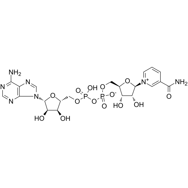
HY-B0445
NAD+Endogenous Metabolite -

HY-10583
Y-27632 dihydrochlorideROCK Inhibitor -

HY-P3506
RetatrutideGCGR/GIPR/GLP-1R Agonist -
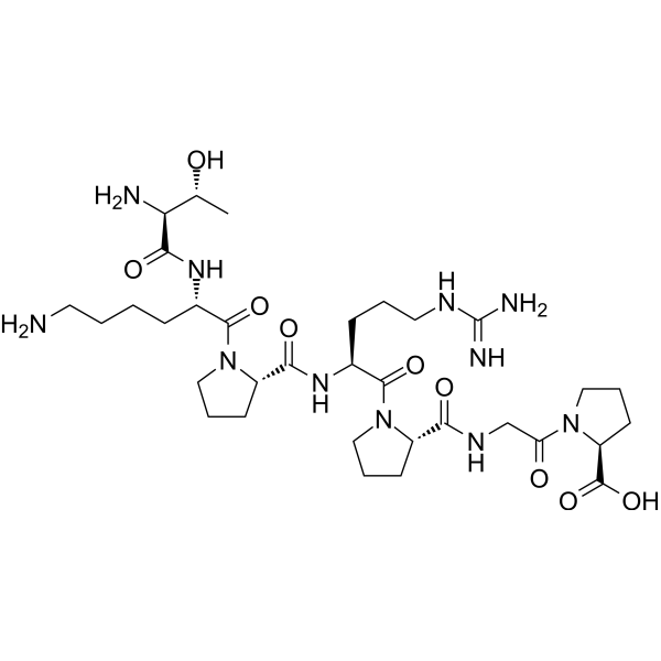
HY-105042
SelankGABAA receptor Modulator -

HY-B0215
AcetylcysteineMucolytic Agent -
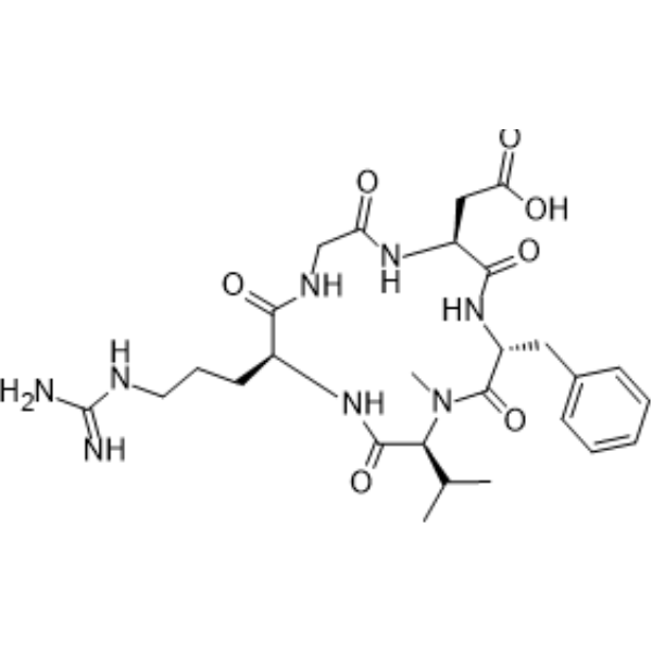
HY-16141
CilengitideIntegrin Inhibitor -
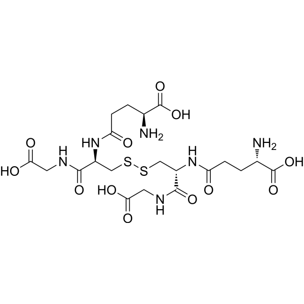
HY-D0844
Glutathione oxidizedAntioxidant -
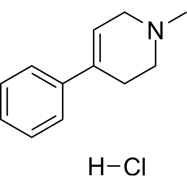
HY-15608
MPTP hydrochlorideDopaminergic Neurotoxin
Publications Citing Use of MedChemExpress (MCE) Copper tripeptide
More

Biological Activity
Description
In Vitro
Copper tripeptide (1 nM; 0-96 hours) affects irradiated fibroblasts such that their population-doubling times approximated that of controls[1]. Copper tripeptide (1 nM; 0-120 hours) significantly produces more basic fibroblast growth factors than normal controls at a 24-hour interval of irradiated fibroblasts[1].
MedChemExpress (MCE) has not independently confirmed the accuracy of these methods. They are for reference only. Further protocols information, click here.
Clinical Trial
| NCT Number | Sponsor | Condition | Start Date |
Phase

|
|---|---|---|---|---|
| NCT01329991 | Plexxikon| | 2011-05 | PHASE1 |


Chemical Information
-
CAS No. 89030-95-5
-
Appearance Solid
-
Molecular Weight 401.91
-
Formula C14H22CuN6O4
-
Color Blue to purple
-
Synonyms
GHK-Cu
-
Sequence
Gly-His-Lys-Cu
-
Sequence Shortening
GHK-Cu
-
Structure Classification
-
Initial Source
-
Shipping
Room temperature in continental US; may vary elsewhere.
-
Storage
Sealed storage, away from moisture and light
Powder -80°C 2 years -20°C 1 year * In solvent : -80°C, 6 months; -20°C, 1 month (sealed storage, away from moisture and light)


Publications (1)
-
Journal Impact Factor
-
Most Recent
-
J Appl Toxicol
Copper induces oxidative stress and apoptosis of hippocampal neuron via pCREB/BDNF/ and Nrf2/HO-1/NQO1 pathway. [Abstract]2022 Apr;42(4):694-705. PMID: 34676557


Solvent & Solubility
In Vitro:
H2O : 50 mg/mL (124.41 mM; Need ultrasonic)
Please refer to the solubility information to select the appropriate solvent. Once prepared, please aliquot and store the solution to prevent product inactivation from repeated freeze-thaw cycles.
Storage method and period of stock solution: -80°C, 6 months; -20°C, 1 month (sealed storage, away from moisture and light). When stored at -80°C, please use it within 6 months. When stored at -20°C, please use it within 1 month.
* Note: If you choose water as the stock solution, please dilute it to the working solution, then filter and sterilize it with a 0.22 μm filter before use.
Please refer to the solubility information to select the appropriate solvent. Once prepared, please aliquot and store the solution to prevent product inactivation from repeated freeze-thaw cycles.
Storage method and period of stock solution: -80°C, 6 months; -20°C, 1 month (sealed storage, away from moisture and light). When stored at -80°C, please use it within 6 months. When stored at -20°C, please use it within 1 month.
* Note: If you choose water as the stock solution, please dilute it to the working solution, then filter and sterilize it with a 0.22 μm filter before use.
Concentration (start) × Volume (start) = Concentration (final) × Volume (final)

Protocols
-
Cell migration
Cell migration is a method that plays an important role in wound healing, cell differentiation, embryonic development, etc.
-
Endothelial Tube Formation Assay
Endothelial tube formation assay evaluates the ability of endothelial cells to attach, migrate, align, and organize into capillary-like networks when cultured on gelled basement membrane extract or Matrigel; the readout is the morphology and quantity of tube-like networks, which reflects an in vitro endothelial morphogenesis step related to angiogenesis. Basement membrane extract/Matrigel provides laminin-rich extracellular matrix cues that support endothelial differentiation into capillary-like structures, but it can contain biologically active growth factors, so growth-factor-reduced matrix is preferred when testing defined angiogenic stimulators or inhibitors.
-
Fibers: Elastic Fiber Staining
Elastic fiber staining is a histochemical technique used to selectively visualize elastin-rich structures such as elastic fibers in connective tissues (e. g. , blood vessels, lung, dermis) based on the affinity of specific dyes or oxidation products for elastin-associated amino acid residues and cross-linked elastic matrix components. Classical methods such as Verhoeff-Van Gieson (VVG), resorcin-fuchsin (Weigert-type stains), or aldehyde fuchsin rely on differential binding of dye complexes to elastic fibers, allowing them to be distinguished from collagen and other extracellular matrix components by contrast staining (typically black or deep purple elastic fibers against red collagen counterstain). These methods are widely used in histopathology to evaluate elastic fiber integrity, fragmentation, or remodeling in vascular diseases, pulmonary pathology, and connective tissue disorders.
-
Cell-Exclusion Zone Migration Assay
The Cell-Exclusion Zone (CEZ) migration assay is an in vitro 2D cell migration method in which a defined cell-free area is created using removable physical barriers such as silicone stoppers, allowing cells to be seeded around the barrier and subsequently migrate into the cleared zone after barrier removal. This approach enables quantification of collective cell migration by monitoring repopulation of the initially cell-free region over time using microscopy-based imaging. Compared with scratch-based wound healing assays, barrier-based exclusion methods are designed to avoid mechanical damage to the extracellular matrix and reduce injury-induced effects on boundary cells, thereby improving interpretability of migration behavior in vitro. The assay readout is typically the progressive reduction in the cell-free area or the number of cells invading the exclusion zone, reflecting coordinated cell motility relevant to physiological processes such as wound healing, epithelial repair, and ca


Purity & Documentation
-
Data Sheet (274 KB)
-
SDS (396 KB)
- English - EN (396 KB)
- Français - FR (396 KB)
- Deutsch - DE (396 KB)
- Norwegian - NO (396 KB)
- Español - ES (396 KB)
- Swedish - SV (396 KB)
- Italian - IT (396 KB)
- Korean - KR (396 KB)
- Portuguese - PT (396 KB)
-
Handling Instructions (2659 KB)
References


Complete Stock Solution Preparation Table
Please refer to the solubility information to select the appropriate solvent. Once prepared, please aliquot and store the solution to prevent product inactivation from repeated freeze-thaw cycles.
Storage method and period of stock solution: -80°C, 6 months; -20°C, 1 month (sealed storage, away from moisture and light). When stored at -80°C, please use it within 6 months. When stored at -20°C, please use it within 1 month.
| Optional Solvent | Concentration Solvent Mass | 1 mg | 5 mg | 10 mg | 25 mg |
|---|---|---|---|---|---|
| H2O | 1 mM | 2.4881 mL | 12.4406 mL | 24.8812 mL | 62.2030 mL |
| 5 mM | 0.4976 mL | 2.4881 mL | 4.9762 mL | 12.4406 mL | |
| 10 mM | 0.2488 mL | 1.2441 mL | 2.4881 mL | 6.2203 mL | |
| 15 mM | 0.1659 mL | 0.8294 mL | 1.6587 mL | 4.1469 mL | |
| 20 mM | 0.1244 mL | 0.6220 mL | 1.2441 mL | 3.1101 mL | |
| 25 mM | 0.0995 mL | 0.4976 mL | 0.9952 mL | 2.4881 mL | |
| 30 mM | 0.0829 mL | 0.4147 mL | 0.8294 mL | 2.0734 mL | |
| 40 mM | 0.0622 mL | 0.3110 mL | 0.6220 mL | 1.5551 mL | |
| 50 mM | 0.0498 mL | 0.2488 mL | 0.4976 mL | 1.2441 mL | |
| 60 mM | 0.0415 mL | 0.2073 mL | 0.4147 mL | 1.0367 mL | |
| 80 mM | 0.0311 mL | 0.1555 mL | 0.3110 mL | 0.7775 mL | |
| 100 mM | 0.0249 mL | 0.1244 mL | 0.2488 mL | 0.6220 mL |
* Note: If you choose water as the stock solution, please dilute it to the working solution, then filter and sterilize it with a 0.22 μm filter before use.


Copper tripeptide Related Classifications
HY-P0063 Related Classifications
 Powered by Bioz