



UNC2383

Based on 1 Customer Validation
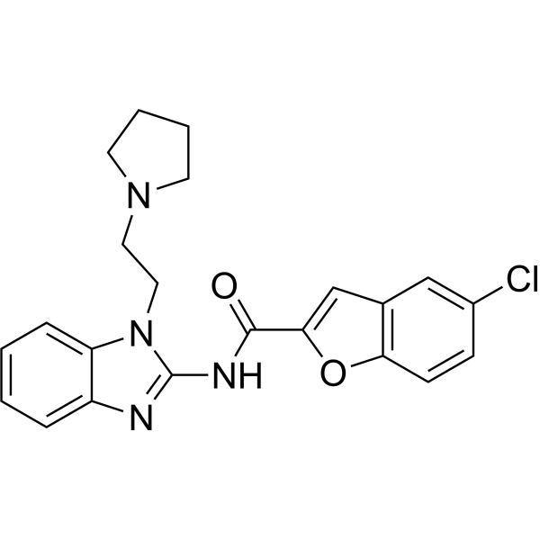
UNC2383 is an oligonucleotide enhancer compound. UNC2383 can enhance the efficacy of antisense oligonucleotides (ASOs) and splice-switching oligonucleotides (SSOs). UNC2383 can be used in research of diseases involving impaired oligonucleotide delivery, such as cystic fibrosis.
For research use only. We do not sell to patients.
- Purity : 98.68%
- CAS No.: 2123481-48-9
- Formula: C22H21ClN4O2
- Molecular Weight:408.88
-
Storage:Powder -20°C, 3 years , 4°C, 2 years ; In solvent -80°C, 6 months , -20°C, 1 month

To place orders, for customer services and technical support, please contact: MedChemExpress USA
Tel: 609-228-6898 E-mail: [email protected] [email protected]


-
 Biological Activity
-
Chemical Information
-
Solvent & Solubility
- Protocols
- Purity & Documentation
- References
-
 Help & FAQs




Biological Activity
Description
In Vitro
UNC2383 (up to 10 μM) enhances SSO effects in HeLa Luc 705 cells[1].
UNC2383 (10 μM; 2 h) post-treatment combined with anti-MDR1 ASO pre-treatment results in a significant decrease in Pgp expression in NIH-3T3-MDR cells[1].
UNC2383 (1.25-10 μM; 2 h) induces a large number of small mCherry-GAL9 structures in Huh7 mCherry-GAL9 cells[2].
MedChemExpress (MCE) has not independently confirmed the accuracy of these methods. They are for reference only.
In Vivo
UNC2383 (5 mg/kg; co-administered with 7.5 mg/kg PPMO654; single) enhances the splice-correcting effect of PPMO654 on EGFP mRNA in the lung of EGFP654 transgenic mice, increasing the level of correctly spliced EGFP mRNA, with no significant impairment of liver or kidney function[3].
MedChemExpress (MCE) has not independently confirmed the accuracy of these methods. They are for reference only.


Chemical Information
-
CAS No. 2123481-48-9
-
Appearance Solid
-
Molecular Weight 408.88
-
Formula C22H21ClN4O2
-
Color White to off-white
-
SMILES
O=C(C(O1)=CC2=C1C=CC(Cl)=C2)NC3=NC4=CC=CC=C4N3CCN5CCCC5
-
Shipping
Room temperature in continental US; may vary elsewhere.
-
Storage
Powder -20°C 3 years 4°C 2 years In solvent -80°C 6 months -20°C 1 month


Solvent & Solubility
In Vitro:
DMSO : 20 mg/mL (48.91 mM; Need ultrasonic; Hygroscopic DMSO has a significant impact on the solubility of product, please use newly opened DMSO)
Please refer to the solubility information to select the appropriate solvent. Once prepared, please aliquot and store the solution to prevent product inactivation from repeated freeze-thaw cycles.
Storage method and period of stock solution: -80°C, 6 months; -20°C, 1 month. When stored at -80°C, please use it within 6 months. When stored at -20°C, please use it within 1 month.
Please refer to the solubility information to select the appropriate solvent. Once prepared, please aliquot and store the solution to prevent product inactivation from repeated freeze-thaw cycles.
Storage method and period of stock solution: -80°C, 6 months; -20°C, 1 month. When stored at -80°C, please use it within 6 months. When stored at -20°C, please use it within 1 month.
Concentration (start) × Volume (start) = Concentration (final) × Volume (final)


Purity & Documentation
-
Data Sheet (275 KB)
-
SDS (251 KB)
- English - EN (251 KB)
- Français - FR (251 KB)
- Deutsch - DE (251 KB)
- Norwegian - NO (251 KB)
- Español - ES (251 KB)
- Swedish - SV (251 KB)
- Italian - IT (251 KB)
- Korean - KR (251 KB)
- Portuguese - PT (251 KB)
-
Handling Instructions (2659 KB)
References
[1]. Ling Wang, et al. A Novel Family of Small Molecules that Enhance the Intracellular Delivery and Pharmacological Effectiveness of Antisense and Splice Switching Oligonucleotides. ACS Chem Biol. 2017 Aug 18;12(8):1999-2007. [Content Brief]
[2]. Munson MJ, et al. A high-throughput Galectin-9 imaging assay for quantifying nanoparticle uptake, endosomal escape and functional RNA delivery. Commun Biol. 2021 Feb 16;4(1):211. [Content Brief]
[3]. Dang Y, et al. Enhanced delivery of peptide-morpholino oligonucleotides with a small molecule to correct splicing defects in the lung. Nucleic Acids Res. 2021 Jun 21;49(11):6100-6113. [Content Brief]


Complete Stock Solution Preparation Table
Please refer to the solubility information to select the appropriate solvent. Once prepared, please aliquot and store the solution to prevent product inactivation from repeated freeze-thaw cycles.
Storage method and period of stock solution: -80°C, 6 months; -20°C, 1 month. When stored at -80°C, please use it within 6 months. When stored at -20°C, please use it within 1 month.
| Optional Solvent | Concentration Solvent Mass | 1 mg | 5 mg | 10 mg | 25 mg |
|---|---|---|---|---|---|
| DMSO | 1 mM | 2.4457 mL | 12.2285 mL | 24.4571 mL | 61.1426 mL |
| 5 mM | 0.4891 mL | 2.4457 mL | 4.8914 mL | 12.2285 mL | |
| 10 mM | 0.2446 mL | 1.2229 mL | 2.4457 mL | 6.1143 mL | |
| 15 mM | 0.1630 mL | 0.8152 mL | 1.6305 mL | 4.0762 mL | |
| 20 mM | 0.1223 mL | 0.6114 mL | 1.2229 mL | 3.0571 mL | |
| 25 mM | 0.0978 mL | 0.4891 mL | 0.9783 mL | 2.4457 mL | |
| 30 mM | 0.0815 mL | 0.4076 mL | 0.8152 mL | 2.0381 mL | |
| 40 mM | 0.0611 mL | 0.3057 mL | 0.6114 mL | 1.5286 mL |


UNC2383 Related Classifications
HY-157261 Related Classifications
 Powered by Bioz