



AM-6494
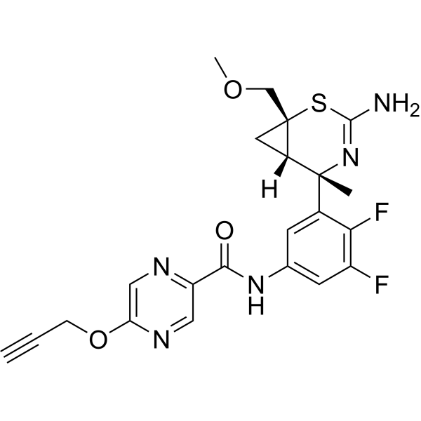
AM-6494 is a potent and orally active BACE1 (efficacious β-site amyloid precursor protein cleaving enzyme 1) inhibitor (IC50=0.4 nM) with in vivo selectivity over BACE2 (IC50=18.6 nM). AM-6494 is a click chemistry reagent, it contains an Alkyne group and can undergo copper-catalyzed azide-alkyne cycloaddition (CuAAc) with molecules containing Azide groups.
For research use only. We do not sell to patients.
- CAS No.: 1874232-80-0
- Formula: C22H21F2N5O3S
- Molecular Weight:473.50
-
Storage:
Please store the product under the recommended conditions in the Certificate of Analysis.

To place orders, for customer services and technical support, please contact: MedChemExpress USA
Tel: 609-228-6898 E-mail: [email protected] [email protected]


-
 Biological Activity
-
Chemical Information
- Protocols
- Purity & Documentation
- References
-
 Help & FAQs




Biological Activity
Description
IC50 & Target
|
BACE1 |
Cellular Effect
|
Cell Line

|
Type | Value | Description | References |
|---|---|---|---|---|
| CHO | IC50 |
137 μM
Compound: 20; AM-6494
|
Inhibition of recombinant CatD (unknown origin) expressed in CHO cells using FRET peptide substrate preincubated for 1 hr followed by substrate addition and measured after 1 hr by FRET assay
Inhibition of recombinant CatD (unknown origin) expressed in CHO cells using FRET peptide substrate preincubated for 1 hr followed by substrate addition and measured after 1 hr by FRET assay
|
[PMID: 31589043] |
| HEK293 | IC50 |
1.2 nM
Compound: 20; AM-6494
|
Inhibition of BACE in HEK293 cells harboring APP Swedish mutant assessed as reduction in amyloid beta 40 level measured after overnight incubation by sandwich ELISA
Inhibition of BACE in HEK293 cells harboring APP Swedish mutant assessed as reduction in amyloid beta 40 level measured after overnight incubation by sandwich ELISA
|
[PMID: 31589043] |
| HEK293 | IC50 |
569 nM
Compound: 20; AM-6494
|
Inhibition of recombinant human BACE2 expressed in HEK293 cells co-transfected with N-terminal HA-tagged/C-terminal Myc tagged Mbeta chain of human PMEL17 measured after overnight incubation by Gaussia luciferase reporter gene assay
Inhibition of recombinant human BACE2 expressed in HEK293 cells co-transfected with N-terminal HA-tagged/C-terminal Myc tagged Mbeta chain of human PMEL17 measured after overnight incubation by Gaussia luciferase reporter gene assay
|
[PMID: 31589043] |


Chemical Information
-
CAS No. 1874232-80-0
-
Molecular Weight 473.50
-
Formula C22H21F2N5O3S
-
SMILES
O=C(C1=NC=C(OCC#C)N=C1)NC2=CC(F)=C(F)C([C@@]3(C)N=C(N)S[C@@]4(COC)C[C@@]34[H])=C2
-
Shipping
Room temperature in continental US; may vary elsewhere.
-
Storage
Please store the product under the recommended conditions in the Certificate of Analysis.

Protocols
-
EdU Incorporation Assay (Click Chemistry-Based DNA Synthesis Measurement)
The EdU incorporation assay measures DNA synthesis by adding the thymidine analog 5-ethynyl-2′-deoxyuridine to cells or tissues, where it is incorporated into newly synthesized DNA during S phase. Incorporated EdU is detected by copper-catalyzed azide-alkyne cycloaddition, in which a fluorescent azide covalently reacts with the ethynyl group on EdU, allowing S-phase cells to be detected by fluorescence microscopy, flow cytometry, or high-content imaging. EdU detection does not require DNA denaturation or anti-BrdU antibody access, which preserves sample structure and improves compatibility with immunostaining and multiparameter cytometry compared with BrdU-based detection. EdU can be cytotoxic in a cell-type- and exposure-dependent manner, so pulse duration, concentration, and continuous-labeling designs should be validated for each cell type.
-
Cell Cytotoxicity Assay
Cytotoxicity assays are usually based on the assessment of cell membrane damage, which can also be indirectly detected by measuring cell viability. Detection methods include MTT assay, CKK-8 assay, LDH assay and ATP assay, etc.
-
Amyloid: Congo Red Amyloid Staining
Congo red amyloid staining is a histochemical method used to detect extracellular amyloid deposits in tissue sections based on the affinity of Congo red dye for β-pleated sheet-rich protein aggregates. When bound to amyloid, Congo red produces characteristic apple-green birefringence under polarized light microscopy, which is widely regarded as a diagnostic feature of amyloid deposition in histopathology. The diagnostic principle relies on the combination of dye binding (congophilia) and optical anisotropy under polarized illumination, which distinguishes amyloid from most non-amyloid eosinophilic extracellular deposits in routine histological evaluation. Amyloid identification by Congo red staining remains a cornerstone in diagnostic pathology despite the availability of adjunct methods such as immunohistochemistry and mass spectrometry, particularly because of its ability to localize deposits directly within tissue architecture. The specificity of Congo red-positive deposits is incre


Purity & Documentation
References

Calculators
Concentration (start) × Volume (start) = Concentration (final) × Volume (final)


AM-6494 Related Classifications
HY-128774 Related Classifications
 Powered by Bioz