



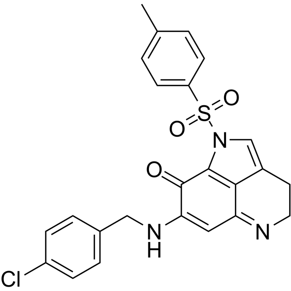
MA242 free base
Based on 1 publication(s) in Google Scholar
MA242 free base is a specific dual inhibitor of MDM2 and NFAT1. MA242 free base directly binds both MDM2 and NFAT1 with high affinity, induces their protein degradation, and inhibits NFAT1-mediated transcription of MDM2. MA242 free base induces apoptosis in pancreatic cancer cell lines regardless of p53 status.
For research use only. We do not sell to patients.
- CAS No.: 1049704-17-7
- Formula: C24H20ClN3O3S
- Molecular Weight:465.95
-
Storage:
Please store the product under the recommended conditions in the Certificate of Analysis.

To place orders, for customer services and technical support, please contact: MedChemExpress USA
Tel: 609-228-6898 E-mail: [email protected] [email protected]


-
 Biological Activity
-
Chemical Information
- Protocols
- Purity & Documentation
- References
-
 Help & FAQs


-
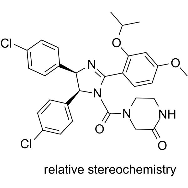
HY-50696
Nutlin-3p53-MDM2 Inhibitor -
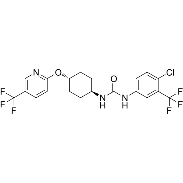
HY-112780
UC2288P21 Attenuator -
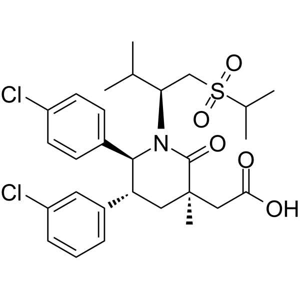
HY-12296
NavtemadlinMDM2-p53 Interaction Inhibitor -
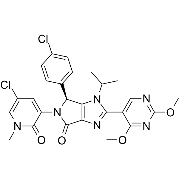
HY-18658
Siremadlinp53-MDM2 Inhibitor -
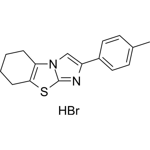
HY-16702A
Pifithrin-β hydrobromideMDM-2/p53 p53 Inhibitor -
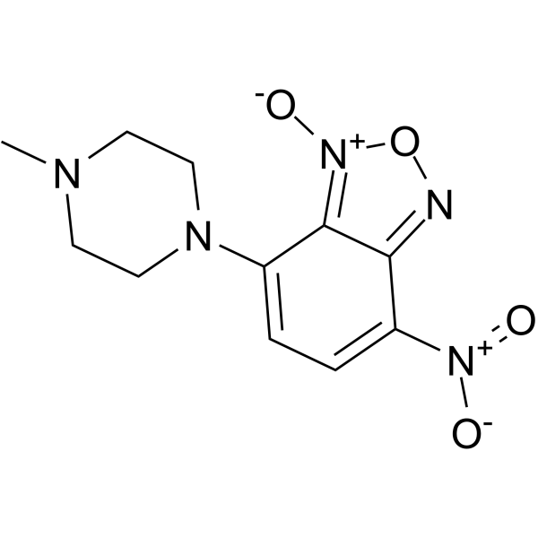
HY-14714
NSC-207895p53 Activator -
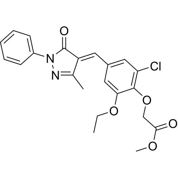
HY-16664
SJ-172550MDM-2/p53 MDM2 Inhibitor -
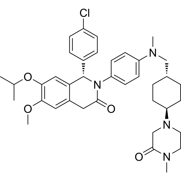
HY-15954
NVP-CGM097MDM2 Inhibitor -
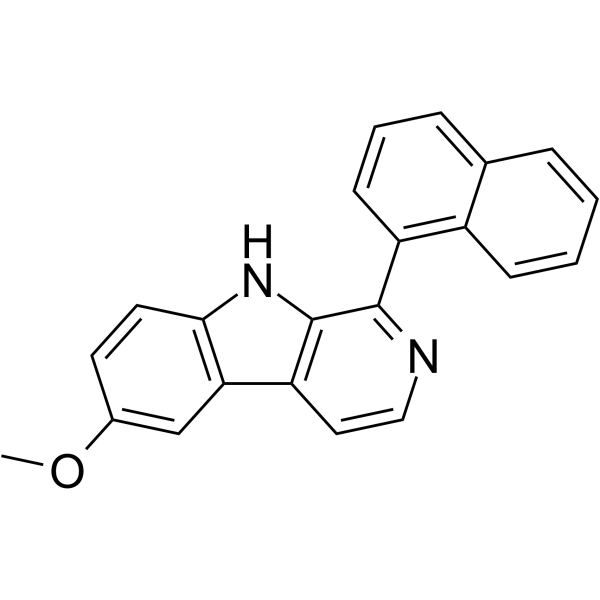
HY-110182
SP-141MDM2 Inhibitor -
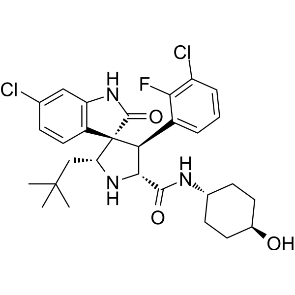
HY-17493
MI-773MDM2-p53 Inhibitor -
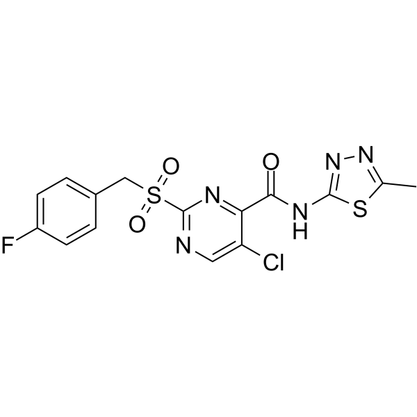
HY-128784
PK11007Anti-p53 Agent -
HY-19726
NSC59984MDM-2/p53 p53 Activator
Publications Citing Use of MedChemExpress (MCE) MA242 free base
More

Biological Activity
Description
IC50 & Target
MDM2, NFAT1[1]
In Vitro
MA242 (0.05-5 μM; 72 hours) free base significantly inhibits pancreatic cancer cell growth, with IC50s ranging from 0.1 to 0.4 μM, regardless of the p53 status of the cells. However, MA242 free base shows minimal effects on the growth of normal HPDE cells (IC50=5.81 μM), indicating that MA242 has selective effects against cancer cells[1].
MA242 (0.1-0.5 μM; 24 hours) free base significantly decreases the MDM2 and NFAT1 protein levels at a low concentration in all three cell lines[1].
MA242 free base decreases cell proliferation and induces apoptosis in pancreatic cancer cell lines regardless of p53 status[1].
MA242 free base alone or in combination with Gemcitabine inhibits pancreatic tumor growth and metastasis without any host toxicity[1].
MA242 free base exerts cytotoxicity against hepatocellular carcinoma (HCC) cells by inhibiting the NFAT1-MDM2 pathway in vitro, independent of p53. MA242 showed selective cytotoxicity against HCC cells, with IC50 values ranging from 0.1-0.31 μM[2].
MedChemExpress (MCE) has not independently confirmed the accuracy of these methods. They are for reference only. Further protocols information, click here.
-
Cell Line:The human pancreatic cancer HPAC, Panc-1, AsPC-1, Mia-Paca-2 and BxPC-3 cell lines; The human pancreatic ductal epithelium (HPDE) cell line
-
Concentration:0.05, 0.5, and 5 μM
-
Incubation Time:72 hours
-
Result:The IC50s are 0.14, 0.14, 0.15, 0.25, 0.40, and 5.81 μM for Panc-1, Mia-Paca-2, AsPC-1, BxPC-3, HPAC, and HPDE cells, respectively.
-
Cell Line:The human pancreatic cancer HPAC, Panc-1, and AsPC-1 cell lines
-
Concentration:0, 0.1, 0.2, and 0.5 μM
-
Incubation Time:24 hours
-
Result:Decreased the expression of MDM2 and NFAT1.
In Vivo
There were no significant differences in the average body weights between the vehicle- and MA242 free base-treated mice in either of the models, did not have significant host toxicity at these effective doses[1].
MedChemExpress (MCE) has not independently confirmed the accuracy of these methods. They are for reference only.
-
Animal Model:Female 4-6-week-old athymic nude mice (nu/nu, 4-6 weeks) bearing AsPC-1-Luc or Panc-1-Luc tumor[1]
-
Dosage:2.5 or 5 mg/kg for Panc-1 tumor-bearing mice; 10 mg/kg for AsPC-1 tumor-bearing mice
-
Administration:IP; 2.5 or 5 mg/kg/d, 5 d/wk for five weeks for Panc-1 tumor-bearing mice;
IP; 10 mg/kg/d, 5 d/wk for three weeks for AsPC-1 tumor-bearing mice -
Result:Resulted in 56.1% and 82.5% inhibition of tumor growth in nude mice bearing Panc-1 orthotopic tumors, respectively.
Significantly suppressed the growth of AsPC-1 orthotopic tumors by 89.5% (P < 0.01) compared with the tumors in control animals.
Led to almost complete tumor regression in MD242-treated mice in both models.


Chemical Information
-
CAS No. 1049704-17-7
-
Molecular Weight 465.95
-
Formula C24H20ClN3O3S
-
SMILES
O=C1C(NCC2=CC=C(C=C2)Cl)=CC3=NCCC4=CN(S(=O)(C5=CC=C(C)C=C5)=O)C1=C34
-
Shipping
Room temperature in continental US; may vary elsewhere.
-
Storage
Please store the product under the recommended conditions in the Certificate of Analysis.


Publications (1)
-
Journal Impact Factor
-
Most Recent
-
Biochem Pharmacol
Curcumol enhances the sensitivity of doxorubicin in triple-negative breast cancer via regulating the miR-181b-2-3p-ABCC3 axis. [Abstract]2020 Apr:174:113795. PMID: 31926937

Protocols
-
Apoptosis
Apoptosis, also called programmed cell death, is generally characterized by distinct morphological characteristics.
-
TUNEL staining for apoptotic DNA fragmentation
TUNEL staining detects DNA strand breaks by using terminal deoxynucleotidyl transferase to add labeled nucleotides to exposed 3′-OH DNA termini, generating either microscopic staining in fixed cells or tissue sections, or fluorescence/cytometric signal in cell suspensions. TUNEL positivity reflects DNA fragmentation but should not be interpreted alone as definitive apoptosis, because TUNEL can also label necrotic, autolytic, mechanically damaged, or DNA-repair-associated DNA breaks.
-
Annexin V plus membrane-impermeant dye apoptosis staining
Annexin V-based apoptosis assays rely on the detection of phosphatidylserine (PS) externalization from the inner leaflet of the plasma membrane to the outer leaflet, an early biochemical hallmark of apoptosis. Fluorescently labeled Annexin V binds PS in a calcium-dependent manner, enabling identification of early apoptotic cells by flow cytometry or fluorescence microscopy. When combined with a membrane-impermeant DNA-binding dye (e. g. , propidium iodide), this approach allows discrimination between viable (Annexin V−/dye−), early apoptotic (Annexin V+/dye−), and late apoptotic or necrotic (Annexin V+/dye+) cell populations by assessing membrane integrity and PS exposure.
-
Apoptosis Solutions
Apoptosis is a regulated, generally non-lytic cell-death pathway that removes unwanted, damaged, infected, or abnormal cells through coordinated morphological changes, caspase activation, DNA fragmentation, and membrane remodeling. The intrinsic apoptosis pathway is controlled mainly by mitochondrial outer membrane permeabilization, BCL-2 family proteins, cytochrome c release, apoptosome formation, caspase-9 activation, and downstream executioner caspase-3/7 activation. The extrinsic apoptosis pathway is initiated by death receptors such as Fas, TNFR, and TRAIL receptors, which recruit adaptor proteins and activate caspase-8 before engaging executioner caspases or mitochondrial amplification through BID cleavage. Apoptosis is linked to many phenotypes, including cancer cell killing, tissue homeostasis, immune regulation, neurodegeneration, infection response, and treatment-induced cytotoxicity; unresolved questions include how apoptosis interacts with necroptosis, pyroptosis, ferroptos


Purity & Documentation
References
[1]. Wei Wang, et al. Discovery and Characterization of Dual Inhibitors of MDM2 and NFAT1 for Pancreatic Cancer Therapy. Cancer Res. 2018 Oct 1;78(19):5656-5667. [Content Brief]
[2]. Wei Wang, et al. MDM2-NFAT1 dual inhibitor, MA242: Effective against hepatocellular carcinoma, independent of p53. Cancer Lett. 2019 Sep 10;459:156-167. [Content Brief]

Calculators
Concentration (start) × Volume (start) = Concentration (final) × Volume (final)


MA242 free base Related Classifications
HY-112816A Related Classifications
 Powered by Bioz