



LH1306
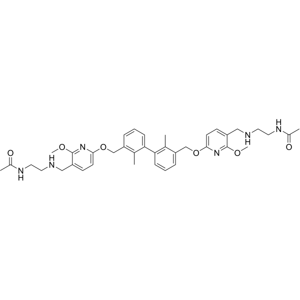
LH1306 is a C2-symmetric inhibitor of PD-1/PD-L1 protein-protein interaction (PPI), with an IC50 value of 25 nM. LH1306 can be used in anti-tumor research.
Nur für Forschungszwecke. Wir verkaufen nicht an Patienten.
- CAS. Nr.: 2182653-84-3
- Formel: C38H48N6O6
- Molecular Weight:684.82
-
Speicherung:
Please store the product under the recommended conditions in the Certificate of Analysis.

To place orders, for customer services and technical support, please contact: MedChemExpress USA
Tel: 609-228-6898 E-mail: [email protected] [email protected]


-
 Biologische Aktivität
-
Chemical Information
- Protokoll
- Reinheit & Dokumentation
- Verweise
-
 Help & FAQs


-
HY-132212
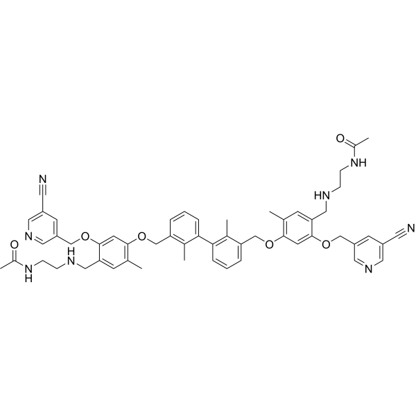
LH1307Inhibitor of PD-1/PD-L1 Protein-Protein Interaction -
HY-134884
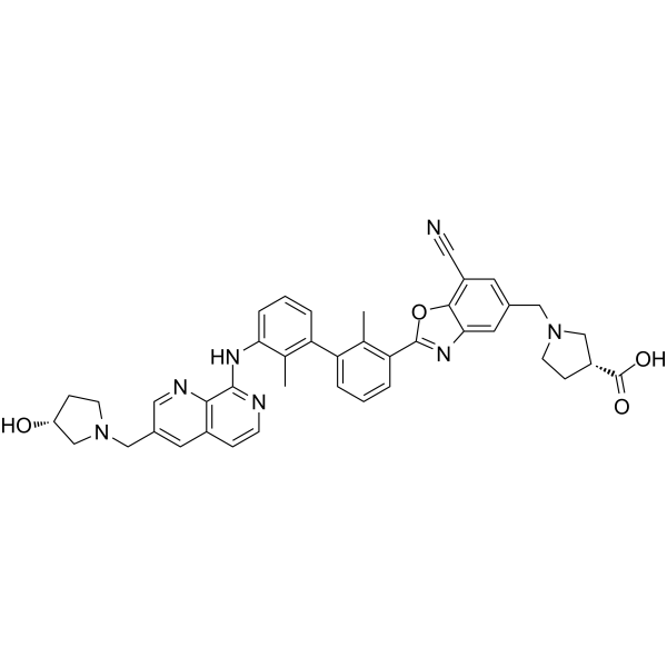
INCB086550PD-L1 Inhibitor -
HY-102011
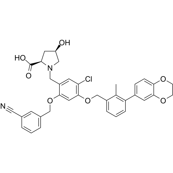
BMS-1166PD-1/PD-L1 Interaction Inhibitor -
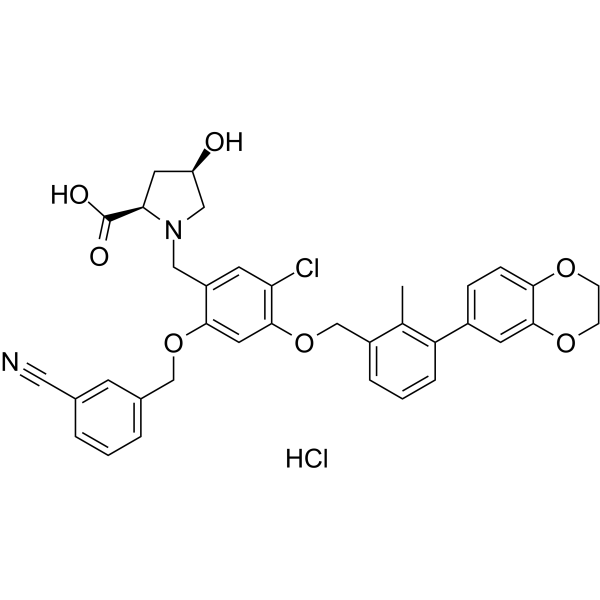
HY-102011A
BMS-1166 hydrochloridePD-1/PD-L1 Interaction Inhibitor -
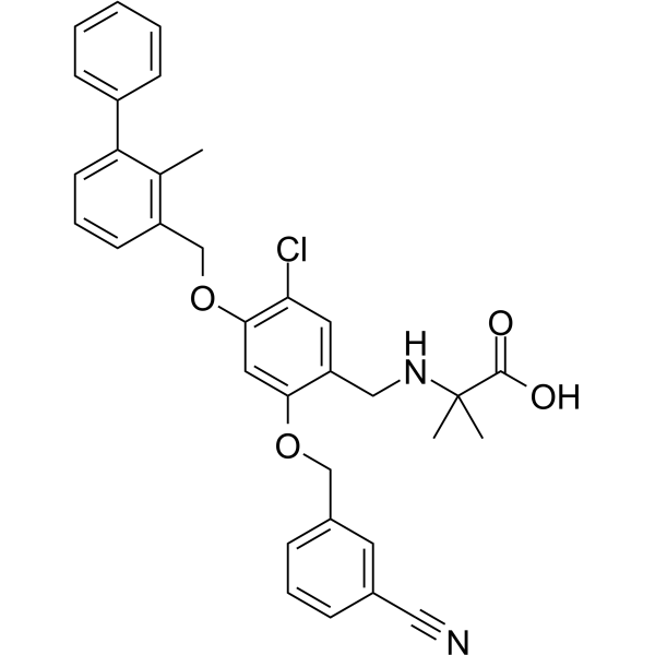
HY-131347
PD-1/PD-L1-IN-NP19PD-1/PD-L1 Inhibitor -
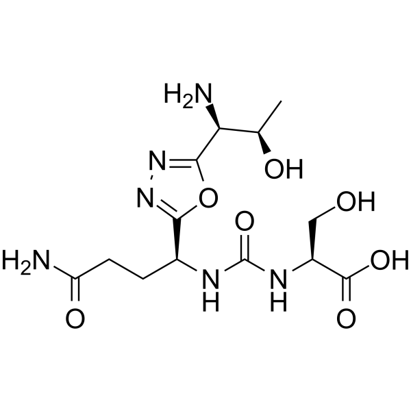
HY-101097
PD-1-IN-17PD-1 Inhibitor -
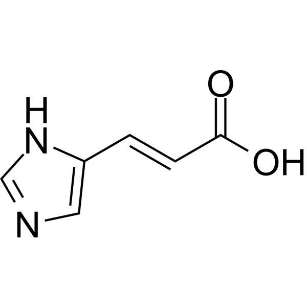
HY-113008B
trans-Urocanic acidNK Cell Inhibitor -
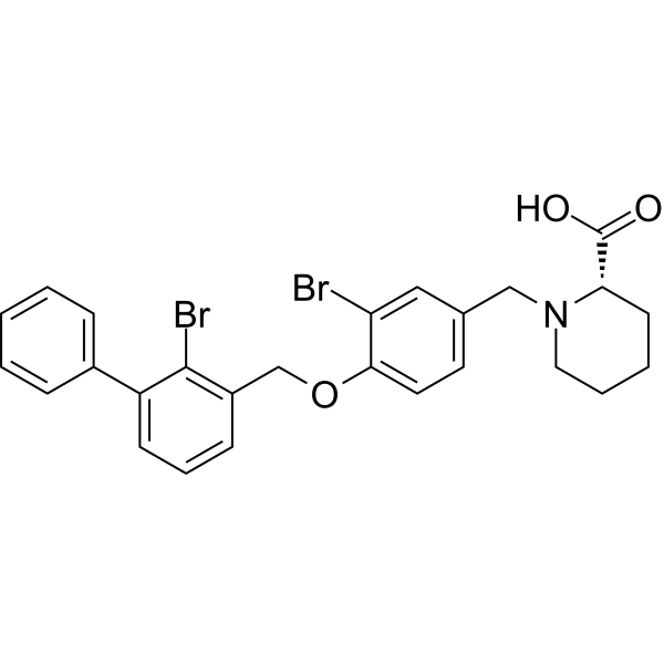
HY-163041
PD-1/PD-L1-IN-36PD-1/PD-L1 Inhibitor -
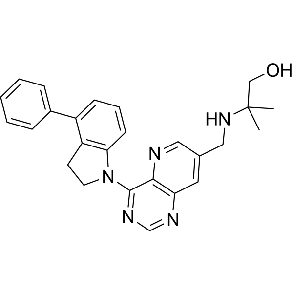
HY-155959
PD-1/PD-L1-IN-33PD-1/PD-L1 Inhibitor -

HY-102011R
BMS-1166 (Standard)Reference Standard -
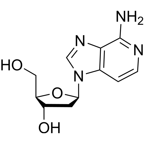
HY-126877
3-Deaza-2'-deoxyadenosineAntiviral agent -

HY-113008BR
trans-Urocanic acid (Standard)NK Cell Inhibitor


Biologische Aktivität
Beschreibung
IC50 & Target
IC50: 25 nM (PD-1/PD-L1)[1].
Cellular Effect
|
Cell Line

|
Type | Value | Description | References |
|---|---|---|---|---|
| CHO-K1 | GI50 |
3.1 μM
Compound: 2a
|
Cytotoxicity against CHOK1 cells stably expressing human PDL1 co-cultured with human Jurkat T cell expressing human PD1 assessed as growth inhibition measured after 24 hrs by XTT assay
Cytotoxicity against CHOK1 cells stably expressing human PDL1 co-cultured with human Jurkat T cell expressing human PD1 assessed as growth inhibition measured after 24 hrs by XTT assay
|
[PMID: 31298541] |
| HEK293 | IC50 |
25 nM
Compound: 2a
|
Inhibition of human Fc-tagged PD1 N-terminal domain (Leu25 to Gln167 residues) expressed in HEK293 cells/human His-tagged PDL1 (Phe19 to Arg238 residues) expressed in HEK293 cells protein-protein interaction after 1 hr by APC-labeled anti-His antibody/Eu-
Inhibition of human Fc-tagged PD1 N-terminal domain (Leu25 to Gln167 residues) expressed in HEK293 cells/human His-tagged PDL1 (Phe19 to Arg238 residues) expressed in HEK293 cells protein-protein interaction after 1 hr by APC-labeled anti-His antibody/Eu-
|
[PMID: 31298541] |
| Jurkat | EC50 |
334 nM
Compound: 2a
|
Inhibition of PD1/PDL1 interaction in human Jurkat cells co-cultured with human U2OS cells expressing PDL1 assessed as reduction in SHP1 recruitment preincubated with U2OS cells for 60 mins followed by Jurkat cell addition and measured after 2 hrs by Path
Inhibition of PD1/PDL1 interaction in human Jurkat cells co-cultured with human U2OS cells expressing PDL1 assessed as reduction in SHP1 recruitment preincubated with U2OS cells for 60 mins followed by Jurkat cell addition and measured after 2 hrs by Path
|
[PMID: 31298541] |
| Jurkat | EC50 |
4214 nM
Compound: 2a
|
Inhibition of human PD1 stably expressed in human Jurkat T cells co-expressing NFAT-induced luciferase/human PDL1 stably expressed in CHOK1 cells co-expressing aAPC protein-protein interaction assessed as increase in NFAT signaling measured after 6 hrs by
Inhibition of human PD1 stably expressed in human Jurkat T cells co-expressing NFAT-induced luciferase/human PDL1 stably expressed in CHOK1 cells co-expressing aAPC protein-protein interaction assessed as increase in NFAT signaling measured after 6 hrs by
|
[PMID: 31298541] |
| Jurkat | GI50 |
3.1 μM
Compound: 2a
|
Cytotoxicity against human Jurkat T cell expressing human PD1 co-cultured with CHOK1 cells stably expressing human PDL1 assessed as growth inhibition measured after 24 hrs by XTT assay
Cytotoxicity against human Jurkat T cell expressing human PD1 co-cultured with CHOK1 cells stably expressing human PDL1 assessed as growth inhibition measured after 24 hrs by XTT assay
|
[PMID: 31298541] |


Chemical Information
-
CAS. Nr. 2182653-84-3
-
Molecular Weight 684.82
-
Formel C38H48N6O6
-
SMILES
CC(NCCNCC(C(OC)=N1)=CC=C1OCC2=CC=CC(C3=CC=CC(COC4=CC=C(C(OC)=N4)CNCCNC(C)=O)=C3C)=C2C)=O
-
Versand
Room temperature in continental US; may vary elsewhere.
-
Speicherung
Please store the product under the recommended conditions in the Certificate of Analysis.


Reinheit & Dokumentation
Verweise

Calculators
Konzentration (Stammlösung) × Volumen (Stammlösung) = Konzentration (Ziellösung) × Volumen (Ziellösung)


LH1306 Related Classifications
HY-132223 Related Classifications
 Powered by Bioz