



Silver(I) sulfide

Based on 1 Customer Validation
Silver (I) sulfide is a biochemical reagent. Silver (I) sulfide nanoparticles exert Antibacterial effects against a variety of resistant bacteria, including Gram-positive and Gram-negative bacteria. Silver (I) sulfide can be used in the research of bacterial infections, such as infections caused by Staphylococcus aureus, Streptococcus agalactiae, Escherichia coli and Pseudomonas species.
Nur für Forschungszwecke. Wir verkaufen nicht an Patienten.
- CAS. Nr.: 21548-73-2
- Formel: Ag2S
- Molecular Weight:247.80
-
Speicherung:
RT, protect from light, stored under nitrogen.
In solvent -80°C, 1 year , -20°C, 6 months


To place orders, for customer services and technical support, please contact: MedChemExpress USA
Tel: 609-228-6898 E-mail: [email protected] [email protected]


-
 Biologische Aktivität
-
Chemical Information
- Protokoll
- Reinheit & Dokumentation
- Verweise
-
 Help & FAQs


-

HY-Y0975
Silver(I) fluorideDrug Intermediate -

HY-W051140
Silver sulfateEthylene Inhibitor -
HY-N3025
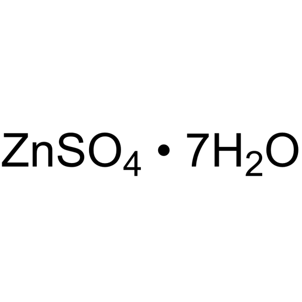
Zinc sulfate heptahydrateDietary Supplement -

HY-W019930
Silver iodideBiochemical Assay Reagents -
HY-B1497
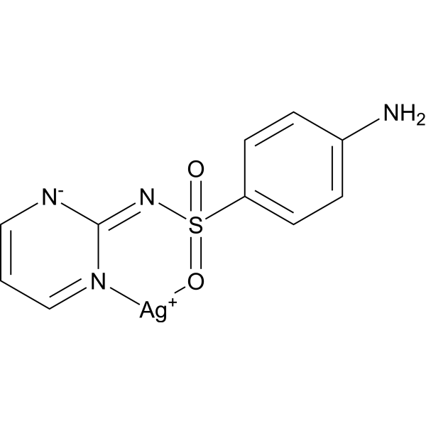
Silver sulfadiazineBacterial Inhibitor -

HY-W019929
Silver chlorideAlcohol Oxidase Modulator -
HY-B1521
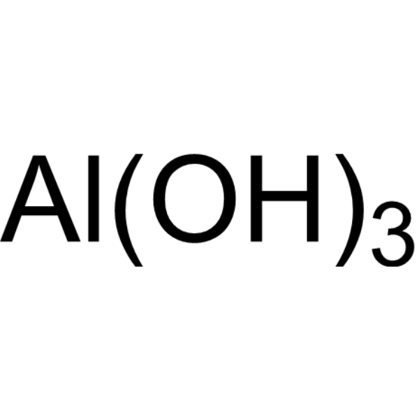
Aluminum HydroxideAntacid Aegnt -
HY-Y1738
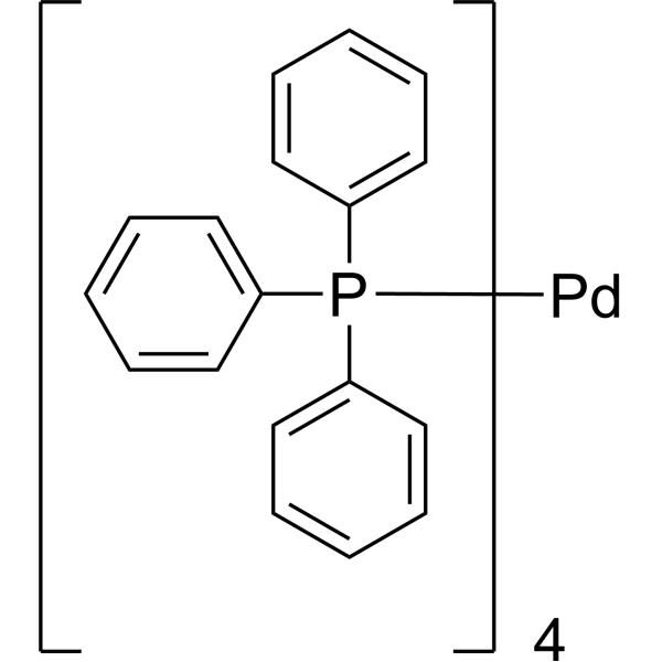
Tetrakis(triphenylphosphine)palladiumCatalyst -

HY-B1608
Chromium chlorideTrivalent Chromium Compound


Biologische Aktivität
Beschreibung


Chemical Information
-
CAS. Nr. 21548-73-2
-
Appearance Solid
-
Molecular Weight 247.80
-
Formel Ag2S
-
Color Brown to black
-
SMILES
[Ag][Ag]=S
-
Versand
Room temperature in continental US; may vary elsewhere.
-
Speicherung
RT, protect from light, stored under nitrogen
In solvent -80°C 1 year -20°C 6 months

Protokoll
-
Research Protocol for Infectious Diseases
Infectious-disease experiments test how pathogens interact with host barriers, innate immune receptors, inflammatory signaling, pathogen replication, and tissue injury; pattern-recognition receptors such as TLRs, RIG-I-like receptors, NOD-like receptors, and inflammasomes detect microbial molecules and activate NF-κB, interferon, and cytokine responses. The central hypothesis is that infection severity reflects the balance between pathogen burden and host response: protective inflammation restricts pathogen growth, whereas excessive or mislocalized inflammation contributes to tissue damage and disease phenotype. Unresolved questions include which host pathways are protective versus pathogenic, why some infection models fail to translate to human disease, and which combined readouts best predict clinically relevant infection outcomes.
-
Gram Staining of Tissue Sections
Gram staining of tissue sections is a histochemical technique used to differentiate Gram-positive and Gram-negative bacteria within histological specimens based on differences in bacterial cell wall structure and dye retention, adapted from classical bacteriological Gram staining into tissue-compatible “histological Gram stain” variants. In tissue applications, modifications of the Brown-Hopps and Brown-Brenn methods are commonly used to improve differentiation of microorganisms embedded within host connective tissue and to reduce overstaining or loss of Gram-negative signal, which are known limitations of earlier approaches. The principle relies on crystal violet-iodine complex retention in Gram-positive organisms and subsequent decolorization and counterstaining steps that allow contrast visualization of Gram-negative organisms against tissue background.


Reinheit & Dokumentation
-
Data Sheet (275 KB)
-
SDS (252 KB)
- Français - FR (252 KB)
- Deutsch - DE (252 KB)
- Norwegian - NO (252 KB)
- Español - ES (252 KB)
- Swedish - SV (252 KB)
- Italian - IT (252 KB)
- Korean - KR (252 KB)
- Portuguese - PT (252 KB)
-
Handling Instructions (2659 KB)
Verweise

Calculators
Konzentration (Stammlösung) × Volumen (Stammlösung) = Konzentration (Ziellösung) × Volumen (Ziellösung)


Silver(I) sulfide Related Classifications
HY-W093378 Related Classifications
 Powered by Bioz