



VRT-325

Based on 1 Customer Validation
VRT-325 is a CFTR modulator. VRT-325 inhibits disulfide cross-linking between cysteines in transmembrane segments 6 and 7 of CFTR and P-gp. VRT-325 promotes maturation of CFTR and P-gp processing mutants, rescues ΔF508-CFTR folding at the endoplasmic reticulum. VRT-325 binds ΔF508-CFTR nucleotide-binding domain 1, and increases mature ΔF508-CFTR cell surface expression and chloride conductance. VRT-325 can be used for the research of cystic fibrosis[1][3].
Nur für Forschungszwecke. Wir verkaufen nicht an Patienten.
- Reinheit : 99.58%
- CAS. Nr.: 815592-21-3
- Formel: C27H34N4O4S
- Molecular Weight:510.65
-
Speicherung:Powder -20°C, 3 years , 4°C, 2 years ; In solvent -80°C, 6 months , -20°C, 1 month


To place orders, for customer services and technical support, please contact: MedChemExpress USA
Tel: 609-228-6898 E-mail: [email protected] [email protected]


-
 Biologische Aktivität
-
Chemical Information
-
Lösungsmittel & Löslichkeit
- Protokoll
- Reinheit & Dokumentation
- Verweise
-
 Help & FAQs


-
HY-108575
Chromanol 293BIKs Blocker -
HY-150090
SRI-41315CFTR Activator -
HY-106203
CrinecerfontCRF1 Receptor Antagonist -
HY-109027
CavosonstatGSNOR Inhibitor -
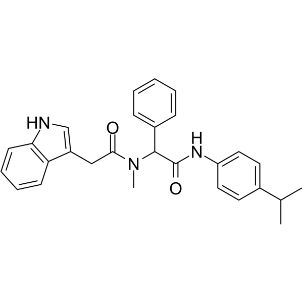
HY-103369
PG01CFTR Potentiator -
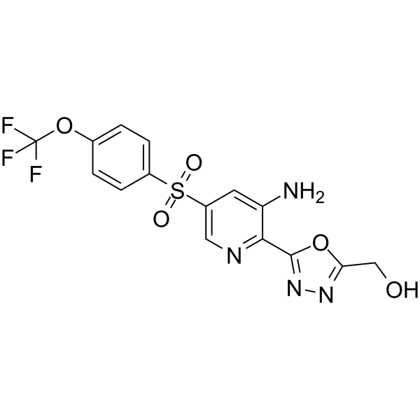
HY-109152
NavocaftorCystic Fibrosis Transmembrane Regulator -
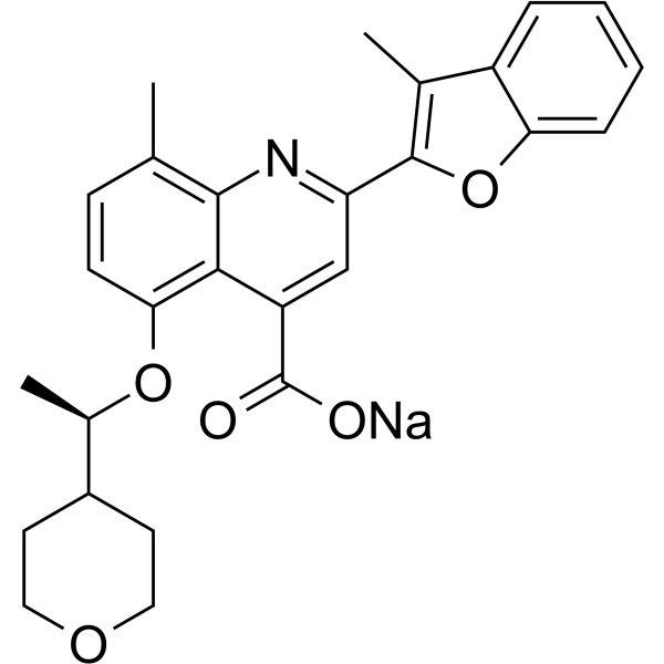
HY-109187A
Posenacaftor sodiumCFTR Modulator -
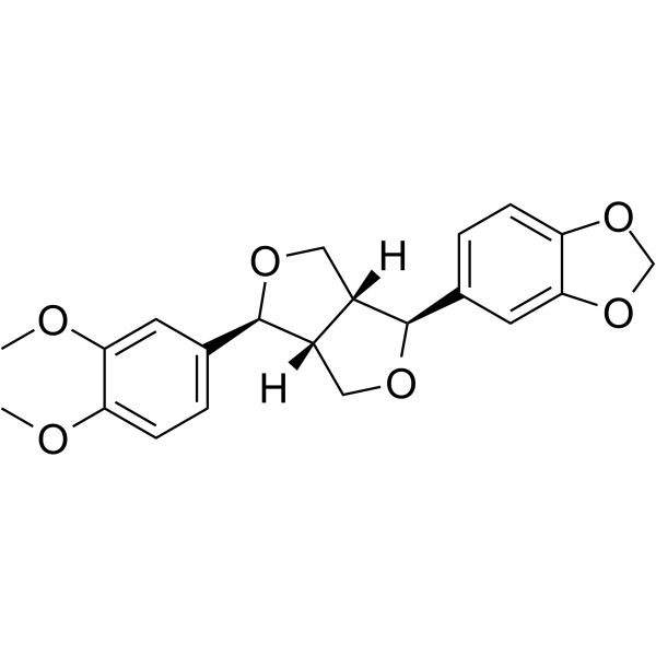
HY-N5101
KobusinCalmodulin inhibitor -
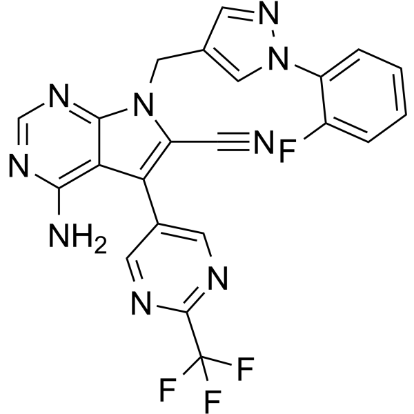
HY-136939
CFTR corrector 6CFTR Activator -
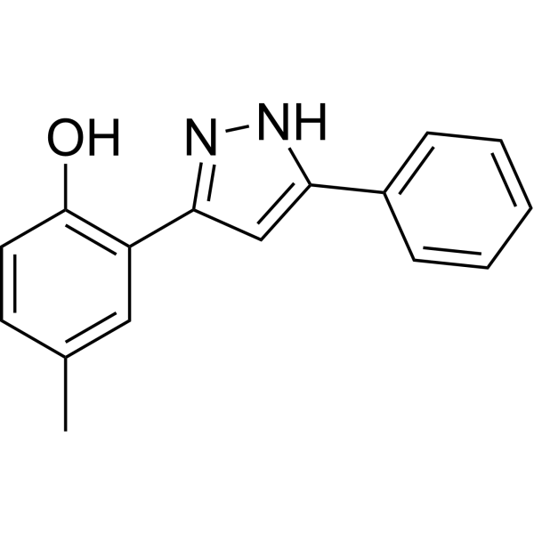
HY-114725
VRT-532CFTR Modulator -
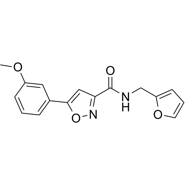
HY-147315
CFTR corrector 9CFTR Modulator -
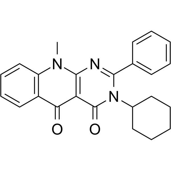
HY-150089
SRI-37240PTCs Inhibitor


Biologische Aktivität
Beschreibung
In Vitro
VRT-325 (30 min) directly interacts with I340C(TM6)/S877C(TM7) Cys-less/V510A CFTR in HEK 293 cells[1].
VRT-325 (5 µM; 24 h) partially rescues mature ΔF508-CFTR protein expression in CFBE-ΔF508 cells[2].
VRT-325 (5 µM; 24 h) rescues ΔF508-CFTR channel function in CFBE-ΔF508 cells[2].
VRT-325 (10 µM; 24 h) rescues ΔF508-CFTR channel function in primary human cystic fibrosis airway epithelial cells[2].
VRT-325 corrects Delta F508-CFTR misfolding and trafficking with an EC50 of approximately 2 μM[3].
MedChemExpress (MCE) has not independently confirmed the accuracy of these methods. They are for reference only.
-
Cell Line:CFBE-ΔF508 cystic fibrosis airway epithelial cells
-
Concentration:5 µM
-
Incubation Time:24 h
-
Result:Increased mature CFTR band C expression to ~350% of the lysogeny broth (LB) control level.
Reduced mature CFTR band C expression to ~20% of the LB control level when combined with PsaDM, representing a significant decrease compared to VRT-325 treatment alone.


Chemical Information
-
CAS. Nr. 815592-21-3
-
Appearance Solid
-
Molecular Weight 510.65
-
Formel C27H34N4O4S
-
Color White to off-white
-
SMILES
CC(N1CCN(CC1)S(=O)(C2=CC=C(C=C2)OC)=O)C3=NC(OC4CCCCC4)=C5C=CC=CC5=N3
-
Versand
Room temperature in continental US; may vary elsewhere.
-
Speicherung
Powder -20°C 3 years 4°C 2 years In solvent -80°C 6 months -20°C 1 month


Lösungsmittel & Löslichkeit
In Vitro:
DMSO : 45 mg/mL (88.12 mM; Need ultrasonic and warming; Hygroscopic DMSO has a significant impact on the solubility of product, please use newly opened DMSO)
Please refer to the solubility information to select the appropriate solvent. Once prepared, please aliquot and store the solution to prevent product inactivation from repeated freeze-thaw cycles.
Storage method and period of stock solution: -80°C, 6 months; -20°C, 1 month. When stored at -80°C, please use it within 6 months. When stored at -20°C, please use it within 1 month.
Please refer to the solubility information to select the appropriate solvent. Once prepared, please aliquot and store the solution to prevent product inactivation from repeated freeze-thaw cycles.
Storage method and period of stock solution: -80°C, 6 months; -20°C, 1 month. When stored at -80°C, please use it within 6 months. When stored at -20°C, please use it within 1 month.
Konzentration (Stammlösung) × Volumen (Stammlösung) = Konzentration (Ziellösung) × Volumen (Ziellösung)


Reinheit & Dokumentation
-
Data Sheet (275 KB)
-
SDS (394 KB)
- English - EN (394 KB)
- Français - FR (394 KB)
- Deutsch - DE (394 KB)
- Norwegian - NO (394 KB)
- Español - ES (394 KB)
- Swedish - SV (394 KB)
- Italian - IT (394 KB)
- Korean - KR (394 KB)
- Portuguese - PT (394 KB)
-
Handling Instructions (2659 KB)
Verweise
[1]. Wang Y, et al. Correctors promote maturation of cystic fibrosis transmembrane conductance regulator (CFTR)-processing mutants by binding to the protein. J Biol Chem. 2007;282(46):33247-33251. [Content Brief]
[2]. Trinh NT, et al. Deleterious impact of Pseudomonas aeruginosa on cystic fibrosis transmembrane conductance regulator function and rescue in airway epithelial cells. Eur Respir J. 2015;45(6):1590-1602. [Content Brief]
[3]. Han X, et al. Recommended Tool Compounds for Modifying the Cystic Fibrosis Transmembrane Conductance Regulator Channel Variants. ACS Pharmacol Transl Sci. 2024;7(4):933-950. Published 2024 Mar 14. [Content Brief]


Complete Stock Solution Preparation Table
Please refer to the solubility information to select the appropriate solvent. Once prepared, please aliquot and store the solution to prevent product inactivation from repeated freeze-thaw cycles.
Storage method and period of stock solution: -80°C, 6 months; -20°C, 1 month. When stored at -80°C, please use it within 6 months. When stored at -20°C, please use it within 1 month.
| Optional Solvent | Concentration Solvent Mass | 1 mg | 5 mg | 10 mg | 25 mg |
|---|---|---|---|---|---|
| DMSO | 1 mM | 1.9583 mL | 9.7914 mL | 19.5829 mL | 48.9572 mL |
| 5 mM | 0.3917 mL | 1.9583 mL | 3.9166 mL | 9.7914 mL | |
| 10 mM | 0.1958 mL | 0.9791 mL | 1.9583 mL | 4.8957 mL | |
| 15 mM | 0.1306 mL | 0.6528 mL | 1.3055 mL | 3.2638 mL | |
| 20 mM | 0.0979 mL | 0.4896 mL | 0.9791 mL | 2.4479 mL | |
| 25 mM | 0.0783 mL | 0.3917 mL | 0.7833 mL | 1.9583 mL | |
| 30 mM | 0.0653 mL | 0.3264 mL | 0.6528 mL | 1.6319 mL | |
| 40 mM | 0.0490 mL | 0.2448 mL | 0.4896 mL | 1.2239 mL | |
| 50 mM | 0.0392 mL | 0.1958 mL | 0.3917 mL | 0.9791 mL | |
| 60 mM | 0.0326 mL | 0.1632 mL | 0.3264 mL | 0.8160 mL | |
| 80 mM | 0.0245 mL | 0.1224 mL | 0.2448 mL | 0.6120 mL |


VRT-325 Related Classifications
HY-119229 Related Classifications
 Powered by Bioz