



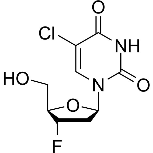
Raluridine

Based on 1 Customer Validation
Raluridine is a purine nucleoside analog. Purine nucleoside analogs have broad antitumor activity targeting indolent lymphoid malignancies. Anticancer mechanisms in this process rely on inhibition of DNA synthesis, induction of apoptosis, etc.
For research use only. We do not sell to patients.
- Purity : 99.53%
- CAS No.: 119644-22-3
- Formula: C9H10ClFN2O4
- Molecular Weight:264.64
-
Storage:
4°C, protect from light
* In solvent : -80°C, 6 months; -20°C, 1 month (protect from light)

To place orders, for customer services and technical support, please contact: MedChemExpress USA
Tel: 609-228-6898 E-mail: [email protected] [email protected]


-
 Biological Activity
-
Chemical Information
-
Solvent & Solubility
- Protocols
- Purity & Documentation
- References
-
 Help & FAQs




Biological Activity
Description
Cellular Effect
|
Cell Line

|
Type | Value | Description | References |
|---|---|---|---|---|
| CCRF-CEM | CC50 |
188 μM
Compound: 1h; FddClU
|
Cytotoxicity in human CEM/0 cells assessed as reduction in cell viability incubated for 4 to 5 days
Cytotoxicity in human CEM/0 cells assessed as reduction in cell viability incubated for 4 to 5 days
|
[PMID: 32515595] |
| MT4 | ED50 |
0.38 μM
Compound: 11 (FddClUrd)
|
Effective dose achieving 50% protection of MT-4 cells against the cytopathic effect of HIV
Effective dose achieving 50% protection of MT-4 cells against the cytopathic effect of HIV
|
[PMID: 2342078] |
| MT4 | ED50 |
0.38 μM
Compound: 17a
|
Inhibitory activity against HIV-1 replication in MT-4 cells
Inhibitory activity against HIV-1 replication in MT-4 cells
|
[PMID: 2754700] |
Clinical Trial
| NCT Number | Sponsor | Condition | Start Date |
Phase
|
|---|---|---|---|---|
| NCT01329991 | 2011-05 | PHASE1 |


Chemical Information
-
CAS No. 119644-22-3
-
Appearance Solid
-
Molecular Weight 264.64
-
Formula C9H10ClFN2O4
-
Color White to off-white
-
SMILES
OC[C@@H]1[C@@H](F)C[C@H](N2C(NC(C(Cl)=C2)=O)=O)O1
-
Synonyms
935U83
-
Shipping
Room temperature in continental US; may vary elsewhere.
-
Storage
4°C, protect from light
* In solvent : -80°C, 6 months; -20°C, 1 month (protect from light)


Solvent & Solubility
In Vitro:
DMSO : 250 mg/mL (944.68 mM; Need ultrasonic; Hygroscopic DMSO has a significant impact on the solubility of product, please use newly opened DMSO)
Please refer to the solubility information to select the appropriate solvent. Once prepared, please aliquot and store the solution to prevent product inactivation from repeated freeze-thaw cycles.
Storage method and period of stock solution: -80°C, 6 months; -20°C, 1 month (protect from light). When stored at -80°C, please use it within 6 months. When stored at -20°C, please use it within 1 month.
Please refer to the solubility information to select the appropriate solvent. Once prepared, please aliquot and store the solution to prevent product inactivation from repeated freeze-thaw cycles.
Storage method and period of stock solution: -80°C, 6 months; -20°C, 1 month (protect from light). When stored at -80°C, please use it within 6 months. When stored at -20°C, please use it within 1 month.
Concentration (start) × Volume (start) = Concentration (final) × Volume (final)


Purity & Documentation
-
Data Sheet (270 KB)
-
SDS (252 KB)
- English - EN (252 KB)
- Français - FR (252 KB)
- Deutsch - DE (252 KB)
- Norwegian - NO (252 KB)
- Español - ES (252 KB)
- Swedish - SV (252 KB)
- Italian - IT (252 KB)
- Korean - KR (252 KB)
- Portuguese - PT (252 KB)
-
Handling Instructions (2659 KB)
References


Complete Stock Solution Preparation Table
Please refer to the solubility information to select the appropriate solvent. Once prepared, please aliquot and store the solution to prevent product inactivation from repeated freeze-thaw cycles.
Storage method and period of stock solution: -80°C, 6 months; -20°C, 1 month (protect from light). When stored at -80°C, please use it within 6 months. When stored at -20°C, please use it within 1 month.
| Optional Solvent | Concentration Solvent Mass | 1 mg | 5 mg | 10 mg | 25 mg |
|---|---|---|---|---|---|
| DMSO | 1 mM | 3.7787 mL | 18.8936 mL | 37.7872 mL | 94.4680 mL |
| 5 mM | 0.7557 mL | 3.7787 mL | 7.5574 mL | 18.8936 mL | |
| 10 mM | 0.3779 mL | 1.8894 mL | 3.7787 mL | 9.4468 mL | |
| 15 mM | 0.2519 mL | 1.2596 mL | 2.5191 mL | 6.2979 mL | |
| 20 mM | 0.1889 mL | 0.9447 mL | 1.8894 mL | 4.7234 mL | |
| 25 mM | 0.1511 mL | 0.7557 mL | 1.5115 mL | 3.7787 mL | |
| 30 mM | 0.1260 mL | 0.6298 mL | 1.2596 mL | 3.1489 mL | |
| 40 mM | 0.0945 mL | 0.4723 mL | 0.9447 mL | 2.3617 mL | |
| 50 mM | 0.0756 mL | 0.3779 mL | 0.7557 mL | 1.8894 mL | |
| 60 mM | 0.0630 mL | 0.3149 mL | 0.6298 mL | 1.5745 mL | |
| 80 mM | 0.0472 mL | 0.2362 mL | 0.4723 mL | 1.1808 mL | |
| 100 mM | 0.0378 mL | 0.1889 mL | 0.3779 mL | 0.9447 mL |


Raluridine Related Classifications
HY-124092 Related Classifications
 Powered by Bioz