



GRB2 Antibody (YA747)
(Synonyms: ASH, GRB2, Growth factor receptor-bound protein 2, Adapter protein GRB2, Protein Ash, SH2/SH3 adapter GRB2)
Based on 1 Customer Validation
GRB2 Antibody (YA747) is a Mouse-derived and non-conjugated IgG2b monoclonal antibody, targeting to GRB2.
-
Host:
Mouse
-
Isotype:
IgG
-
Application:
WB, IHC-P, ICC/IF
-
Reactivity :
Human, Rat
-
Formulation:
Supplied in 1*PBS (pH7.4), 0.2% BSA and 50% Glycerol. Preservative: 0.05% Sodium Azide.
-
Conjugation:
Non-conjugated


To place orders, for customer services and technical support, please contact: MedChemExpress USA
Tel: 609-228-6898 E-mail: [email protected] [email protected]





Applications
| Application |
WB

WB: Western Blot
|
ICC/IF
ICC/IF: Immunocytochemistry/
Immunofluorescence |
IHC-P
IHC-P: Immunohistochemistry-Paraffin
|
|---|---|---|---|
| Dilution Ratio | 1:500-1:1000 | 1:50-1:200 | 1:50-1:200 |


Product Details
GRB2 Antibody (YA747) is a Mouse-derived and non-conjugated IgG2b monoclonal antibody, targeting to GRB2.
-
Host Mouse

-
Clonality Monoclonal

-
Species ReactivityHuman, Rat
-
Observed Molecular WeightObserved band size: 25 kDa
Note: Due to possible protein modifications or aggregation, the molecular weight should be confirmed by actual measurement, and the predicted value is for reference only.

-
Calculated Molecular Weight Predicted band size: 25 kDa

Entrez Gene: 2885 Human ; 14784 Mouse ; 81504 Rat
SwissProt: P62993 Human ; Q60631 Mouse ; P62994 Rat
OMIM: 108355 Human
Synthetic peptide corresponding to Human GRB2.AA range:21-217.
Endogenous
Protein G affinity purified.
Non-conjugated
Unmodified
IgG


Product Properties
-
Appearance
Solution
-
Formulation
Supplied in 1*PBS (pH7.4), 0.2% BSA and 50% Glycerol. Preservative: 0.05% Sodium Azide.
-
Concentration
Batch-dependent, Please check the COA for the concentration of each lot. Check Lot Concentration
-
Storage & Stability
Stored at -20°C for 1 year. Avoid repeated freeze / thaw cycles.
-
Versand
Shipping with blue ice.


Verification Images
-
 Western blot analysis was performed on protein extracts (25 μg) from Jurkat (lane 2), HeLa (lane 3) and Ramos (lane 4) using GRB2 antibody. Proteins were transferred onto a 0.45 μm PVDF membrane using the Trans-Blot® Turbo™ system for 13 min. The membrane was then blocked with 5% nonfat milk in TBST (HY-K1025) for 1 h at room temperature. The primary antibody (1:1000) and loading control antibody GAPDH Antibody (HRP) (HY-P80954A) (1:5000) were diluted in 5% nonfat milk in TBST and incubated with the membrane overnight at 4°C. After washing, the membrane of primary antibody was incubated with HRP-conjugated goat anti-rabbit/mouse IgG secondary antibody (HY-P8001/HY-P8004) (1:5000) diluted in 5% nonfat milk in TBST for 1 h at room temperature. Protein bands were visualized using an Ultra High Sensitivity ECL detection kit (HY-K1005).
 -
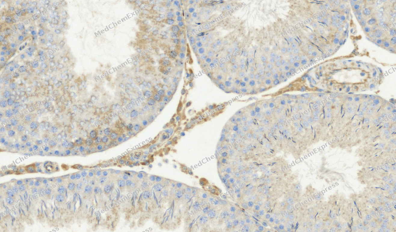 Immunohistochemical analysis of paraffin-embedded Rat testis tissue using GRB2 Antibody. The section was pre-treated using heat mediated antigen retrieval with Tris-EDTA buffer (pH 9.0) for 8 minutes. The tissues were blocked in QuickBlock for 20 minutes at room temperature, washed with ddH2O and PBS, and then probed with the primary antibody (HY-P80147, 1/200) in 4℃ overnight. The detection was performed using an HRP conjugated compact polymer system. DAB was used as the chromogen. Tissues were counterstained with hematoxylin and mounted with DPX.
 -
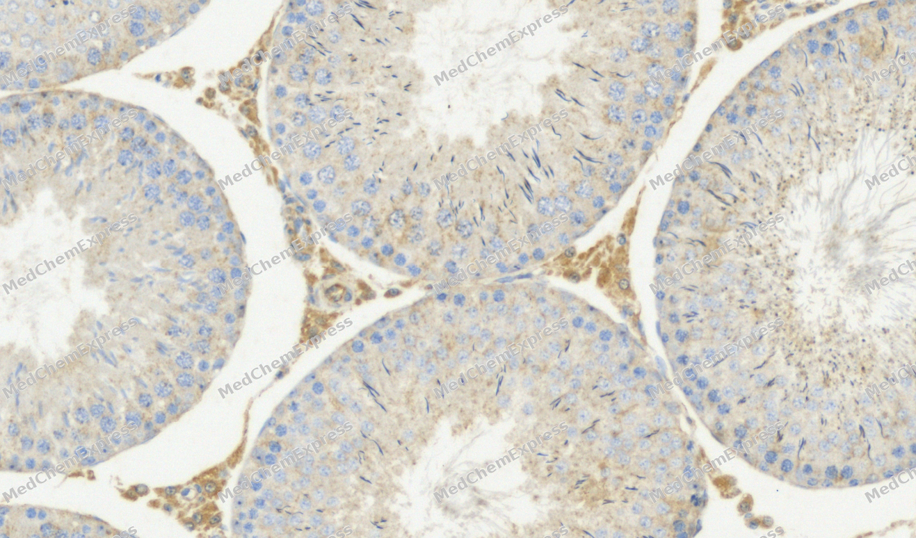 Immunohistochemical analysis of paraffin-embedded Rat testis tissue using GRB2 Antibody. The section was pre-treated using heat mediated antigen retrieval with Tris-EDTA buffer (pH 9.0) for 8 minutes. The tissues were blocked in QuickBlock for 20 minutes at room temperature, washed with ddH2O and PBS, and then probed with the primary antibody (HY-P80147, 1/200) in 4℃ overnight. The detection was performed using an HRP conjugated compact polymer system. DAB was used as the chromogen. Tissues were counterstained with hematoxylin and mounted with DPX.
 -
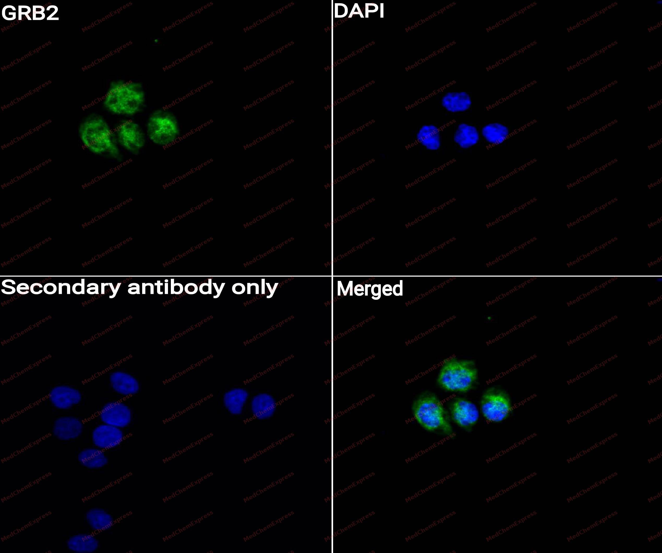 Immunocytochemistry analysis of HepG2 cells labeling GRB2 with GRB2 Antibody (HY-P80147) at 1/50 dilution. Cells were fixed in 4% paraformaldehyde for 15 minutes at room temperature, permeabilized with 0.1% Triton X-100 for 10 minutes at room temperature, then blocked with QuickBlock™ Blocking Buffer for Immunol Staining for 10 min at room temperature. Cells were then incubated with GRB2 Antibody (HY-P80147) at 1/50 dilution in QuickBlock™ Blocking Buffer for Immunol Staining at 4 ℃. Alexa Fluor® 488-conjugated AffiniPure Goat Anti-Rabbit IgG H&L(HY-P8002, Green) was used as the secondary antibody at 1/1,000 dilution. PBS instead of the primary antibody was used as the secondary antibody only control. The Nuclear counterstain was DAPI (Blue).
 -
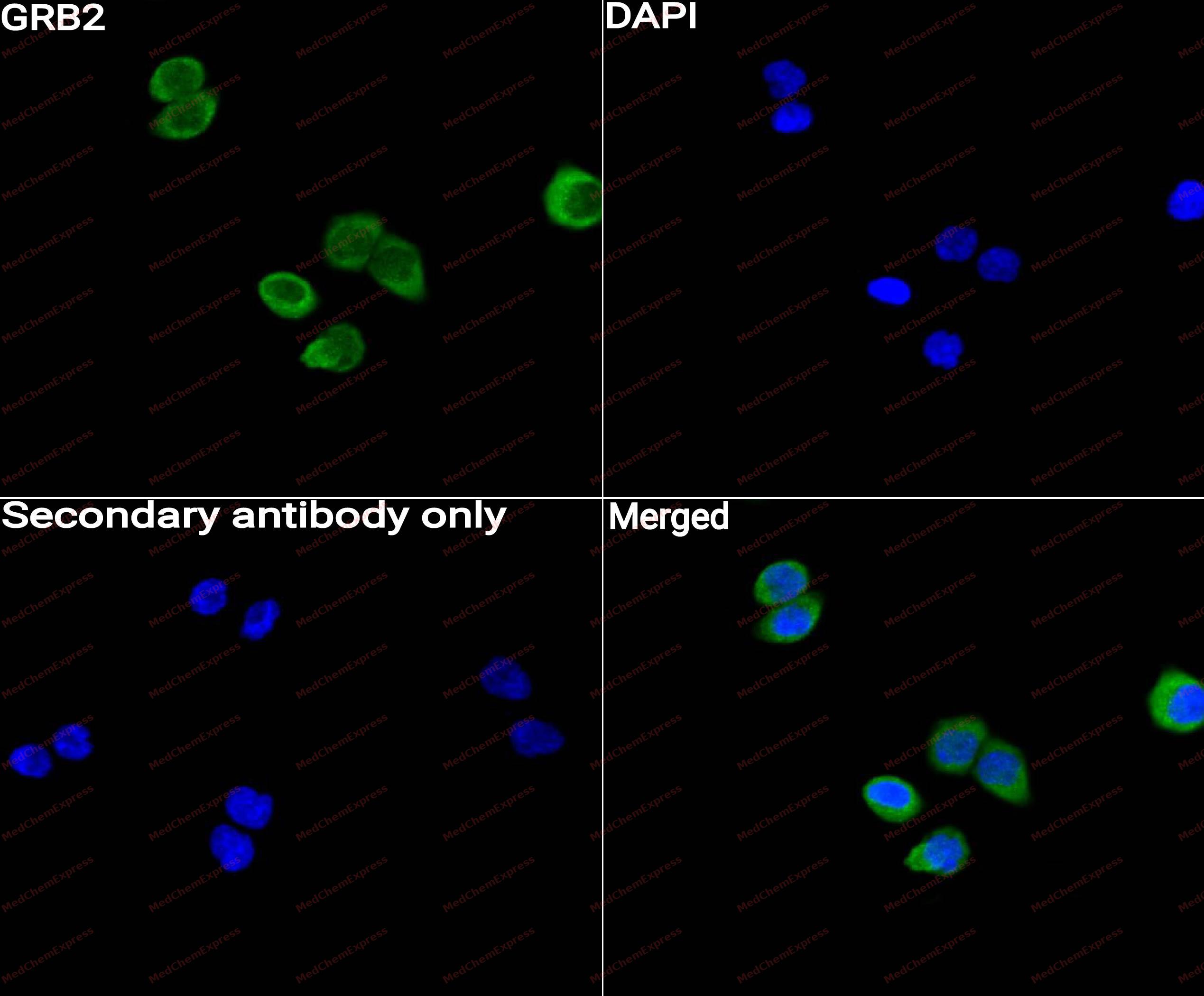 Immunocytochemistry analysis of MCF-7 cells labeling GRB2 with GRB2 Antibody (HY-P80147) at 1/50 dilution. Cells were fixed in 4% paraformaldehyde for 15 minutes at room temperature, permeabilized with 0.1% Triton X-100 for 10 minutes at room temperature, then blocked with QuickBlock™ Blocking Buffer for Immunol Staining for 10 min at room temperature. Cells were then incubated with GRB2 Antibody (HY-P80147)at 1/50 dilution in QuickBlock™ Blocking Buffer for Immunol Staining at 4 ℃. Alexa Fluor® 488-conjugated AffiniPure Goat Anti-Rabbit IgG H&L(HY-P8002,Green) was used as the secondary antibody at 1/1,000 dilution. PBS instead of the primary antibody was used as the secondary antibody only control. The Nuclear counterstain was DAPI (Blue).


Background
-
Function
GRB2 is a Non-enzymatic adapter protein that plays a pivotal role in precisely regulated signaling cascades from cell surface receptors to cellular responses, including signaling transduction and gene expression. Thus, participates in many biological processes including regulation of innate and adaptive immunity, autophagy, DNA repair or necroptosis. Controls signaling complexes at the T-cell antigen receptor to facilitate the activation, differentiation, and function of T-cells. Mechanistically, engagement of the TCR leads to phosphorylation of the adapter protein LAT, which serves as docking site for GRB2. In turn, GRB2 establishes a a connection with SOS1 that acts as a guanine nucleotide exchange factor and serves as a critical regulator of KRAS/RAF1 leading to MAPKs translocation to the nucleus and activation. Functions also a role in B-cell activation by amplifying Ca(2+) mobilization and activation of the ERK MAP kinase pathway upon recruitment to the phosphorylated B-cell antigen receptor (BCR). Plays a role in switching between autophagy and programmed necrosis upstream of EGFR by interacting with components of necrosomes including RIPK1 and with autophagy regulators SQSTM1 and BECN1. Regulates miRNA biogenesis by forming a functional ternary complex with AGO2 and DICER1. Functions in the replication stress response by protecting DNA at stalled replication forks from MRE11-mediated degradation. Mechanistically, inhibits RAD51 ATPase activity to stabilize RAD51 on stalled replication forks. Additionally, directly recruits and later releases MRE11 at DNA damage sites during the homology-directed repair (HDR) process; Does not bind to phosphorylated epidermal growth factor receptor (EGFR) but inhibits EGF-induced transactivation of a RAS-responsive element. Acts as a dominant negative protein over GRB2 and by suppressing proliferative signals, may trigger active programmed cell death. Mechanistically, inhibits RAS-ERK signaling and downstream cell proliferation by competing with GRB2 for SOS1 binding and thus by regulating SOS1 membrane recruitment[1][2][3][4][5][6][7][8][9][10][11][12][13][14].
-
Subcellular Localization
Nucleus; Cytoplasm; Endosome; Golgi apparatus
-
Subunit
Homodimer (PubMed:36864087). Associates (via SH2 domain) with activated EGF and PDGF receptors (tyrosine phosphorylated) (PubMed:10026169, PubMed:19836242, PubMed:35831301). Interacts with PDGFRA (tyrosine phosphorylated); the interaction may be indirect (By similarity). Also associates to other cellular Tyr-phosphorylated proteins such as SIT1, IRS1, IRS2, IRS4, SHC and LNK; probably via the concerted action of both its SH2 and SH3 domains (PubMed:11433379, PubMed:8388384, PubMed:8491186, PubMed:9553137, PubMed:19109239). It also seems to interact with RAS in the signaling pathway leading to DNA synthesis. Interacts with SOS1 (PubMed:11726515, PubMed:25870599, PubMed:7664271, PubMed:8493579). Forms a complex with MUC1 and SOS1, tH2O2gh interaction of the SH3 domains with SOS1 and the SH2 domain with phosphorylated MUC1 (PubMed:7664271). Interacts with phosphorylated MET (PubMed:11063574, PubMed:11827484). Interacts with phosphorylated TOM1L1 (By similarity). Interacts with the phosphorylated C-terminus of SH2B2 (PubMed:9233773). Interacts with phosphorylated SIT1, LAX1, LAT, LAT2 and LIME1 upon TCR and/or BCR activation (PubMed:12359715, PubMed:12486104, PubMed:12514734, PubMed:25870599, PubMed:9489702). Interacts with NISCH, PTPNS1 and REPS2 (PubMed:11912194, PubMed:9062191, PubMed:9422736). Interacts with syntrophin SNTA1 (By similarity). Interacts (via SH3 domains) with REPS1 (By similarity). Interacts (via SH3 domains) with PIK3C2B (PubMed:11533253). Interacts with CBL and CBLB (PubMed:10022120, PubMed:10086340). Interacts with AJUBA and CLNK (By similarity). Interacts (via SH2 domain) with TEK/TIE2 (tyrosine phosphorylated) (By similarity). Interacts with SHB, INPP5D/SHIP1, SKAP1 and SKAP2 (PubMed:10942756, PubMed:12171928, PubMed:8723348, PubMed:9108392, PubMed:9484780). Interacts with PTPN11 (By similarity). Interacts with PRNP (By similarity). Interacts with RALGPS1 (PubMed:10747847). Interacts with HCST (PubMed:16582911). Interacts with KDR (By similarity). Interacts with FLT1 (tyrosine-phosphorylated) (By similarity). Interacts with GAPT and PTPRE (PubMed:10980613, PubMed:18559951). Interacts (via SH2 domain) with KIF26A (PubMed:19914172). Interacts (via SH3 2) with GAB2 (PubMed:19523899). Interacts with ADAM15 (PubMed:18296648). Interacts with THEMIS2 (By similarity). Interacts (via SH2 domain) with AXL (phosphorylated) (PubMed:19815557, PubMed:9178760). Interacts (via SH2 domain) with KIT (phosphorylated) (PubMed:15526160, PubMed:16129412). Interacts with PTPRJ and BCR (PubMed:12475979, PubMed:15302586). Interacts with PTPN23 (PubMed:21179510). Interacts with FLT4 (tyrosine phosphorylated) (PubMed:15102829). Interacts with EPHB1 and SHC1; activates the MAPK/ERK cascade to regulate cell migration (PubMed:12925710, PubMed:8798570). Part of a complex including TNK2, GRB2, LTK and one receptor tyrosine kinase (RTK) such as AXL and PDGFRL, in which GRB2 promotes RTK recruitment by TNK2 (PubMed:19815557, PubMed:9178760). Interacts (via SH2 domain) with CSF1R (tyrosine phosphorylated) (PubMed:8262059). Interacts with ERBB4 (PubMed:10867024). Interacts with NTRK1 (phosphorylated upon ligand-binding) (PubMed:15488758). Interacts with PTK2/FAK1 (tyrosine phosphorylated) (PubMed:9148935). Interacts with PTK2B/PYK2 (tyrosine phosphorylated) (PubMed:20521079). Interacts (via SH3 domains) with GAREM1 isoform 1 (via proline-rich domain and tyrosine phosphorylated); the interaction occurs upon EGF stimulation (PubMed:19509291). Interacts with DAB2 (By similarity). Interacts with TESPA1 (PubMed:22561606). Interacts with PLCG1, LAT and THEMIS upon TCR activation in thymocytes; the association is weaker in the absence of TESPA1 (By similarity). Interacts with CD28 (PubMed:24098653). Interacts with RAB13; may recruit RAB13 to the leading edge of migrating endothelial cells where it can activate RHOA (By similarity). Interacts with ASAP3 (phosphorylated form) (PubMed:22027826). Interacts (via SH2 domain) with PTPRH (phosphorylated form) (By similarity). Interacts with PTPRO (phosphorylated form) (By similarity). Interacts with PTPRB (phosphorylated form) (By similarity). Interacts (via SH3 domain 2) with PRR14 (via proline-rich region) (PubMed:27041574). Interacts with FCRL6 (tyrosine phosphorylated form) (PubMed:20933011). Interacts with RHEX (via tyrosine-phosphorylated form) (PubMed:25092874). Interacts with DENND2B (PubMed:29030480). Interacts with SPRY2 (PubMed:17974561). Interacts with LRRC8A (By similarity). Interacts with PEAK1 (PubMed:35687021). Interacts with CD28 (By similarity). Interacts with FCRL1 (By similarity). Interacts with PCNA (PubMed:38459011). Interacts with CD19 (PubMed:29523808). Interacts with BECN1 (PubMed:38182563). Interacts with RAD51; the interaction inhibits RAD51 ATPase to stabilize RAD51-DNA complex at stalled replication forks. Interacts with MRE11; this interaction recruits MRE11 to the DNA damage sites (PubMed:34348893). Interacts with RIPK1 ans SQSTM1; these interactions play a critical role in regulating programmed necrosis (PubMed:35831301). Interacts with AGO2; this interaction is important for the formation of a ternary complex containing GRB2, AGO2 and DICER1 (PubMed:37328606). Interacts with TIGIT; this interaction inhibits PI3K and MAPK signaling cascades (PubMed:23154388). Interacts with CD226; this interaction leads to activation of VAV1, PI3K and PLCG1 (By similarity)
-
SwissProt ID
-
Synonyms
ASH, GRB2, Growth factor receptor-bound protein 2, Adapter protein GRB2, Protein Ash, SH2/SH3 adapter GRB2
-
Research Field
Signal Transduction


Documentation
-
Data Sheet (263 KB)
-
SDS (252 KB)
- English - EN (252 KB)
- Français - FR (252 KB)
- Deutsch - DE (252 KB)
- Norwegian - NO (252 KB)
- Español - ES (252 KB)
- Swedish - SV (252 KB)
- Italian - IT (252 KB)
- Korean - KR (252 KB)
- Portuguese - PT (252 KB)
-
User Guide for Antibodies (1077 KB)
References
[1]. Li S, et al. Tyrosine phosphorylation of Grb2 by Bcr/Abl and epidermal growth factor receptor: a novel regulatory mechanism for tyrosine kinase signaling. EMBO J. 2001 Dec 3;20(23):6793-804. [Content Brief]
[2]. Zhou J, et al. RNF173 suppresses RAF/MEK/ERK signaling to regulate invasion and metastasis via GRB2 ubiquitination in Hepatocellular Carcinoma. Cell Commun Signal. 2023 Aug 25;21(1):224. [Content Brief]
[3]. Hou B, et al. Grb2 interacts with necrosome components and is involved in rasfonin-induced necroptosis. Cell Death Discov. 2022 Jul 13;8(1):319. [Content Brief]
[4]. Montero-Vergara J, et al. GRB2 is a BECN1 interacting protein that regulates autophagy. Cell Death Dis. 2024 Jan 5;15(1):14. [Content Brief]
[5]. Sandouk A, et al. GRB2 dimerization mediated by SH2 domain-swapping is critical for T cell signaling and cytokine production. Sci Rep. 2023 Mar 2;13(1):3505. [Content Brief]
[6]. Zhang W, et al. LAT: the ZAP-70 tyrosine kinase substrate that links T cell receptor to cellular activation. Cell. 1998 Jan 9;92(1):83-92. [Content Brief]
[7]. Wu L, et al. SKAP55 recruits to lipid rafts and positively mediates the MAPK pathway upon T cell receptor activation. J Biol Chem. 2002 Oct 25;277(43):40420-7. [Content Brief]
[8]. Bilal MY, et al. GRB2 Nucleates T Cell Receptor-Mediated LAT Clusters That Control PLC-γ1 Activation and Cytokine Production. Front Immunol. 2015;6:141. [Content Brief]
[9]. Engels N, et al. The immunoglobulin tail tyrosine motif upgrades memory-type BCRs by incorporating a Grb2-Btk signalling module. Nat Commun. 2014 Nov 21;5:5456. [Content Brief]
[10]. Vanshylla K, et al. Grb2 and GRAP connect the B cell antigen receptor to Erk MAP kinase activation in human B cells. Sci Rep. 2018 Mar 9;8(1):4244. [Content Brief]
[11]. Stainthorp AK, et al. Regulation of microRNA expression by the adaptor protein GRB2. Sci Rep. 2023 Jun 16;13(1):9784. [Content Brief]
[12]. Ye Z, et al. GRB2 stabilizes RAD51 at reversed replication forks suppressing genomic instability and innate immunity against cancer. Nat Commun. 2024 Mar 8;15(1):2132. [Content Brief]
[13]. Ye Z, et al. GRB2 enforces homology-directed repair initiation by MRE11. Sci Adv. 2021 Aug;7(32):. [Content Brief]
[14]. Seiler C, et al. The Grb2 splice variant, Grb3-3, is a negative regulator of RAS activation. Commun Biol. 2022 Sep 28;5(1):1029. [Content Brief]

 Powered by Bioz
