



Fe2+ Indicator Probes
- 1
- 2
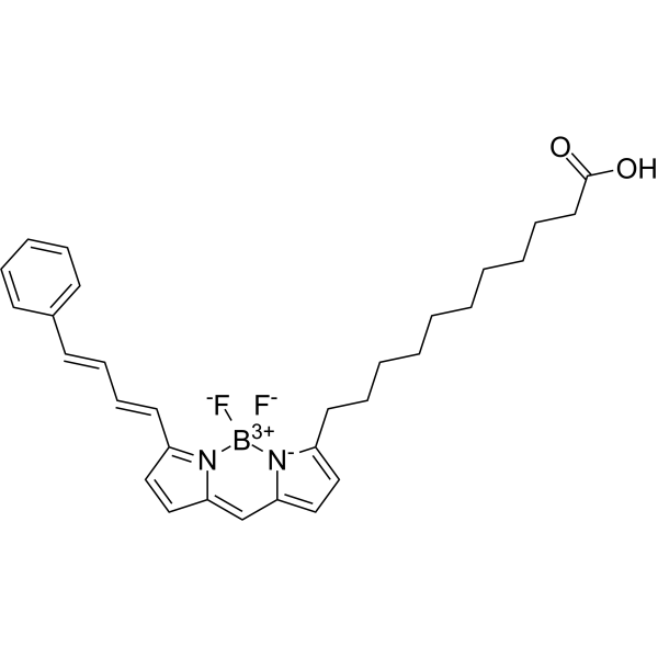
- Formula: C30H35BF2N2O2
- Molecular Weight: 504.42
BODIPY 581/591 C11 is a BODIPY borofluoroprene derivative with good light stability and low fluorescence artifacts. BODIPY 581/591 C11 can be used for study lipid peroxidation and antioxidant properties in living cells, or detect ferroptosis by reaction with hydroxyl radicals. BODIPY 581/591 C11 is emitted at 591 nm (reduced prototype), or redshifted to 510 nm (oxidized type). The excitation wavelengths were 581 nm (reduced prototype) and 500 nm (oxidized type).
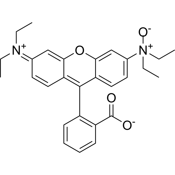
- Formula: C28H30N2O4
- Molecular Weight: 458.55
RhoNox-1 is a fluorescent probe for the specific detection of divalent iron ions, and when RhoNox-1 reacts with Fe2+. RhoNox-1 can generate an irreversible orange (red) fluorescent product (Ex/Em:540/575 nm) . FeRhoNox-1 can enter the cell well, suitable for the detection of Fe2+ in living cells, and tends to be localized in the Golgi apparatus.
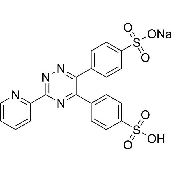
- Formula: C20H13N4NaO6S2
- Molecular Weight: 492.46
Ferrozine is a spectrophotometric reagent for iron ions, can react with divalent Fe to form a stable magenta complex species. The complex has an absorption peak at 562 nm. Ferrozine-based colorimetric assays can quantify iron in cells
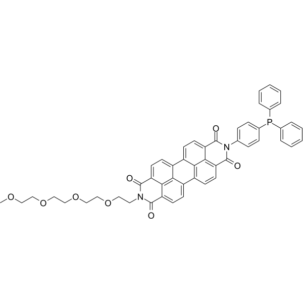
- Formula: C51H41N2O8P
- Molecular Weight: 840.85
LPd peroxida probe-1, a marker of ferroptosis, is a useful fluorescent probe for investigating the roles of lipid peroxidation in a variety of cell pathophysiologies. LPd peroxida probe-1 reduces lipid hydroperoxides to lipid alcohols and is used for imaging lipid hydroperoxides in living cells.

- Formula: C38H44N4O8
- Molecular Weight: 684.78
RhoNox-4 is a ferroptosis inhibitor and a fluorescence-activating indicator specific to Fe2+. RhoNox-4 selectively reacts with labile Fe2+ and oxidizes it to Fe3+, depleting labile Fe2+ involved in lipid peroxidation and inhibiting ferroptosis induced by ferroptosis inducers. RhoNox-4 is cell-permeable, accumulates in the endoplasmic reticulum and/or Golgi apparatus, and can be used for live-cell imaging as well as high-throughput screening of subcellular iron status modulators, making it applicable for iron biology research.

- Formula: C24H24N2O3
- Molecular Weight: 388.46
HMRhoNox-M (LysoRhoNox) is a selective fluorescent probe for detecting Fe2+ (ferrous iron ions), with excitation/emission wavelengths of Ex/Em = 550/575 nm. HMRhoNox-M exists in a non-fluorescent spirocyclic closed conformation under physiological pH conditions. Fe2+ triggers the deoxidation of its N-oxide group, shifting the equilibrium toward the fluorescent quinone-type (TMHMR) open conformation. HMRhoNox-M shows selectivity for Fe2+ over other metal ions, as well as biologically relevant reactive oxygen species, reactive sulfur species, reactive nitrogen species and reducing agents. HMRhoNox-M can penetrate cell membranes and localize to lysosomes, enabling the imaging of intracellular Fe2+ fluctuations, transferrin-mediated labile iron accumulation, and lysosomal Fe2+ changes associated with ferroptosis.

- Formula: C67H71N5O27
- Molecular Weight: 1378.30
FlCFe1 is a ratiometric ferrous ion probe that enables imaging of iron distribution in 3D cell models such as tumor spheroids. Its detection mechanism relies on its structure as a ratiometric analog of calcein AM. It has an excitation wavelength of 405 nm, with emission wavelengths of 490 nm (coumarin emission signal) and 530 nm (fluorescein emission signal).

- Formula: C89H138N8O22
- Molecular Weight: 1672.09
Mem-RhoNox is a fluorescent probe used to detect labile Fe (II) on the plasma membrane surface and inside endosomes during iron uptake in live cells. Mem-RhoNox can anchor to the extracellular surface of the plasma membrane; upon contact with Fe (II), its N-oxide group undergoes a deoxygenation reaction, converting weakly fluorescent Mem-RhoNox into strongly fluorescent Mem-Rhodamine. During transferrin-mediated endocytosis, Mem-RhoNox enters endosomes together with the transferrin-transferrin receptor complex, and reacts with Fe (II) released by transferrin to enable detection of endosomal Fe (II).
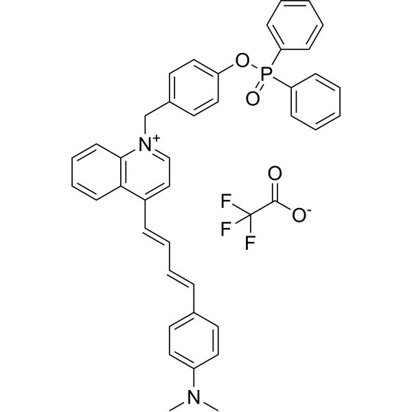
- Formula: C42H36F3N2O4P
- Molecular Weight: 720.72
MQA-P is a multifunctional near-infrared (NIR) fluorescent probe that simultaneously detects ONOO-, viscosity, and polarity within mitochondria. MQA-P exhibits significant response to ONOO-, λem=645 nm; and NIR channel at λem>704 nm Medium is highly sensitive to viscosity/polarity. MQA-P possesses excited-state intramolecular charge transfer (ESICT) properties that are highly sensitive to polarity by designing the N,N-dimethylamino group as the electron donor and the quinoline cation unit as the electron acceptor. MQA-P is used for ferroptosis or cancer diagnosis in vitro and in vivo via dual-channel images.

- Formula: C20H19N3O
- Molecular Weight: 317.38
DCA-Mln is a Fluorescent probe for ferric iron (Fe³⁺) detection. The probe works via an internal charge transfer (ICT) process from its diethylamino group to its dicyano group, which produces bright pink fluorescence in its unbound state; when Fe³⁺ is present, two molecules of DCA-Mln chelate with one Fe³⁺ ion, with one cyano group of each DCA-Mln participating in the complexation, and this binding triggers a photoinduced electron transfer (PET) process that provides a nonradiative deactivation pathway, resulting in fluorescence quenching; additionally, the probe exhibits a visible color change from purple to peach pink upon Fe³⁺ binding, enabling dual-channel detection. The probe has an excitation wavelength of 570 nm and an emission wavelength of 670 nm, and it shows a rapid response, with fluorescence stabilizing within 15 seconds of Fe³⁺ addition. DCA-Mln is cell-membrane-permeable, nontoxic at the imaging concentration of 10 μM, and can be used for Fe³⁺ detection in actual water samples, Fe³⁺ imaging in living cells, and as an anti-counterfeiting ink[1].

- Formula: C62H73N7O5
- Molecular Weight: 996.29
Mito-RhFe is a Fluorescent probe for mitochondrial labile Fe³⁺ monitoring via imaging and flow cytometry. This probe is a rhodamine-based construct with a spirolactam fluorescence signaling group and an N2-hydroxyethyldiethylenetriamine chelator; its delocalized positive charge enables mitochondria-targeting ability in live cells, and it exhibits fine cell membrane permeability. In its native state, it exists in the non-fluorescent spirolactam form, but upon binding to Fe³⁺, it undergoes a ring-opening conversion to the fluorescent rhodamine form, triggering a turn-on fluorescent response; this process is reversible, as the addition of the metal chelator TPEN removes Fe³⁺ and converts the probe back to its non-fluorescent spirolactam form, and re-addition of Fe³⁺ restores fluorescence. The probe shows high selectivity for Fe³⁺ over most other metal cations present in living systems, with a ~90-fold fluorescence enhancement upon binding to 20 equiv of Fe³⁺. Mito-RhFe has excitation/emission wavelengths of Ex/Em = 540/578 nm, with an ~8 nm bathochromic shift in emission upon Fe³⁺ binding, and it can also be excited at 543 nm for confocal imaging with emission detected at 570-620 nm[1].

- Formula: C47H68N4O5Si
- Molecular Weight: 797.15
Gol-SiRhoNox is a Golgi apparatus-targeted fluorescent probe with high selectivity for Fe (II), with a limit of detection of 50 nM. Gol-SiRhoNox integrates a myristoyl motif for Golgi apparatus targeting and a silicon-fused rhodamine N-oxide as the Fe (II)-sensitive moiety. Gol-SiRhoNox is non-fluorescent in its native state; Fe (II)-mediated deoxygenation of the N-oxide triggers fluorescence recovery. Its fluorescent product, Gol-SiR, has excitation/emission wavelengths of 630/665 nm and a maximum absorption wavelength of 655 nm.

- Formula: C18H12N4OS
- Molecular Weight: 332.38
NtHzBtd is a fluorescent probe for detecting Fe3+. NtHzBtd is applicable to the selective fluorescent detection of Fe3+ ions and live cell imaging studies. NtHzBtd can selectively coordinate with Fe3+ to form a 1:1 complex, triggering chelation enhanced quenching (CHEQ) and intramolecular charge transfer (ICT) processes, which result in fluorescence turn-off, thereby enabling sensitive detection of Fe3+ and live cell fluorescence imaging. After binding to Fe3+, NtHzBtd reduces fluorescence intensity, exhibits a rapid response property, with a limit of detection of 0.036 μM and a response time of approximately 55 s. The detection wavelengths are Ex/Em = 334/401 nm (solution system).

- Formula: C48H42F6N2O7P2
- Molecular Weight: 934.79
Ac-MtFluNox is a mitochondria-targeted Fluorescent probe that can be used for the selective detection of labile Fe (II). Ac-MtFluNox can enter living cells, where intracellular esterases cleave its acetyl protecting group to generate activated MtFluNox. Ac-MtFluNox functions through an Fe (II)-mediated deoxygenation mechanism of its amine N-oxide group, which eliminates the fluorescence quenching effect and oxidizes Fe (II) to Fe (III) to deplete the catalytically active Fe (II) pool. A FITC filter set (excitation 465-500 nm, emission 516-556 nm) is used for live-cell imaging. Ac-MtFluNox can be applied to studies related to myocardial ferroptosis.

- Formula: C29H21BN4O3
- Molecular Weight: 484.31
IOPBA is a fluorescent probe that can be used for the detection and bioimaging of trivalent iron ions Fe3+ and fluoride ions F- in living cells. IOPBA emits blue fluorescence in its native state, but upon the introduction of Fe3+, it forms a Fe3+-IOPBA π-complex, where Fe3+ ions coordinate with two phenyl groups of IOPBA, resulting in fluorescence quenching. IOPBA returns to its free state and recovers its original fluorescence after F- sequesters Fe3+ to form the more stable [FeF6]3- and dissociates the Fe3+-IOPBA complex. The excitation wavelength of IOPBA is 342 nm, and its emission wavelength is 458 nm.

- Formula: C46H56N6O5S
- Molecular Weight: 805.04

- Formula: C20H13N4NaO6S2
- Molecular Weight: 492.46
Ferrozine (solution) is a spectrophotometric reagent for iron ions, can react with divalent Fe to form a stable magenta complex species. The complex has an absorption peak at 562 nm. Ferrozine-based colorimetric assays can quantify iron in cells
Solvent and concentration: ddH2O: 10 mM
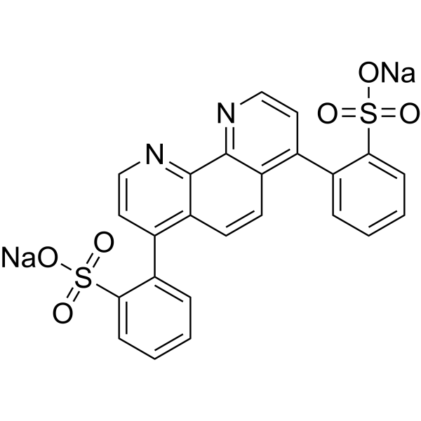
- Formula: C24H14N2Na2O6S2
- Molecular Weight: 536.49
Bathophenanthroline disulfonic acid sodium is a multifunctional dye. Dyes are important tools in biological experiments. They can help researchers observe and analyze cell structures, track biomolecules, evaluate cell functions, distinguish cell types, detect biomolecules, study tissue pathology and monitor microorganisms. Their applications range from basic scientific research to clinical A wide range of diagnostics. Dyes are also widely used in traditional fields such as textile dyeing, as well as in emerging fields such as functional textile processing, food pigments and dye-sensitized solar cells.
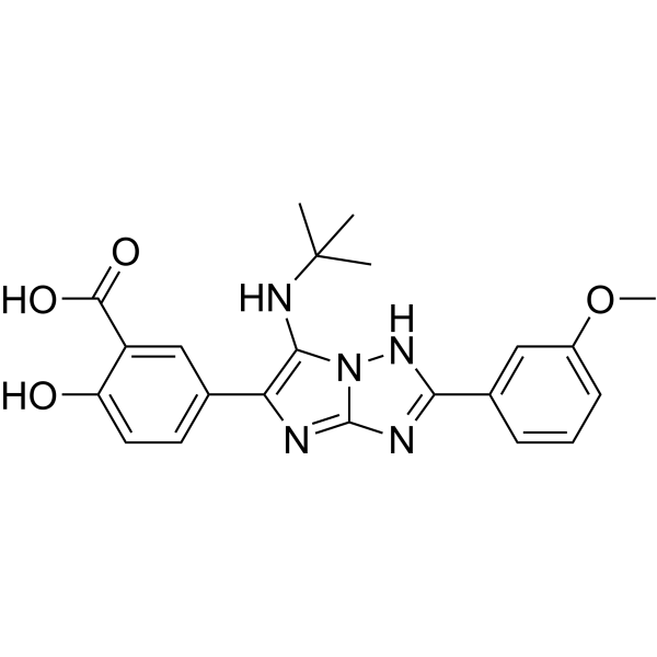
- Formula: C22H23N5O4
- Molecular Weight: 421.45
SHP1-IN-1 (compound 5p) is a fluorescent probe for the protein tyrosine phosphatase SHP1 containing the Src homology 2 domain. SHP1-IN-1 has SHP1 inhibitory activity, selectivity for Fe3+ ions and good fluorescence properties. SHP1-IN-1 exhibits aggregation post-quenching (ACQ) effect, good interference immunity and low detection limit (5.55 μM).
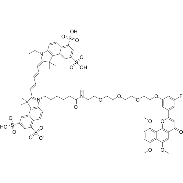
- Formula: C71H76FN3O22S4
- Molecular Weight: 1470.63
CYP1B1-IN-6 (compound 19) is a fluorescence molecular probes which inhibits CYP1B1 activity. CYP1B1-IN-6 can identify tumor sites in fluorescence imaging and photoacoustic imaging modes.
- 1
- 2



