



Cathepsin D Antibody (YA9975)
(Synonyms: CPSD, CTSD, Cathepsin D)Cathepsin D Antibody (YA9975) is a Rabbit-derived and non-conjugated IgG Monoclonal antibody, targeting to Cathepsin D Antibody.
-
Host:
Rabbit
-
Isotype:
IgG
-
Application:
WB, IHC-P, ICC/IF, IP, FC
-
Reactivity :
Human, Mouse, Rat
-
Formulation:
Supplied in PBS, 50% glycerol, 0.05% BSA, and 0.05% Proclin300.
-
Conjugation:
Non-conjugated


To place orders, for customer services and technical support, please contact: MedChemExpress USA
Tel: 609-228-6898 E-mail: [email protected] [email protected]





Applications
| Application |
WB

WB: Western Blot
|
IHC-P
IHC-P: Immunohistochemistry-Paraffin
|
ICC/IF
ICC/IF: Immunocytochemistry/
Immunofluorescence |
IP
IP: Immunoprecipitation
|
FC
FC: Flow Cytometry
|
|---|---|---|---|---|---|
| Dilution Ratio | 1:1000-2000 | 1:100-200 | 1:100-200 | 1:10-50 | 1:10-50 |


Product Details
Cathepsin D Antibody (YA9975) is a Rabbit-derived and non-conjugated IgG Monoclonal antibody, targeting to Cathepsin D Antibody.
-
Host Rabbit

-
Clonality Monoclonal,Recombinant

-
Species ReactivityHuman, Mouse, Rat
-
Observed Molecular WeightObserved band size: 33 kDa
Note: Due to possible protein modifications or aggregation, the molecular weight should be confirmed by actual measurement, and the predicted value is for reference only.

-
Calculated Molecular Weight Predicted band size: 45 kDa

Synthetic peptide corresponding to human Cathepsin D.
Endogenous
affinity purified
Non-conjugated
Unmodified
IgG


Product Properties
-
Appearance
Solution
-
Formulation
Supplied in PBS, 50% glycerol, 0.05% BSA, and 0.05% Proclin300.
-
Storage & Stability
Stored at -20°C for 1 year. Avoid repeated freeze / thaw cycles.
-
Shipping
Shipping with blue ice.


Verification Images
-
 Western blot analysis of extracts from Human liver (lane 1(20μg)) 、MCF7 (lane 2(20μg)) 、Mouse brain (lane 3(20μg)) 、Rat brain (lane 4(20μg)) and Rat liver (lane 5(20μg)) using Cathepsin D Antibody. Proteins were transferred to a PVDF membrane and blocked with 5% nonfat dry milk in TBST for 1.5 hour at room temperature. The primary antibody (HY-P810731, 1/1000) and Loading control antibody (β Tubulin, 1/10000) was used in 5% nonfat dry milk in TBST at 4℃ overnight. Goat Anti-Rabbit IgG-HRP Secondary Antibody (1/10,000) was used for 1 hour at room temperature.
 -
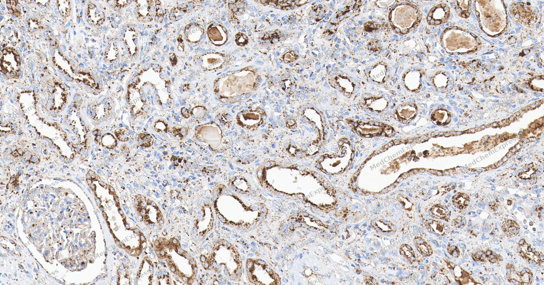 Immunohistochemical analysis of paraffin-embedded human kidney tissue using Cathepsin D Antibody. The section was pre-treated using heat mediated antigen retrieval with Tris/EDTA buffer (pH 9.0) for 20 minutes. The tissues were probed with the primary antibody (HY-P810731, 1/150) overnight at 4℃. The detection was performed using an HRP conjugated compact polymer system (HY-P83652). DAB was used as the chromogen. Tissues were counterstained with hematoxylin and mounted with neutral balsam.
 -
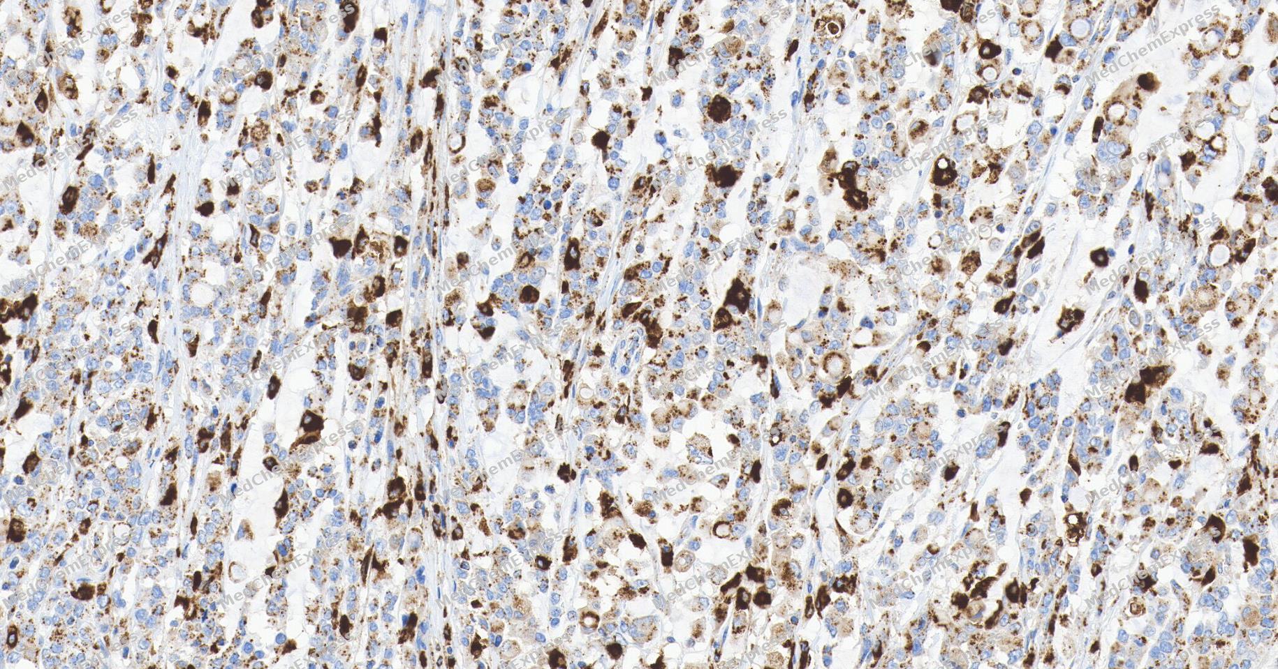 Immunohistochemical analysis of paraffin-embedded human gastric cancer tissue using Cathepsin D Antibody. The section was pre-treated using heat mediated antigen retrieval with Tris/EDTA buffer (pH 9.0) for 20 minutes. The tissues were probed with the primary antibody (HY-P810731, 1/150) overnight at 4℃. The detection was performed using an HRP conjugated compact polymer system (HY-P83652). DAB was used as the chromogen. Tissues were counterstained with hematoxylin and mounted with neutral balsam.
 -
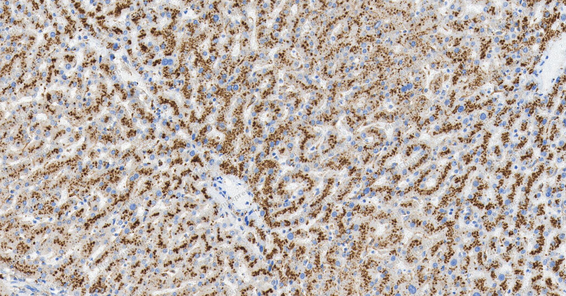 Immunohistochemical analysis of paraffin-embedded human liver tissue using Cathepsin D Antibody. The section was pre-treated using heat mediated antigen retrieval with Tris/EDTA buffer (pH 9.0) for 20 minutes. The tissues were probed with the primary antibody (HY-P810731, 1/150) overnight at 4℃. The detection was performed using an HRP conjugated compact polymer system (HY-P83652). DAB was used as the chromogen. Tissues were counterstained with hematoxylin and mounted with neutral balsam.
 -
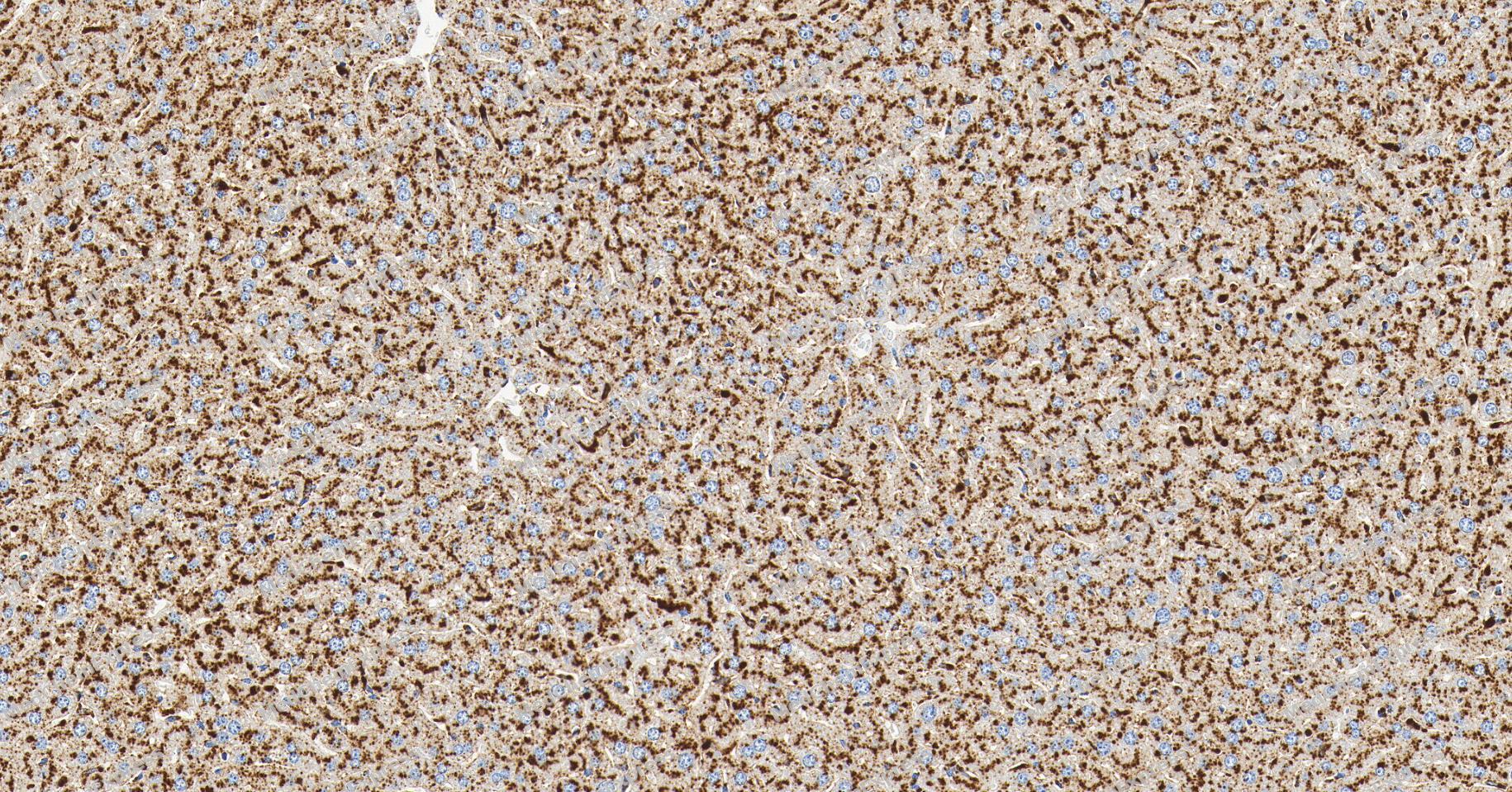 Immunohistochemical analysis of paraffin-embedded mouse liver tissue using Cathepsin D Antibody. The section was pre-treated using heat mediated antigen retrieval with Tris/EDTA buffer (pH 9.0) for 20 minutes. The tissues were probed with the primary antibody (HY-P810731, 1/150) overnight at 4℃. The detection was performed using an HRP conjugated compact polymer system (HY-P83652). DAB was used as the chromogen. Tissues were counterstained with hematoxylin and mounted with neutral balsam.
 -
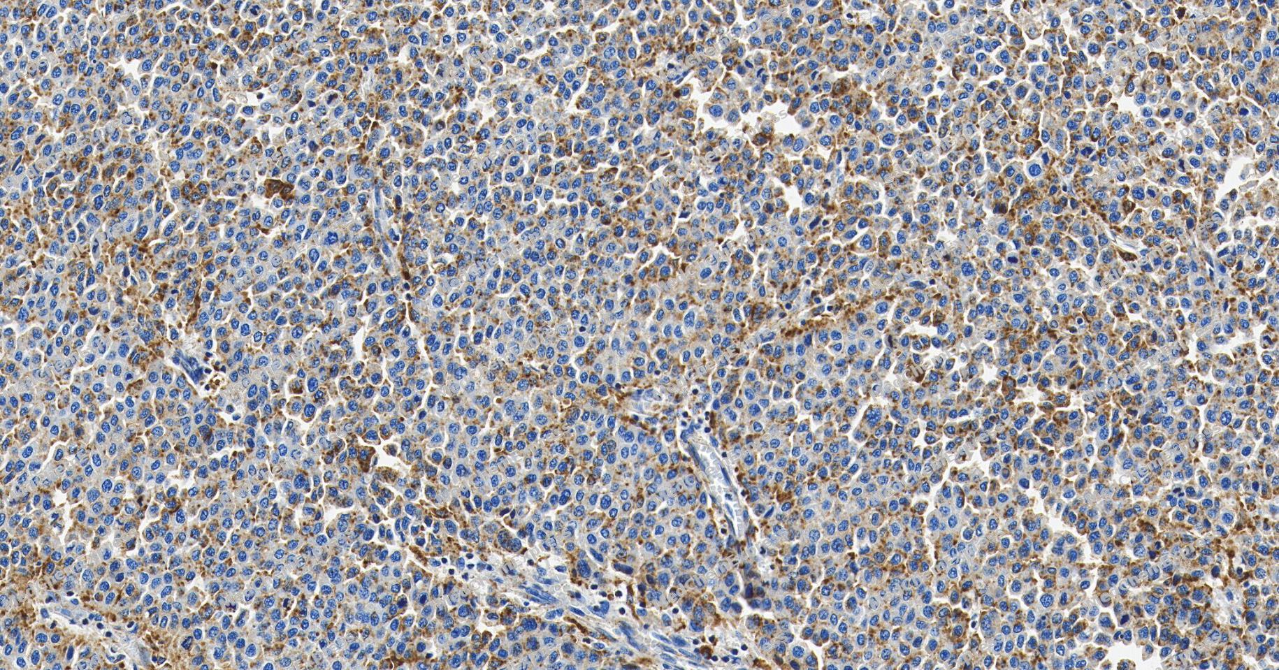 Immunohistochemical analysis of paraffin-embedded human liver cancer tissue using Cathepsin D Antibody. The section was pre-treated using heat mediated antigen retrieval with Tris/EDTA buffer (pH 9.0) for 20 minutes. The tissues were probed with the primary antibody (HY-P810731, 1/150) overnight at 4℃. The detection was performed using an HRP conjugated compact polymer system (HY-P83652). DAB was used as the chromogen. Tissues were counterstained with hematoxylin and mounted with neutral balsam.
 -
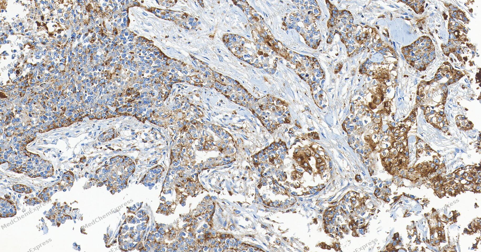 Immunohistochemical analysis of paraffin-embedded human breast cancer tissue using Cathepsin D Antibody. The section was pre-treated using heat mediated antigen retrieval with Tris/EDTA buffer (pH 9.0) for 20 minutes. The tissues were probed with the primary antibody (HY-P810731, 1/150) overnight at 4℃. The detection was performed using an HRP conjugated compact polymer system (HY-P83652). DAB was used as the chromogen. Tissues were counterstained with hematoxylin and mounted with neutral balsam.
 -
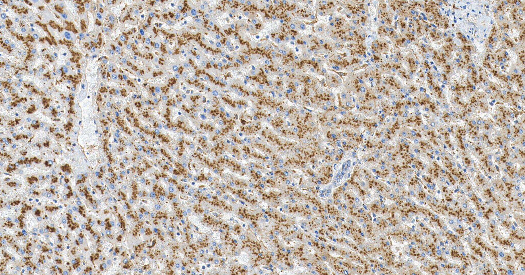 Immunohistochemical analysis of paraffin-embedded human liver tissue using Cathepsin D Antibody. The section was pre-treated using heat mediated antigen retrieval with Tris/EDTA buffer (pH 9.0) for 20 minutes. The tissues were probed with the primary antibody (HY-P810731, 1/300) overnight at 4℃. The detection was performed using an HRP conjugated compact polymer system (HY-P83652). DAB was used as the chromogen. Tissues were counterstained with hematoxylin and mounted with neutral balsam.


Background
-
Function
Cathepsin D is an Acid protease active in intracellular protein breakdown. Plays a role in APP processing following cleavage and activation by ADAM30 which leads to APP degradation. Involved in the pathogenesis of several diseases such as breast cancer and possibly Alzheimer disease[1].
-
Subcellular Localization
Lysosome; Melanosome; Secreted, extracellular space
-
Expression
Tissue_Specificity: Expressed in the aorta extracellular space (at protein level). Expressed in liver (at protein level). -
Isoforms & Post-Translational Modification
Cathepsin D has an amino acid length of 412, molecular weight is 44552 Da.
N- and O-glycosylated. -
Subunit
Consists of a light chain and a heavy chain.
-
SwissProt ID
-
Synonyms
CPSD, CTSD, Cathepsin D


Documentation
References

 Powered by Bioz
