



AI-3
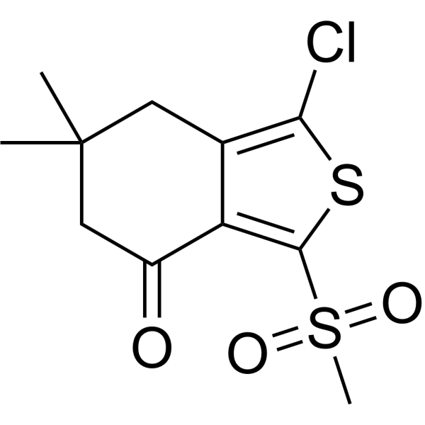
AI-3 is a potent ARE (antioxidant response element) activator. AI-3 increases the NQO1 at the transcript levels and protein expression levels. AI-3 has the potential for the research of oxidative stress related diseases.
For research use only. We do not sell to patients.
- CAS No.: 882288-28-0
- Formula: C11H13ClO3S2
- Molecular Weight:292.80
-
Storage:
Please store the product under the recommended conditions in the Certificate of Analysis.

To place orders, for customer services and technical support, please contact: MedChemExpress USA
Tel: 609-228-6898 E-mail: [email protected] [email protected]


-
 Biological Activity
-
Chemical Information
- Protocols
- Purity & Documentation
- References
-
 Help & FAQs


-
HY-17363
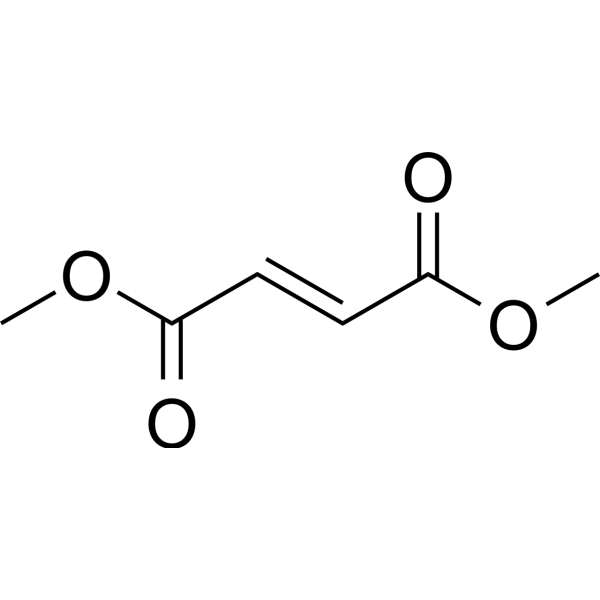
Dimethyl fumarateNrf2 Activator -
HY-19734
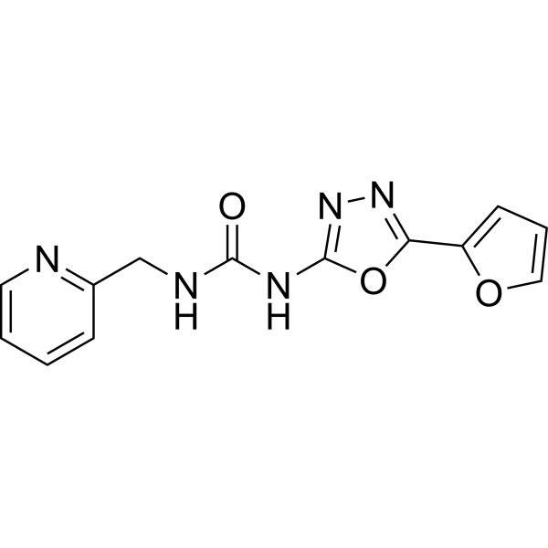
NK-252Nrf2 Activator -

HY-17363R
Dimethyl fumarate (Standard)Nrf2 Activator -
HY-112675
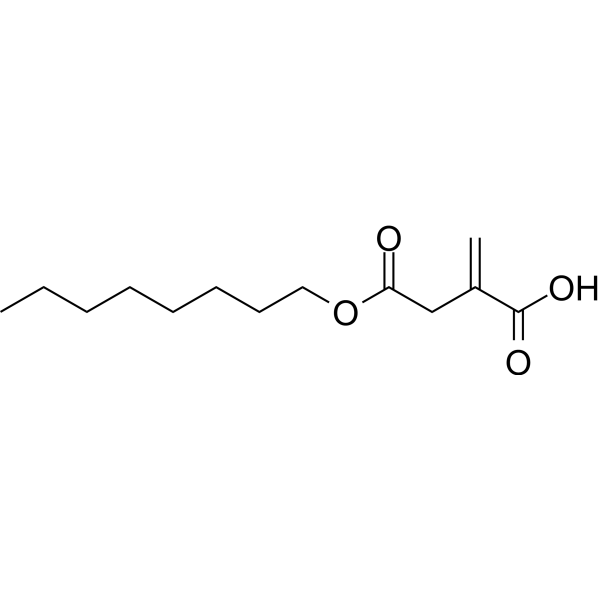
4-Octyl itaconateNrf2 Activator -

HY-130495
CDDO-TFEA -
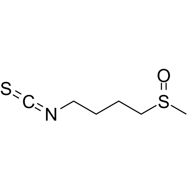
HY-13755
SulforaphaneKeap1/Nrf2/ARE Inducer -
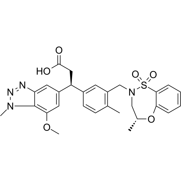
HY-101140
KI696KEAP1/NRF2 Protein-protein Interaction Inhibitor -

HY-P1328
TAT-14Nrf2 Activator -
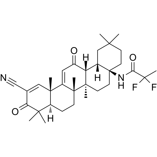
HY-12212
OmaveloxoloneNrf2 Activator -
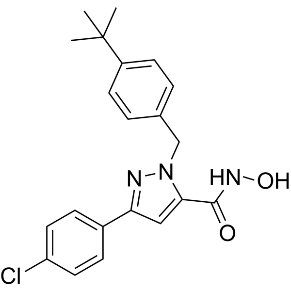
HY-101025
Nrf2-IN-1Nrf2 Inhibitor -
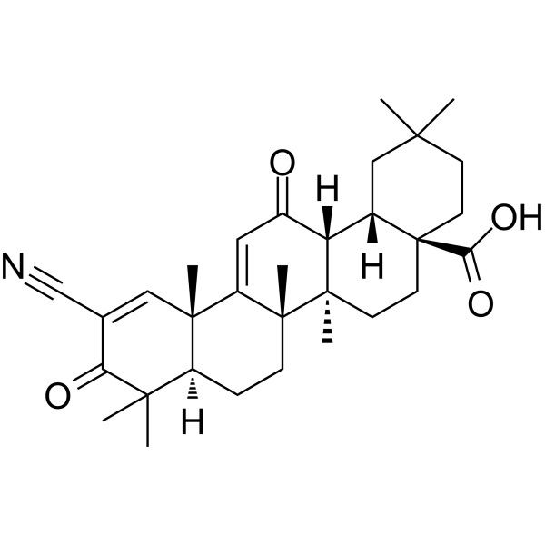
HY-14909
BardoxoloneNrf2 Activator -
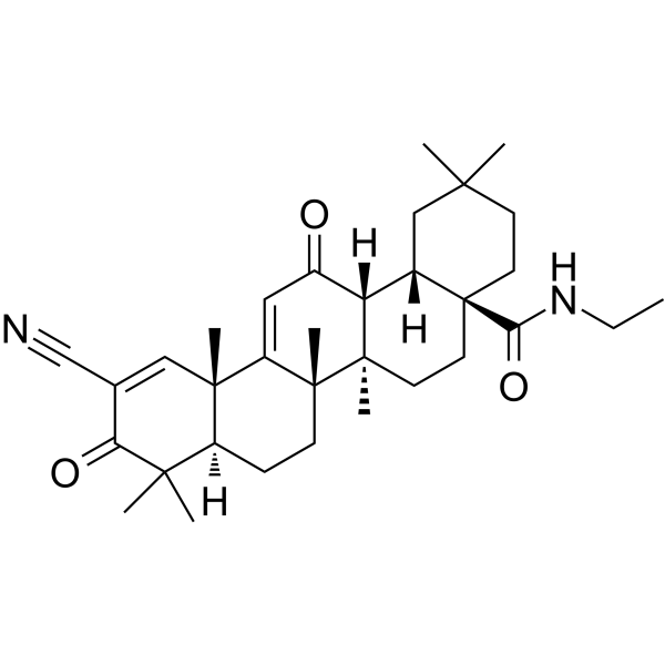
HY-12213
CDDO-EAKeap1-Nrf2 Activator


Biological Activity
Description
IC50 & Target
ARE[1]
In Vitro
AI-3 (0.1, 0.3, 1, 3.2, 10, 32 μM; 12, 24 h) increases the NQO1 at the transcript levels and protein expression levels in a dose-dependent manner in IMR-32 cells[1].
AI-3 (10 μM; 2-24 h) induces GSH synthesis starting from 4 h up to 24 h[1].
AI-3 requires Nrf2 and PI3K for its bioactivities in IMR-32 cells[1].
MedChemExpress (MCE) has not independently confirmed the accuracy of these methods. They are for reference only. Further protocols information, click here.
-
Cell Line:IMR-32 cells
-
Concentration:0.1, 0.3, 1, 3.2, 10, 32 µM
-
Incubation Time:24 h
-
Result:Induced NQO1 protein expression starting from 0.3 μM.


Chemical Information
-
CAS No. 882288-28-0
-
Molecular Weight 292.80
-
Formula C11H13ClO3S2
-
SMILES
O=C1C2=C(S(=O)(C)=O)SC(Cl)=C2CC(C)(C1)C
-
Shipping
Room temperature in continental US; may vary elsewhere.
-
Storage
Please store the product under the recommended conditions in the Certificate of Analysis.

Protocols
-
RNA extraction experimental
By lysing cells, releasing RNA, and removing impurities such as proteins and DNA, high-purity RNA products are finally obtained. The commonly used traditional method is the guanidine isothiocyanate/phenol/chloroform method (Trizol), which is suitable for a variety of animal materials including animal tissues, microorganisms, cultured cells, etc., and most plant materials.
-
Western Blot
Western blotting (WB) is a commonly used experimental method in molecular biology, biochemistry, and immunogenetics for identifying and quantifying target proteins. It combines gel electrophoresis with immunoassay, enabling researchers to analyze protein expression, post-translational modifications, and molecular weight.
-
ROS/oxidative-stress fluorescent staining
ROS/oxidative-stress fluorescent staining uses cell-permeant fluorogenic probes that become fluorescent after oxidation inside cells or tissues; commonly used examples include DCFH-DA/DCFDA for broad cellular oxidant detection, DHE for superoxide-related signal detection, MitoSOX for mitochondrial superoxide-related signal detection, and CellROX probes for oxidative-stress-associated fluorescence readouts. The assay detects probe oxidation rather than a single ROS species unless the probe and analysis method have been chemically validated for that species. DCFH-DA enters cells, is deacetylated by intracellular esterases to DCFH, and produces fluorescent DCF after oxidation, so the readout is used as an operational measure of total cellular oxidative stress rather than a species-specific ROS measurement. DHE and MitoSOX can report superoxide-related oxidation, but red fluorescence alone can include non-specific ethidium-like oxidation products; HPLC or optimized spectral approaches are
-
Protocol For Protein Expression And Purification
Recombinant protein expression in Escherichia coli followed by purification of a His-tagged soluble protein by immobilized metal affinity chromatography (IMAC), with optional MBP fusion and TEV tag removal when the construct includes these elements. The biological readout is production of the encoded target protein, detected as an inducible band at the expected molecular mass by SDS-PAGE and quantified by total protein assay or chromatographic absorbance; the purification readout is enrichment of the target protein in elution fractions after selective binding of polyhistidine residues to immobilized Ni2+/metal-chelate resin and elution by imidazole-containing buffer. Expression is driven by an inducible bacterial expression system, commonly T7/lac-based, in which IPTG or lactose/auto-induction activates transcription and translation of the cloned gene; lower induction temperature, lower inducer concentration, induction timing, and solubility-enhancing fusion tags can influence the frac


Purity & Documentation
References

Calculators
Concentration (start) × Volume (start) = Concentration (final) × Volume (final)


AI-3 Related Classifications
HY-107736 Related Classifications
 Powered by Bioz