



Crystal Violet-d6
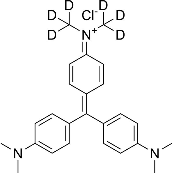
Crystal Violet-d6 is the deuterium labeled Crystal Violet (HY-B0324A). Crystal Violet, also known as Gentian violet, methyl violet 10B, is a triphenyl-methane, an alkaline dye that binds to DNA in the nucleus of a cell, staining it a deep purple. It is often used for Gram staining to classify bacteria, or for cell or histological staining.
연구목적의 판매만을 진행합니다. 환자를 대상으로 한 판매는 하지 않습니다.
- CAS No.: 1266676-01-0
- 화학식: C25H24D6ClN3
- 분자량:414.02
-
보관:
Please store the product under the recommended conditions in the Certificate of Analysis.

To place orders, for customer services and technical support, please contact: MedChemExpress USA
Tel: 609-228-6898 E-mail: [email protected] [email protected]


-
 Biological Activity
-
Chemical Information
- Protocol
- 순도&문서
- References
-
 Help & FAQs




Biological Activity
제품 설명
In Vitro
Stable heavy isotopes of hydrogen, carbon, and other elements have been incorporated into drug molecules, largely as tracers for quantitation during the drug development process. Deuteration has gained attention because of its potential to affect the pharmacokinetic and metabolic profiles of drugs[75].
MedChemExpress (MCE) has not independently confirmed the accuracy of these methods. They are for reference only. Further protocols information, click here.
신청
1. This compound can be used as a tracer
2. This compound can be used as an internal standard for quantitative analysis by NMR, GC-MS, or LC-MS.


Chemical Information
-
CAS No. 1266676-01-0
-
Unlabeled CAS 548-62-9
-
분자량 414.02
-
화학식 C25H24D6ClN3
-
SMILES
CN(C)C(C=C1)=CC=C1/C(C2=CC=C(C=C2)N(C)C)=C3C=C/C(C=C/3)=[N+](C([2H])([2H])[2H])/C([2H])([2H])[2H].[Cl-]
-
Synonyms
Basic Violet 3-d6; Gentian Violet-d6; Methyl Violet 10B-d6
-
선적
Room temperature in continental US; may vary elsewhere.
-
보관
Please store the product under the recommended conditions in the Certificate of Analysis.

Protocol
-
Gram Staining of Tissue Sections
Gram staining of tissue sections is a histochemical technique used to differentiate Gram-positive and Gram-negative bacteria within histological specimens based on differences in bacterial cell wall structure and dye retention, adapted from classical bacteriological Gram staining into tissue-compatible “histological Gram stain” variants. In tissue applications, modifications of the Brown-Hopps and Brown-Brenn methods are commonly used to improve differentiation of microorganisms embedded within host connective tissue and to reduce overstaining or loss of Gram-negative signal, which are known limitations of earlier approaches. The principle relies on crystal violet-iodine complex retention in Gram-positive organisms and subsequent decolorization and counterstaining steps that allow contrast visualization of Gram-negative organisms against tissue background.


순도&문서
References

Calculators
Concentration (start) × Volume (start) = Concentration (final) × Volume (final)


Crystal Violet-d6 Related Classifications
HY-B0324AS Related Classifications
 Powered by Bioz