
CSRM617
Based on 1 publication(s) in Google Scholar
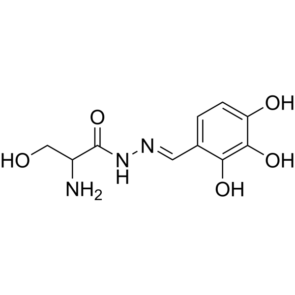
CSRM617 is a selective small-molecule inhibitor of the transcription factor ONECUT2 (OC2, a master regulator of androgen receptor) with a Kd of 7.43 uM in SPR assays, binding to OC2-HOX domain directly. CSRM617 induces apoptosis by appearance of cleaved Caspase-3 and PARP. CSRM617 is well tolerated in the prostate cancer mouse model
연구목적의 판매만을 진행합니다. 환자를 대상으로 한 판매는 하지 않습니다.
- CAS No.: 787504-88-5
- 화학식: C10H13N3O5
- 분자량:255.23
-
보관:
Please store the product under the recommended conditions in the Certificate of Analysis.
Publications Citing Use of MedChemExpress (MCE) CSRM617
More
Biological Activity
제품 설명
In Vitro
CSRM617 (0.01-100 μM; 48 hours) inhibits cell growth in several PC cell lines: PC-3, 22RV1, LNCaP, C4-2 cells[1]. CSRM617 (10-20 μM; 48 hours) induces apoptosis in 22Rv1 cells results in cell death in a concentration-dependent fashion[1]. CSRM617 (20 μM; 72 hours) induces apoptosis in 22Rv1 cells by appearance of cleaved Caspase-3 and PARP[1].
MedChemExpress (MCE) has not independently confirmed the accuracy of these methods. They are for reference only. Further protocols information, click here.
Chemical Information
-
CAS No. 787504-88-5
-
분자량 255.23
-
화학식 C10H13N3O5
-
SMILES
O=C(C(CO)N)N/N=C/C1=CC=C(C(O)=C1O)O
-
선적
Room temperature in continental US; may vary elsewhere.
-
보관
Please store the product under the recommended conditions in the Certificate of Analysis.
Publications (1)
-
Journal Impact Factor
-
Most Recent
-
Cancers (Basel)
Tumor-Associated Fibroblast-Derived Exosomal circDennd1b Promotes Pituitary Adenoma Progression by Modulating the miR-145-5p/ONECUT2 Axis and Activating the MAPK Pathway. [Abstract]2023 Jun 27;15(13):3375. PMID: 37444485
Protocol
-
Nuclear Protein Extraction (High-Salt/Hypotonic Fractionation)
The high-salt/hypotonic fractionation method for nuclear protein extraction is based on the differential solubility of cellular components. Cytoplasmic proteins are extracted first using a hypotonic buffer that causes cell swelling and membrane rupture, followed by centrifugation to separate the cytoplasmic supernatant from the nuclear pellet. The nuclear pellet is then subjected to high-salt extraction (e. g. , 0. 4 M (NH4)2SO4 or 1 M NaCl) to solubilize tightly bound nuclear matrix proteins, including transcription factors, histones, and structural proteins associated with chromatin and the nuclear scaffold. This approach allows for the isolation of both soluble cytoplasmic proteins and salt-resistant nuclear proteins while minimizing cross-contamination.
-
Apoptosis
Apoptosis, also called programmed cell death, is generally characterized by distinct morphological characteristics.
-
TUNEL staining for apoptotic DNA fragmentation
TUNEL staining detects DNA strand breaks by using terminal deoxynucleotidyl transferase to add labeled nucleotides to exposed 3′-OH DNA termini, generating either microscopic staining in fixed cells or tissue sections, or fluorescence/cytometric signal in cell suspensions. TUNEL positivity reflects DNA fragmentation but should not be interpreted alone as definitive apoptosis, because TUNEL can also label necrotic, autolytic, mechanically damaged, or DNA-repair-associated DNA breaks.
-
Annexin V plus membrane-impermeant dye apoptosis staining
Annexin V-based apoptosis assays rely on the detection of phosphatidylserine (PS) externalization from the inner leaflet of the plasma membrane to the outer leaflet, an early biochemical hallmark of apoptosis. Fluorescently labeled Annexin V binds PS in a calcium-dependent manner, enabling identification of early apoptotic cells by flow cytometry or fluorescence microscopy. When combined with a membrane-impermeant DNA-binding dye (e. g. , propidium iodide), this approach allows discrimination between viable (Annexin V−/dye−), early apoptotic (Annexin V+/dye−), and late apoptotic or necrotic (Annexin V+/dye+) cell populations by assessing membrane integrity and PS exposure.
-
Research Protocol for Endocrine Diseases
Endocrine diseases often arise from disrupted hormone production, hormone signaling, or target-tissue responsiveness; for diabetes-focused endocrine disease models, insulin signaling regulates glucose uptake, hepatic glucose output, lipid metabolism, and β-cell compensation. Type 2 diabetes develops through interacting defects in insulin resistance, β-cell dysfunction, adipose inflammation, hepatic glucose overproduction, altered incretin signaling, and ectopic lipid metabolism. A major unresolved question is whether endocrine dysfunction is driven primarily by target-tissue insulin resistance, intrinsic β-cell failure, immune/inflammatory stress, or combined multi-organ failure that differs by disease stage.
-
Apoptosis Solutions
Apoptosis is a regulated, generally non-lytic cell-death pathway that removes unwanted, damaged, infected, or abnormal cells through coordinated morphological changes, caspase activation, DNA fragmentation, and membrane remodeling. The intrinsic apoptosis pathway is controlled mainly by mitochondrial outer membrane permeabilization, BCL-2 family proteins, cytochrome c release, apoptosome formation, caspase-9 activation, and downstream executioner caspase-3/7 activation. The extrinsic apoptosis pathway is initiated by death receptors such as Fas, TNFR, and TRAIL receptors, which recruit adaptor proteins and activate caspase-8 before engaging executioner caspases or mitochondrial amplification through BID cleavage. Apoptosis is linked to many phenotypes, including cancer cell killing, tissue homeostasis, immune regulation, neurodegeneration, infection response, and treatment-induced cytotoxicity; unresolved questions include how apoptosis interacts with necroptosis, pyroptosis, ferroptos
-
How to Choose the Right Model Animal
Choosing the right model animal is a validity-driven decision in which the species, strain, sex, age, genetic background, disease-induction method, outcome measures, and welfare burden must match the scientific question rather than laboratory tradition or convenience. A model should be selected by judging face validity, construct validity, and predictive validity: whether it resembles the human phenotype, whether it reproduces relevant mechanisms, and whether results are likely to predict human biology or treatment response. Animal studies often fail to translate because of species differences, weak disease resemblance, poor experimental design, inadequate reporting, publication bias, and underuse of randomization, blinding, and sample-size justification. Unresolved questions include how to rank competing models objectively, how much human-disease complexity must be reproduced for a given objective, and when non-animal systems such as organoids, ex vivo tissue, or computational models
순도&문서
References
Calculators
Concentration (start) × Volume (start) = Concentration (final) × Volume (final)


