



FPR2 agonist 2

Based on 1 Customer Validation
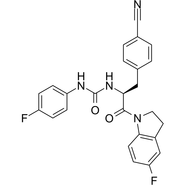
FPR2 agonist 2 is a potent and CNS-penetrant FPR2 agonist with an EC50 of 0.13 μM, 1.1 μM for FPR2 and FPR1, respectively. FPR2 agonist 2 inhibits the production of pro-inflammatory cytokines, counterbalances the changes in mitochondrial function, and inhibits caspase-3 activity.
연구목적의 판매만을 진행합니다. 환자를 대상으로 한 판매는 하지 않습니다.
- Purity : 98.51%
- CAS No.: 2829263-20-7
- 화학식: C25H20F2N4O2
- 분자량:446.45
-
보관:Powder -20°C, 3 years , 4°C, 2 years ; In solvent -80°C, 6 months , -20°C, 1 month

To place orders, for customer services and technical support, please contact: MedChemExpress USA
Tel: 609-228-6898 E-mail: [email protected] [email protected]


-
 Biological Activity
-
Chemical Information
-
용액&용해도
- Protocol
- 순도&문서
- References
-
 Help & FAQs




Biological Activity
제품 설명
IC50 & Target
EC50: 0.13 μM (FPR2); 1.1 μM (FPR1)[1]
In Vitro
FPR2 agonist 2 (compound (S)-11l) (1-100 μM; 48 h) exhibits low cytotoxicity with an EC50 value of 20.8 μM in N9 cells[1].
FPR2 agonist 2 (FPR1/FPR2 HL60 cells) shows agonist activity with EC50s of 0.13 μM, 1.1 μM (IC50s of 0.085 μM, Not determined) for FPR2 and FPR1, respectively[1].
FPR2 agonist 2 (0.1 μM) effectively blocks LPS-induced cell death and NO production and effectively suppresses the effect of LPS stimulation[1].
FPR2 agonist 2 (0.1 μM) counterbalances the changes in mitochondrial function, and inhibits caspase-3 activity[1].
MedChemExpress (MCE) has not independently confirmed the accuracy of these methods. They are for reference only.
-
Cell Line:N9 cells
-
Concentration:1-100 µM
-
Incubation Time:48 h
-
Result:Exhibited low cytotoxicity with an EC50 value of 20.8 µM in N9 cells.
In Vivo
MedChemExpress (MCE) has not independently confirmed the accuracy of these methods. They are for reference only.
-
Animal Model:25-30 g, male CD-1 mice[1]
-
Dosage:
-
Administration:1 mg/kg for i.v.; 10 mg/kg for i.p. (dissolved in 5% DMSO, 10% solutol HS 15, and 85% sterile water)
-
Result:Showed the ability to permeate the blood−brain barrier and to accumulate in the brain.


Chemical Information
-
CAS No. 2829263-20-7
-
Appearance Solid
-
분자량 446.45
-
화학식 C25H20F2N4O2
-
Color White to off-white
-
SMILES
FC1=CC=C(C=C1)NC(N[C@H](C(N2CCC3=C2C=CC(F)=C3)=O)CC4=CC=C(C=C4)C#N)=O
-
선적
Room temperature in continental US; may vary elsewhere.
-
보관
Powder -20°C 3 years 4°C 2 years In solvent -80°C 6 months -20°C 1 month


용액&용해도
In Vitro:
DMSO : 50 mg/mL (111.99 mM; Need ultrasonic; Hygroscopic DMSO has a significant impact on the solubility of product, please use newly opened DMSO)
Please refer to the solubility information to select the appropriate solvent. Once prepared, please aliquot and store the solution to prevent product inactivation from repeated freeze-thaw cycles.
Storage method and period of stock solution: -80°C, 6 months; -20°C, 1 month. When stored at -80°C, please use it within 6 months. When stored at -20°C, please use it within 1 month.
Please refer to the solubility information to select the appropriate solvent. Once prepared, please aliquot and store the solution to prevent product inactivation from repeated freeze-thaw cycles.
Storage method and period of stock solution: -80°C, 6 months; -20°C, 1 month. When stored at -80°C, please use it within 6 months. When stored at -20°C, please use it within 1 month.
Concentration (start) × Volume (start) = Concentration (final) × Volume (final)


순도&문서
-
Data Sheet (275 KB)
-
SDS (251 KB)
- English - EN (251 KB)
- Français - FR (251 KB)
- Deutsch - DE (251 KB)
- Norwegian - NO (251 KB)
- Español - ES (251 KB)
- Swedish - SV (251 KB)
- Italian - IT (251 KB)
- Korean - KR (251 KB)
- Portuguese - PT (251 KB)
-
Handling Instructions (2659 KB)
References


Complete Stock Solution Preparation Table
Please refer to the solubility information to select the appropriate solvent. Once prepared, please aliquot and store the solution to prevent product inactivation from repeated freeze-thaw cycles.
Storage method and period of stock solution: -80°C, 6 months; -20°C, 1 month. When stored at -80°C, please use it within 6 months. When stored at -20°C, please use it within 1 month.
| Optional Solvent | Concentration Solvent Mass | 1 mg | 5 mg | 10 mg | 25 mg |
|---|---|---|---|---|---|
| DMSO | 1 mM | 2.2399 mL | 11.1995 mL | 22.3989 mL | 55.9973 mL |
| 5 mM | 0.4480 mL | 2.2399 mL | 4.4798 mL | 11.1995 mL | |
| 10 mM | 0.2240 mL | 1.1199 mL | 2.2399 mL | 5.5997 mL | |
| 15 mM | 0.1493 mL | 0.7466 mL | 1.4933 mL | 3.7332 mL | |
| 20 mM | 0.1120 mL | 0.5600 mL | 1.1199 mL | 2.7999 mL | |
| 25 mM | 0.0896 mL | 0.4480 mL | 0.8960 mL | 2.2399 mL | |
| 30 mM | 0.0747 mL | 0.3733 mL | 0.7466 mL | 1.8666 mL | |
| 40 mM | 0.0560 mL | 0.2800 mL | 0.5600 mL | 1.3999 mL | |
| 50 mM | 0.0448 mL | 0.2240 mL | 0.4480 mL | 1.1199 mL | |
| 60 mM | 0.0373 mL | 0.1867 mL | 0.3733 mL | 0.9333 mL | |
| 80 mM | 0.0280 mL | 0.1400 mL | 0.2800 mL | 0.7000 mL | |
| 100 mM | 0.0224 mL | 0.1120 mL | 0.2240 mL | 0.5600 mL |


FPR2 agonist 2 Related Classifications
HY-144604 Related Classifications
 Powered by Bioz