



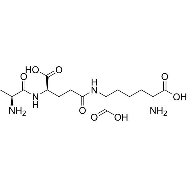
iE-DAP
iE-DAP is a Nod1 agonist. iE-DAP activates NOD1, which in turn activates the NF-κB signaling pathway and MLCK signaling pathway, inducing cellular inflammatory responses and tight junction disruption. iE-DAP downregulates the expression of ZO-1 and Occludin genes. iE-DAP increases the secretion of IL-6, GRO-α, MCP-1, IL-8 and MIP-1β in term human trophoblast cell cultures. iE-DAP triggers preterm birth in pregnant mice, reduces fetal body weight, and induces fetal inflammation. iE-DAP is applicable to research related to mastitis and preterm birth.
연구목적의 판매만을 진행합니다. 환자를 대상으로 한 판매는 하지 않습니다.
- CAS No.: 592520-07-5
- 화학식: C12H21N3O7
- 분자량:319.31
-
보관:
Please store the product under the recommended conditions in the Certificate of Analysis.


To place orders, for customer services and technical support, please contact: MedChemExpress USA
Tel: 609-228-6898 E-mail: [email protected] [email protected]


-
 Biological Activity
-
Chemical Information
- Protocol
- 순도&문서
- References
-
 Help & FAQs




Biological Activity
제품 설명
In Vitro
iE-DAP (10-10000 ng/mL; 1-24 h) upregulates the mRNA expression of IL-1β, IL-6 and IL-8 in BMECs, downregulates the mRNA expression of ZO-1 and Occludin in BMECs, reduces TEER, and increases the paracellular dextran permeability in BMECs[1].
iE-DAP (1000 ng/mL; 12 h) upregulates the protein expression levels of p-MLC2 and MLCK as well as the phosphorylation level of MLC2, downregulates the mRNA and protein expression levels of ZO-1 and Occludin, reduces TEER, increases the paracellular dextran permeability, and disrupts the distribution of ZO-1 in BMECs[1].
iE-DAP (1000 ng/mL; 12 h) upregulates the mRNA expression and secretion levels of IL-1β, IL-6 and IL-8 in BMECs, and activates the NF-κB pathway by increasing the expression of p-p65 protein, the phosphorylation level of p65 and the nuclear translocation of p65[1].
iE-DAP (1000 ng/mL; 12 h) upregulates the expression of NOD1 protein, p-p65 protein, the phosphorylation level of p65, and promotes the nuclear translocation of p65 in BMECs, thereby activating the NOD1/NF-κB pathway[1].
iE-DAP (100 μg/mL; 72 h) potently stimulates human primary third-trimester cytotrophoblasts to secrete IL-6, GRO-α, MCP-1, IL-8 and MIP-1β[2].
MedChemExpress (MCE) has not independently confirmed the accuracy of these methods. They are for reference only.
-
Cell Line:bovine mammary epithelial cells (BMECs)
-
Concentration:1000 ng/mL
-
Incubation Time:1, 3, 6, 12, 24 h
-
Result:Significantly upregulated IL-1β mRNA relative expression at 1 h.
Significantly upregulated IL-6 mRNA relative expression at 1 h.
Significantly upregulated IL-8 mRNA relative expression at 1 h, with all inflammatory cytokines increasing in a time-dependent manner.
Significantly downregulated ZO-1 mRNA relative expression at 6 h.
Significantly downregulated Occludin mRNA relative expression at 12 h, with both tight junction proteins decreasing in a time-dependent manner.
-
Cell Line:bovine mammary epithelial cells (BMECs)
-
Concentration:0, 1, 10, 100, 1000, 10000 ng/mL
-
Incubation Time:12 h
-
Result:Increased the mRNA relative expression and release of IL-1β at 10 ng/mL.
Increased the mRNA relative expression and release of IL-6 and IL-8 at 100 ng/mL.
Increased the gene expression and release of inflammatory cytokines in a concentration-dependent manne.
Reduced the relative mRNA expression of ZO-1、Occludin at 100、1000 ng/mL.
Decreased the gene expression of TJ proteins in a concentration-dependent manne.
Decreased the TEER and increased
paracellular dextran passage at 100 ng/mL.
-
Cell Line:bovine mammary epithelial cells (BMECs)
-
Concentration:1000 ng/mL
-
Incubation Time:12 h
-
Result:Increased p-MLC2 protein expression and MLC2 phosphorylation, reduced protein expression of ZO-1 and Occludin, decreases TEER.
Significantly increased p-p65 protein expression and the p-p65/p65 phosphorylation ratio.
Significantly increased NOD1 protein expression.
Significantly increased MLCK protein expression.
-
Cell Line:bovine mammary epithelial cells (BMECs)
-
Concentration:1000 ng/mL
-
Incubation Time:12 h
-
Result:Caused redistribution of ZO-1 protein. Induced nuclear translocation of NF-κB p65.
In Vivo
MedChemExpress (MCE) has not independently confirmed the accuracy of these methods. They are for reference only.
-
Animal Model:C57BL/6J (adult, 8-12 wk old, pregnant females)[2]
-
Dosage:500 μg; 750 μg; 1000 μg
-
Administration:i.p.; single dose
-
Result:Induced preterm delivery within 24 hours in 100% of treated mice (3/3 mice) at 1000 μg.
Significantly reduced fetal weight compared to controls at 750 μg.
Altered the maternal-fetal interface cytokine profile at 750 μg: increased placental Eotaxin levels, decreased placental MCP-1 levels, increased decidual IL-1α levels, and increased decidual RANTES levels.
Induced a broad fetal proinflammatory response at 750 μg, with significantly elevated levels of Eotaxin, IL-1α, IL-3, IL-10, IL-12 (p40), IL-12 (p70), IL-13, IL-17, G-CSF, GM-CSF, MIP-1α, MIP-1β, KC, TNF-α, and IFN-γ in fetal tissues.
Did not induce preterm delivery at 500 μg.


Chemical Information
-
CAS No. 592520-07-5
-
분자량 319.31
-
화학식 C12H21N3O7
-
Sequence
γ-d-Glu-meso-DAP
-
선적
Room temperature in continental US; may vary elsewhere.
-
보관
Please store the product under the recommended conditions in the Certificate of Analysis.


순도&문서
References
[1]. Wang Y, et al. iE-DAP Induced Inflammatory Response and Tight Junction Disruption in Bovine Mammary Epithelial Cells via NOD1-Dependent NF-κB and MLCK Signaling Pathway. Int J Mol Sci. 2023;24(7):6263. Published 2023 Mar 27. [Content Brief]
[2]. Cardenas I, et al. Nod1 activation by bacterial iE-DAP induces maternal-fetal inflammation and preterm labor. J Immunol. 2011;187(2):980-986. [Content Brief]

Calculators
Concentration (start) × Volume (start) = Concentration (final) × Volume (final)


iE-DAP Related Classifications
HY-P5523 Related Classifications
 Powered by Bioz