



Polygalacturonic acid

Based on 1 Customer Validation
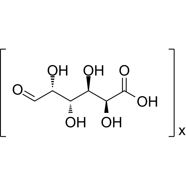
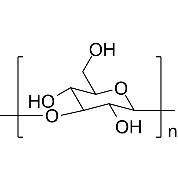
Polygalacturonic acid (Galacturonic acid polymer) is transparent colloid, is a major component of the cell wall. Polygalacturonic acid can be used to prepare silver nanoparticles (AgNPs), as an antioxidant and anti-inflammatory that protect cells from destructive effect of elevated ROS and accelerate wound healing. Polygalacturonic acid nanoparticles also displays anti-bacterial activity.
연구목적의 판매만을 진행합니다. 환자를 대상으로 한 판매는 하지 않습니다.
- Purity : 99.58%
- CAS No.: 25990-10-7
- 화학식: (C6H10O7)x
-
보관:
4°C, protect from light
* In solvent : -80°C, 6 months; -20°C, 1 month (protect from light)

To place orders, for customer services and technical support, please contact: MedChemExpress USA
Tel: 609-228-6898 E-mail: [email protected] [email protected]


-
 Biological Activity
-
Chemical Information
-
용액&용해도
- Protocol
- 순도&문서
- References
-
 Help & FAQs


-
HY-18085
QuercetinFlavonoid Antioxidant -
HY-N6612
D-Glucuronic acidEndogenous Metabolite -
HY-119109
LaminaranImmunotherapy Agent -

HY-N7075
InulinEndogenous Metabolite -

HY-N6653
LentinanNrf2 Activator -

HY-W250113
Zymosan ATLR2 Agonist -

HY-132179
FucoidanAnticoagulant/Cancer Agent -

HY-113511
Glycogen, MusselGlycolytic Intermediates -
HY-131166
CurdlanPolysaccharide -

HY-W145667
MannanPolysaccharide Compound -

HY-N6664
Gum arabicComplex Branched Polysaccharide -

HY-113511C
Glycogen, from bovine liver, ≥85%Biochemical Assay Reagent


Biological Activity
제품 설명
In Vitro
Polygalacturonic acid can be used to format a durable hydrogel, to prolong the effect of drugs. Polygalacturonic acid hydrogel/hyaluronate, conjugated Ibuprofen (HY-78131) for example, prevents epidural fibrosis, the severe scar tissue adhesion of laminectomized male adult rats, and increases the efficiency of local inflammation control and reduces the side-effect of Ibuprofen[1].
Polygalacturonic acid hydrogel/hyaluronate (PGA-HA) (2%, 200 μL; 24 h) shows low cytotoxicity on L929 fibroblasts[1].
Polygalacturonic acid (PGA) preparing (Ag-PGA/HA)-PVA nanoparticles, shows anti-bacterial activity against gram-positive bacterial strains; Bacillus Subtilis and Staphylococcus Aureus, as well as gram-negative bacterial strain; Escherichia Coli[2].
MedChemExpress (MCE) has not independently confirmed the accuracy of these methods. They are for reference only. Further protocols information, click here.
In Vivo
MedChemExpress (MCE) has not independently confirmed the accuracy of these methods. They are for reference only.
-
Animal Model:Full thickness wound model in albino rats (60-day-old, 180 g)[2]
-
Dosage:Ag-Polygalacturonic acid/hyaluronic acid nanoparticles
-
Administration:Applied on dorsal muscle fascia; cleaned the wound daily with alcohol; for 14 days
-
Result:Resulted no sign of abscess formation or hypertrophic scars both in the early phase; 5th day or in the final phase; 14th day, respectively.
Showed significant wound healing from day 8 while treated with (Ag-PGA/HA)-PVA nanofiber and blank (PGA/HA)-PVA nanofiber and induced epithelization on day 14.


Chemical Information
-
CAS No. 25990-10-7
-
Appearance Solid
-
화학식 (C6H10O7)x
-
Color Off-white to light yellow
-
SMILES
O=C[C@H](O)[C@@H](O)[C@@H](O)[C@H](O)C(O)=O.[x]
-
Synonyms
Galacturonic acid polymer
-
Structure Classification
-
Initial Source
-
선적
Room temperature in continental US; may vary elsewhere.
-
보관
4°C, protect from light
* In solvent : -80°C, 6 months; -20°C, 1 month (protect from light)


용액&용해도
In Vitro:
0.1 M NaOH : 10 mg/mL (ultrasonic and warming and adjust pH to 11 with NaOH and heat to 60°C)
H2O : < 0.1 mg/mL (insoluble)

Protocol
-
ROS/oxidative-stress fluorescent staining
ROS/oxidative-stress fluorescent staining uses cell-permeant fluorogenic probes that become fluorescent after oxidation inside cells or tissues; commonly used examples include DCFH-DA/DCFDA for broad cellular oxidant detection, DHE for superoxide-related signal detection, MitoSOX for mitochondrial superoxide-related signal detection, and CellROX probes for oxidative-stress-associated fluorescence readouts. The assay detects probe oxidation rather than a single ROS species unless the probe and analysis method have been chemically validated for that species. DCFH-DA enters cells, is deacetylated by intracellular esterases to DCFH, and produces fluorescent DCF after oxidation, so the readout is used as an operational measure of total cellular oxidative stress rather than a species-specific ROS measurement. DHE and MitoSOX can report superoxide-related oxidation, but red fluorescence alone can include non-specific ethidium-like oxidation products; HPLC or optimized spectral approaches are
-
Cell migration
Cell migration is a method that plays an important role in wound healing, cell differentiation, embryonic development, etc.
-
Research Protocol for Inflammation-related Diseases
The NLRP3 inflammasome is a cytosolic innate immune signaling platform that integrates priming signals and danger-signal activation to promote caspase-1 activation, maturation of IL-1β and IL-18, and gasdermin D-mediated pyroptotic cell death. The core experimental logic is to determine whether inflammatory disease phenotypes are driven by increased NLRP3 expression, ASC-containing inflammasome assembly, caspase-1 cleavage, GSDMD cleavage, and extracellular release of IL-1β/IL-18 rather than by nonspecific cell injury alone. The pathway is strongly linked to inflammation-related disease phenotypes because monosodium urate crystals activate NALP3/NLRP3 inflammasome signaling in gout-like crystal inflammation, cholesterol crystals activate NLRP3 inflammasomes in atherogenesis models, and DSS-induced intestinal inflammation has been reported to involve NLRP3 inflammasome activity. However, experimental colitis studies also show context-dependent protective effects of NLRP3 inflammasome co
-
Cell-Exclusion Zone Migration Assay
The Cell-Exclusion Zone (CEZ) migration assay is an in vitro 2D cell migration method in which a defined cell-free area is created using removable physical barriers such as silicone stoppers, allowing cells to be seeded around the barrier and subsequently migrate into the cleared zone after barrier removal. This approach enables quantification of collective cell migration by monitoring repopulation of the initially cell-free region over time using microscopy-based imaging. Compared with scratch-based wound healing assays, barrier-based exclusion methods are designed to avoid mechanical damage to the extracellular matrix and reduce injury-induced effects on boundary cells, thereby improving interpretability of migration behavior in vitro. The assay readout is typically the progressive reduction in the cell-free area or the number of cells invading the exclusion zone, reflecting coordinated cell motility relevant to physiological processes such as wound healing, epithelial repair, and ca


순도&문서
-
Data Sheet (272 KB)
-
SDS (394 KB)
- English - EN (394 KB)
- Français - FR (394 KB)
- Deutsch - DE (394 KB)
- Norwegian - NO (394 KB)
- Español - ES (394 KB)
- Swedish - SV (394 KB)
- Italian - IT (394 KB)
- Korean - KR (394 KB)
- Portuguese - PT (394 KB)
-
Handling Instructions (2659 KB)
References
[1]. Lin CY, et al. Ibuprofen-conjugated hyaluronate/polygalacturonic acid hydrogel for the prevention of epidural fibrosis. J Biomater Appl. 2016 May;30(10):1589-600. [Content Brief]
[2]. El-Aassar MR, et al. Wound healing of nanofiber comprising Polygalacturonic/Hyaluronic acid embedded silver nanoparticles: In-vitro and in-vivo studies. Carbohydr Polym. 2020 Jun 15;238:116175. [Content Brief]

Calculators
Concentration (start) × Volume (start) = Concentration (final) × Volume (final)


Polygalacturonic acid Related Classifications
HY-N6613 Related Classifications
 Powered by Bioz