



Quilizumab

Based on 1 Customer Validation
Quilizumab (Anti-Human IGHE Recombinant Antibody; 47H4; MEMP1972A) is a monoclonal antibody that targets FcγRIIIa and the M1 prime segment of human membrane-bound IgE. Quilizumab has enhanced FcγRIIIa binding affinity and activity against IgE class-switched B cells, and reduces serum IgE levels by inducing antibody-dependent cell-mediated cytotoxicity and apoptosis through cross-linking of membrane-bound IgE receptors. Quilizumab depletes IgE-producing cells; it also directly binds the membrane-bound M1 prime domain on IgE class-switched cells to inhibit IgE production. Quilizumab is used in research on allergic asthma and allergic diseases (asthma, allergic rhinitis).
연구목적의 판매만을 진행합니다. 환자를 대상으로 한 판매는 하지 않습니다.
- Purity : 99.56%
- CAS No.: 1228538-47-3
- 분자량:146.5 kDa
-
보관:
Please store the product under the recommended conditions in the Certificate of Analysis.


To place orders, for customer services and technical support, please contact: MedChemExpress USA
Tel: 609-228-6898 E-mail: [email protected] [email protected]


-
 Biological Activity
-
Chemical Information
- Protocol
- 순도&문서
- References
-
 Help & FAQs






Biological Activity
제품 설명
Isotype
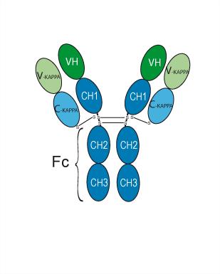
Human IgG1 kappa
Recommend Isotype Controls
Species Reactivity
Human
IC50 & Target
IGHE
In Vitro
Quilizumab (afucosylated anti-M1 prime antibody) after afucosylation modification shows approximately 24-fold and 7-fold increases in binding affinity for human FcγRIIIa-F158 and FcγRIIIa-V158 receptors, respectively[1].
Quilizumab (0.0051-10000 ng/mL; 3 h) mediates potent ADCC activity against Daudi B cells transfected with membrane-bound IgE, exhibiting approximately 10-fold enhanced ADCC potency compared with the fucosylated wild-type anti-M1 prime antibody[1].
Quilizumab (0.001-25 μg/mL; 72 h) induces apoptosis in Daudi B cells transfected with membrane-bound IgE, with pro-apoptotic potency comparable to that of the fucosylated wild-type anti-M1 prime antibody[1].
MedChemExpress (MCE) has not independently confirmed the accuracy of these methods. They are for reference only. Further protocols information, click here.
-
Cell Line:Daudi human B lymphoma cells transfected with human membrane IgE
-
Concentration:0.001, 0.01, 0.1, 1, 10, 25 μg/mL
-
Incubation Time:72 h
-
Result:Induced apoptosis of target cells with no significant differences in maximal percent apoptosis or dose-response compared to the wildtype antibody.
In Vivo
Quilizumab inhibits the production of new allergen-specific IgE induced by allergen stimulation in biological model systems related to allergic asthma[2].
MedChemExpress (MCE) has not independently confirmed the accuracy of these methods. They are for reference only.
-
Animal Model:SCID-beige mice (sublethally irradiated with 3.5 Gy; reconstituted with human PBMCs)[1]
-
Dosage:3, 30, 300 μg
-
Administration:i.p.; days 0, 4, and 7
-
Result:Reduced human IgE in serum at all doses (3, 30, 300 μg) with maximal activity.
Exhibited approximately 100-fold higher potency than the wildtype counterpart, showing comparable activity at 1/100th the dose.
Did not affect human IgM levels.
Clinical Trial
| NCT Number | Sponsor | Condition | Start Date |
Phase

|
|---|---|---|---|---|
| NCT01329991 | Plexxikon| | 2011-05 | PHASE1 |
Accession
Conjugated
Unconjugated
Reconsititution
The product can be reconstituted/diluted with sterile PBS or saline.
Format
-
Human IgG1 kappa
신청
ELISA, FACS, Functional assay
Verified Bioactivity
-
 Immobilized IgE Protein, Human (HEK293, His, HY-P73907) can bind Quilizumab. The EC50 for this effect is 486.8 ng/mL.


Chemical Information
-
CAS No. 1228538-47-3
-
Appearance Liquid
-
분자량 146.5 kDa
-
Color Colorless to light yellow
-
SMILES
[Quilizumab]
-
Synonyms
Anti-Human IGHE Recombinant Antibody; 47H4; MEMP1972A
-
선적
Shipping with dry ice.
-
Formulation
Please refer to the lot-specific COA for specific buffer information.
-
보관
Please store the product under the recommended conditions in the Certificate of Analysis.

Protocol
-
Apoptosis
Apoptosis, also called programmed cell death, is generally characterized by distinct morphological characteristics.
-
TUNEL staining for apoptotic DNA fragmentation
TUNEL staining detects DNA strand breaks by using terminal deoxynucleotidyl transferase to add labeled nucleotides to exposed 3′-OH DNA termini, generating either microscopic staining in fixed cells or tissue sections, or fluorescence/cytometric signal in cell suspensions. TUNEL positivity reflects DNA fragmentation but should not be interpreted alone as definitive apoptosis, because TUNEL can also label necrotic, autolytic, mechanically damaged, or DNA-repair-associated DNA breaks.
-
Cell Cytotoxicity Assay
Cytotoxicity assays are usually based on the assessment of cell membrane damage, which can also be indirectly detected by measuring cell viability. Detection methods include MTT assay, CKK-8 assay, LDH assay and ATP assay, etc.
-
Mammalian live/dead viability and cytotoxicity staining
Live/dead viability and cytotoxicity staining assays are based on the simultaneous detection of intracellular esterase activity in metabolically active (viable) cells and membrane integrity loss in non-viable cells. In commonly used dual-staining approaches, membrane-permeant fluorogenic substrates are converted by intracellular esterases into fluorescent products in live cells, while impermeant DNA-binding dyes selectively enter cells with compromised plasma membranes and label nucleic acids in dead or dying cells, enabling discrimination between viable and non-viable populations by fluorescence microscopy or flow cytometry.
-
Annexin V plus membrane-impermeant dye apoptosis staining
Annexin V-based apoptosis assays rely on the detection of phosphatidylserine (PS) externalization from the inner leaflet of the plasma membrane to the outer leaflet, an early biochemical hallmark of apoptosis. Fluorescently labeled Annexin V binds PS in a calcium-dependent manner, enabling identification of early apoptotic cells by flow cytometry or fluorescence microscopy. When combined with a membrane-impermeant DNA-binding dye (e. g. , propidium iodide), this approach allows discrimination between viable (Annexin V−/dye−), early apoptotic (Annexin V+/dye−), and late apoptotic or necrotic (Annexin V+/dye+) cell populations by assessing membrane integrity and PS exposure.
-
Ovalbumin-Induced Allergic Airway Inflammation
Ovalbumin-induced allergic airway inflammation is a mouse model in which systemic sensitization to ovalbumin, usually with aluminum hydroxide adjuvant, is followed by airway ovalbumin challenge to induce allergic airway inflammation, eosinophil recruitment, mucus production, serum antigen-specific IgE, Th2 cytokine responses, and airway hyperresponsiveness to methacholine. The model is used to study allergen-driven airway inflammation and asthma-like immune responses, but it does not reproduce every feature of human asthma. The main readouts are bronchoalveolar lavage fluid cellularity, lung histopathology, airway hyperresponsiveness, serum OVA-specific IgE, and cytokines such as IL-4, IL-5, and IL-13 in bronchoalveolar lavage fluid or lung samples. Eosinophilia and Th2 cytokines reflect allergic type 2 inflammation, while methacholine responsiveness provides a functional airway-reactivity endpoint.
-
Apoptosis Solutions
Apoptosis is a regulated, generally non-lytic cell-death pathway that removes unwanted, damaged, infected, or abnormal cells through coordinated morphological changes, caspase activation, DNA fragmentation, and membrane remodeling. The intrinsic apoptosis pathway is controlled mainly by mitochondrial outer membrane permeabilization, BCL-2 family proteins, cytochrome c release, apoptosome formation, caspase-9 activation, and downstream executioner caspase-3/7 activation. The extrinsic apoptosis pathway is initiated by death receptors such as Fas, TNFR, and TRAIL receptors, which recruit adaptor proteins and activate caspase-8 before engaging executioner caspases or mitochondrial amplification through BID cleavage. Apoptosis is linked to many phenotypes, including cancer cell killing, tissue homeostasis, immune regulation, neurodegeneration, infection response, and treatment-induced cytotoxicity; unresolved questions include how apoptosis interacts with necroptosis, pyroptosis, ferroptos
-
Recombinant antibody expression and Protein A/G purification
Recombinant antibody expression produces antibody chains from cloned antibody genes in mammalian cells, commonly HEK293-derived cells, so that secreted IgG or Fc-fusion antibodies can be collected from culture supernatant. Protein A/G purification is affinity chromatography in which immobilized Protein A, Protein G, or Protein A/G binds the antibody Fc region; nonbound culture components are washed away, and bound antibody is recovered by changing buffer conditions, commonly acidic elution followed by neutralization.
-
Cell Viability Determination by MTT Colorimetric Assay
The following protocol uses the MTT colorimetric assay as a classic literature-established method for assessing cell viability/metabolic activity in cultured mammalian cells. MTT[3-(4,5-dimethylthiazol-2-yl)-2,5-diphenyltetrazolium bromide] is reduced by metabolically active cells to a colored formazan product; the amount of formazan is quantified spectrophotometrically and provides an indirect measure of metabolically active viable cells. Importantly, MTT reduction reflects cellular oxidoreductase/metabolic activity rather than an absolute direct count of living cells, so changes in cellular metabolism can alter the signal independently of cell number.


순도&문서
-
Data Sheet (276 KB)
-
SDS (252 KB)
- English - EN (252 KB)
- Français - FR (252 KB)
- Deutsch - DE (252 KB)
- Norwegian - NO (252 KB)
- Español - ES (252 KB)
- Swedish - SV (252 KB)
- Italian - IT (252 KB)
- Korean - KR (252 KB)
- Portuguese - PT (252 KB)
-
Inhibitory Antibodies User Guide (603 KB)
References

Calculators
Concentration (start) × Volume (start) = Concentration (final) × Volume (final)


Quilizumab Related Classifications
HY-P99313 Related Classifications
 Powered by Bioz
Keywords
- Quilizumab
- 1228538-47-3
- Anti-Human IGHE Recombinant Antibody
- 47H4
- MEMP1972A
- Drug Derivative
- Fc Receptor (FcR)
- Apoptosis
- Daudi B cells
- FcγRIIIa
- allergic asthma
- allergic rhinitis
- IgE-switched B cells
- human membrane IgE
- apoptosis
- human PBMC-SCID mouse model
- M1 prime segment
- antibody-dependent cell-mediated cytotoxicity
- Inhibitor
- inhibitor
- inhibit