



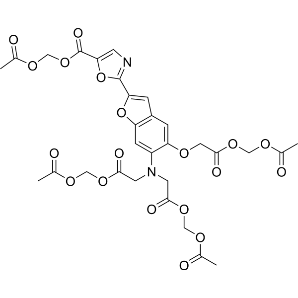
Mag-Fura-2 AM
Calcium is an important part of the human body, usually in the form of calcium, a large number of bones and teeth of the human body, a small amount of blood and tissues. MCE calcium ion detection probe can specifically identify intracellular calcium ions, with high sensitivity, low cytotoxicity, increased AM acetylmethyl ester can enter the cell well, after being sheared by the intracellular esterase stay in the cell to bind to calcium ions, produce strong fluorescence, in addition, Mag-indo-1/AM and Mag-Fluo-4 AM at a certain concentration (usually 5 mM) can effectively identify intracellular magnesium ions.
For research use only. We do not sell to patients.
- CAS No.: 130100-20-8
- Formula: C30H30N2O19
- Molecular Weight:722.56
-
Storage:
Please store the product under the recommended conditions in the Certificate of Analysis.

To place orders, for customer services and technical support, please contact: MedChemExpress USA
Tel: 609-228-6898 E-mail: [email protected] [email protected]


-
 Biological Activity
-
Chemical Information
- Purity & Documentation
- References
-
 Help & FAQs




Biological Activity
Description
In Vitro
Guidelines (Following is our recommended protocol. This protocol only provides a guideline, and should be modified according to your specific needs).
1. Preparation of Mag-Fura-2 AM working solution
1.1 Preparation of the stock solution
Dissolve Mag-Fura-2 AM in DMSO to obtain 10 mM of stock solution.
Note: It is recommended to store the stock solution at -20°C or -80°C, keep away from light and avoid repetitive freeze-thaw cycles.
1.2 Preparation of Mag-Fura-2 AM working solution
Dilute the stock solution in serum-free cell culture medium or PBS to obtain 1-10 μM of working solution.
Note: Please adjust the concentration of Mag-Fura-2 AM working solution according to the actual situation.
2. Cell staining (Suspension cells)
2.1 Centrifuge at 1000 g at 4°C for 3-5 min and then discard the supernatant. Wash twice with PBS, 5 min each time. The cell density is 1×10⁶/mL.
2.2 Add 1 mL of working solution, and then incubate at room temperature for 5-30 min.
2.3 Centrifuge at 400 g at 4°C for 3-4 min and then discard the supernatant.
2.4 Wash twice with PBS, 5 min each time.
2.5 Resuspend cells with serum-free cell culture medium or PBS. Observation by fluorescence microscopy or flow cytometry.
3. Cell staining (Adherent cells)
3.1 Culture adherent cells on sterile coverslips.
3.2 Remove the coverslip from the medium and aspirate excess medium.
3.3 Add 100 μL of working solution, gently shake it to completely cover the cells, and then incubate at room temperature for 5-30 min.
3.4 Wash twice with medium, 5 min each time. Observation by fluorescence microscopy or flow cytometry.
MedChemExpress (MCE) has not independently confirmed the accuracy of these methods. They are for reference only.
Emission (Em)
510
Excitation (Ex)
340


Chemical Information
-
CAS No. 130100-20-8
-
Molecular Weight 722.56
-
Formula C30H30N2O19
-
SMILES
O=C(C1=CN=C(C2=CC3=C(C=C(N(CC(OCOC(C)=O)=O)CC(OCOC(C)=O)=O)C(OCC(OCOC(C)=O)=O)=C3)O2)O1)OCOC(C)=O
-
Shipping
Room temperature in continental US; may vary elsewhere.
-
Storage
Please store the product under the recommended conditions in the Certificate of Analysis.


Purity & Documentation
References

Calculators
Concentration (start) × Volume (start) = Concentration (final) × Volume (final)


Mag-Fura-2 AM Related Classifications
HY-D1701 Related Classifications
 Powered by Bioz