



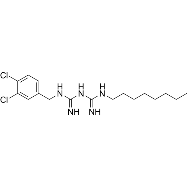
Olanexidine
Olanexidine is an antibacterial agent. Olanexidine is active against a wide range of bacteria, imcluding both Gram-positive and Gram-negative bacteria Olanexidine is also an antiseptic. Olanexidine can be used in the research of infection and inflammation.
Nur für Forschungszwecke. Wir verkaufen nicht an Patienten.
- CAS. Nr.: 146510-36-3
- Formel: C17H27Cl2N5
- Molecular Weight:372.34
-
Speicherung:
Please store the product under the recommended conditions in the Certificate of Analysis.

To place orders, for customer services and technical support, please contact: MedChemExpress USA
Tel: 609-228-6898 E-mail: [email protected] [email protected]


-
 Biologische Aktivität
-
Chemical Information
- Protokoll
- Reinheit & Dokumentation
- Verweise
-
 Help & FAQs




Biologische Aktivität
Beschreibung
In Vitro
Olanexidine (50 μM, 48 h) exhibits broad-spectrum bactericidal activity against Gram-positive cocci and Gram-negative bacteria[1].
Olanexidine (50 μg/mL, 24 h) inhibits chronic inflammatory reactions in oral mucosal cells[2].
Olanexidine (Olanexidine gluconate, 0-15000 μg/mL) inhibits the binding ability of virus-like particles to the binding receptor of human norovirus and increases the aggregation of virus-like particles[3].
MedChemExpress (MCE) has not independently confirmed the accuracy of these methods. They are for reference only.
-
Cell Line:Gram-positive cocci, Enterococcus spp., Gram-positive bacilli, Gram-negative strains
-
Concentration:0-1 mg/mL approximately
-
Incubation Time:30 s
-
Result:Inhibited bacterial activity with MIC values ranging from 6.8 to 1740 μg/mL approximately.
In Vivo
MedChemExpress (MCE) has not independently confirmed the accuracy of these methods. They are for reference only.
-
Animal Model:Male cynomolgus monkeys[4]
-
Dosage:1 %, 1.5%, 2% in saline
-
Administration:Applied to the skin
-
Result:Showed the fast-acting and long-lasting bactericidal effects at 1.5% concentration.


Chemical Information
-
CAS. Nr. 146510-36-3
-
Molecular Weight 372.34
-
Formel C17H27Cl2N5
-
SMILES
N=C(NCC1=CC=C(Cl)C(Cl)=C1)NC(NCCCCCCCC)=N
-
Versand
Room temperature in continental US; may vary elsewhere.
-
Speicherung
Please store the product under the recommended conditions in the Certificate of Analysis.


Reinheit & Dokumentation
Verweise
[1]. Hagi A, et al. Bactericidal Effects and Mechanism of Action of Olanexidine Gluconate, a New Antiseptic. Antimicrob Agents Chemother. 2015 Aug;59(8):4551-9. [Content Brief]
[2]. Nii T, Yumoto H, Hirota K, Miyake Y. Anti-inflammatory effects of olanexidine gluconate on oral epithelial cells. BMC Oral Health. 2019 Nov 8;19(1):239. [Content Brief]
[3]. Imai K, et al. Disinfection efficacy and mechanism of olanexidine gluconate against norovirus. Am J Infect Control. 2022 Jul;50(7):764-771. [Content Brief]
[4]. Nakata H, et al. Effects of olanexidine gluconate on preoperative skin preparation: an experimental study in cynomolgus monkeys. J Med Microbiol. 2017 May;66(5):678-685. [Content Brief]

Calculators
Konzentration (Stammlösung) × Volumen (Stammlösung) = Konzentration (Ziellösung) × Volumen (Ziellösung)


Olanexidine Related Classifications
HY-125654 Related Classifications
 Powered by Bioz