



FAP Protein, Human (HEK293, N-His)
Based on 2 publication(s) in Google Scholar
The FAP protein is a cell surface glycoprotein serine protease that is critical for extracellular matrix degradation and plays multiple roles in tissue remodeling, fibrosis, wound healing, inflammation, and tumor growth. FAP Protein, Human (HEK293, His) is the recombinant human-derived FAP protein, expressed by HEK293 , with N-8*His labeled tag.
- Species: Human
- Source: HEK293
-
Storage:Stored at -20°C for 2 years from date of receipt. After reconstitution, it is stable at 4°C for 1 week or -20°C for longer (with carrier protein). It is recommended to freeze aliquots at -20°C or -80°C for extended storage.


To place orders, for customer services and technical support, please contact: MedChemExpress USA
Tel: 609-228-6898 E-mail: [email protected] [email protected]


-
 Biological Activity
-
Technical Parameters
-
Product Properties
- Documentation
- Help & FAQs




Biological Activity
Description
The FAP protein is a cell surface glycoprotein serine protease that is critical for extracellular matrix degradation and plays multiple roles in tissue remodeling, fibrosis, wound healing, inflammation, and tumor growth. FAP Protein, Human (HEK293, His) is the recombinant human-derived FAP protein, expressed by HEK293 , with N-8*His labeled tag.
Background
FAP protein, a cell surface glycoprotein serine protease, plays a crucial role in extracellular matrix degradation and is involved in diverse cellular processes, including tissue remodeling, fibrosis, wound healing, inflammation, and tumor growth. Both the plasma membrane and soluble forms of FAP exhibit post-proline cleaving endopeptidase activity, demonstrating a preference for Ala/Ser-Gly-Pro-Ser/Asn/Ala consensus sequences on substrates such as alpha-2-antiplasmin SERPINF2 and SPRY2. FAP can degrade gelatin, heat-denatured type I collagen, and various other substrates. Additionally, it possesses dipeptidyl peptidase activity, hydrolyzing prolyl bonds in synthetic dipeptide substrates with a preference for specific amino acid sequences. In association with DPP4, PLAUR, or integrins, the plasma membrane form of FAP participates in pericellular proteolysis of the extracellular matrix, promoting cell adhesion, migration, and invasion. FAP's multifaceted functions extend to tissue remodeling during development and wound healing. In malignant melanoma cancers, FAP enhances cell invasiveness towards the extracellular matrix and promotes glioma cell invasion through the brain parenchyma by degrading the proteoglycan brevican. While contributing to tumor growth progression by increasing angiogenesis, collagen fiber degradation, and apoptosis, FAP paradoxically acts as a tumor suppressor in melanocytic cells through the regulation of cell proliferation and survival in a serine protease activity-independent manner.
Verified Bioactivity
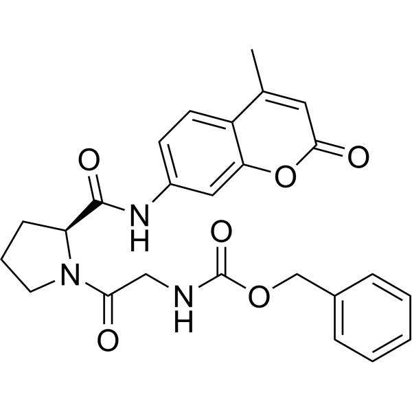
1.Measured by its ability to convert the substrate benzyloxycarbonyl-Gly-Pro-7-amido-4-methylcoumarin (HY-D1670) to Z-Gly-Pro and 7-amino-4-methylcoumarin (HY-D0027) that incubate at room temperature in kinetic mode for 5 minutes. The specific activity is > 1800 pmol/min/μg.
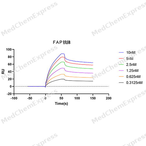
2.FAP Protein, Human (HEK293, N-His) captured on CM5 Sensor Chip, can bind FAP Antibody (YA1442) (HY-P81697) with an affinity constant of 1.27 nM as determined in SPR assay.
MCE Validation Data
-
Bioactivity - SPR
Bioactivity - SPR


Publications (2)
-
Journal Impact Factor
-
Most Recent
-
J Control Release
Structure-guided design and clinical evaluation of 68Ga-GP01 for FAP-targeted PET imaging in solid tumors. [Abstract]2026 Jan 10:389:114475. PMID: 41325906 -
Acta Pharmacol Sin
A FAPI-based small-molecule drug conjugate alleviates rheumatoid arthritis by targeting pathogenic FAPα-expressing fibroblasts. [Abstract]2026 Jan 1. PMID: 41478880


Technical Parameters
-
Species Human
-
Source HEK293
-
Tag N-8*His
-
Accession
Q12884-1 (L26-D760)
-
Gene ID2191
-
Molecular Construction
-
N-term
-
8*His
-
FAP (L26-D760)
Accession # Q12884 -
C-term
-
-
Protein Length
Extracellular Domain
-
Synonyms
FAP; Prolyl Endopeptidase FAP; Fibroblast Activation Protein Alpha; Dipeptidyl Peptidase FAP; Seprase; FAPalpha; DPPIV; SIMP; 170 KDa Melanoma Membrane-Bound Gelatinase; CDNA FLJ60298, Highly Similar To Seprase; Gelatine Degradation Protease FAP; Fibroblast Activation Protein, Alpha; Integral Membrane Serine Protease; Serine Integral Membrane Protease; Post-Proline Cleaving Enzyme; FAPA; Surface-Expressed Protease
-
AA Sequence
LRPSRVHNSEENTMRALTLKDILNGTFSYKTFFPNWISGQEYLHQSADNNIVLYNIETGQSYTILSNRTMKSVNASNYGLSPDRQFVYLESDYSKLWRYSYTATYYIYDLSNGEFVRGNELPRPIQYLCWSPVGSKLAYVYQNNIYLKQRPGDPPFQITFNGRENKIFNGIPDWVYEEEMLATKYALWWSPNGKFLAYAEFNDTDIPVIAYSYYGDEQYPRTINIPYPKAGAKNPVVRIFIIDTTYPAYVGPQEVPVPAMIASSDYYFSWLTWVTDERVCLQWLKRVQNVSVLSICDFREDWQTWDCPKTQEHIEESRTGWAGGFFVSTPVFSYDAISYYKIFSDKDGYKHIHYIKDTVENAIQITSGKWEAINIFRVTQDSLFYSSNEFEEYPGRRNIYRISIGSYPPSKKCVTCHLRKERCQYYTASFSDYAKYYALVCYGPGIPISTLHDGRTDQEIKILEENKELENALKNIQLPKEEIKKLEVDEITLWYKMILPPQFDRSKKYPLLIQVYGGPCSQSVRSVFAVNWISYLASKEGMVIALVDGRGTAFQGDKLLYAVYRKLGVYEVEDQITAVRKFIEMGFIDEKRIAIWGWSYGGYVSSLALASGTGLFKCGIAVAPVSSWEYYASVYTERFMGLPTKDDNLEHYKNSTVMARAEYFRNVDYLLIHGTADDNVHFQNSAQIAKALVNAQVDFQAMWYSDQNHGLSGLSTNHLYTHMTHFLKQCFSLSD
-
Molecular Weight
Approximately 95 kDa, based on SDS-PAGE under reducing conditions, due to the glycosylation.
-
Glycosylation
Yes
-
Purity
≥ 95%, as determined by reducing SDS-PAGE.


Product Properties
Lyophilized powder.
Lyophilized from a 0.22 μm filtered solution of PBS, pH 7.4, 5% trehalose, 5% mannitol and 0.01% Tween 80.
<1 EU/μg, determined by LAL method.
It is not recommended to reconstitute to a concentration less than 100 μg/mL in ddH2O.
Stored at -20°C for 2 years from date of receipt. After reconstitution, it is stable at 4°C for 1 week or -20°C for longer (with carrier protein). It is recommended to freeze aliquots at -20°C or -80°C for extended storage.
Room temperature in continental US; may vary elsewhere.


Documentation
-
Data Sheet (241 KB)
-
SDS (251 KB)
- English - EN (251 KB)
- Français - FR (251 KB)
- Deutsch - DE (251 KB)
- Norwegian - NO (251 KB)
- Español - ES (251 KB)
- Swedish - SV (251 KB)
- Italian - IT (251 KB)
- Korean - KR (251 KB)
- Portuguese - PT (251 KB)
-
Handling Instructions (2659 KB)

Calculators
Concentration (start) × Volume (start) = Concentration (final) × Volume (final)


 Powered by Bioz

