



Fas/CD95 Protein, Mouse (HEK293, Fc)

Based on 1 Customer Validation
Fas receptor is a cell surface death receptor, can bind to Fas ligand to form death-inducing signaling complexes, such as Fas associated death domain proteins (FADD). Fas receptor participates in the caspase cascade and regulate the activation of JNK and p38-K downstream. It is also involved in the signaling cascade of ERK/JNK MAPKs, activating MAPK3/ERK1, MAPK8/JNK and NF-κB, which has been implicated in the pathogenesis of various malignant tumors and immune system diseases. Mouse Fas receptor contain a death domain (222-306 a.a.) that plays a key role in regulating programmed death. Fas/CD95 Protein, Mouse (HEK293, Fc) has a full length of 148 amino acids (Q22-R169), produced in HEK293 cells with C-terminal hFc-tag.
- Species: Mouse
- Source: HEK293
-
Storage:Stored at -20°C for 2 years from date of receipt. After reconstitution, it is stable at 4°C for 1 week or -20°C for longer (with carrier protein). It is recommended to freeze aliquots at -20°C or -80°C for extended storage.


To place orders, for customer services and technical support, please contact: MedChemExpress USA
Tel: 609-228-6898 E-mail: [email protected] [email protected]


-
 Biological Activity
-
Technical Parameters
-
Product Properties
- Documentation
- References
- Help & FAQs




Biological Activity
Description
Fas receptor is a cell surface death receptor, can bind to Fas ligand to form death-inducing signaling complexes, such as Fas associated death domain proteins (FADD)[1][2]. Fas receptor participates in the caspase cascade and regulate the activation of JNK and p38-K downstream. It is also involved in the signaling cascade of ERK/JNK MAPKs, activating MAPK3/ERK1, MAPK8/JNK and NF-κB, which has been implicated in the pathogenesis of various malignant tumors and immune system diseases[3][4]. Mouse Fas receptor contain a death domain (222-306 a.a.) that plays a key role in regulating programmed death. Fas/CD95 Protein, Mouse (HEK293, Fc) has a full length of 148 amino acids (Q22-R169), produced in HEK293 cells with C-terminal hFc-tag.
Background
Fas receptor is the receptor for TNFSF6/FASLG, also known as apoptosis-mediating surface antigen FAS and Apo-1 antigen. It is a cell-surface protein that mediates apoptosis upon ligation with Fas ligand. Fas receptor belongs to tumor necrosis factor receptor superfamily, there are 7 isoforms produced by alternative splicing, some of which are candidates for nonsense-mediated mRNA decay (NMD). The Fas gene is expressed in several tissues in human and mouse, including thymus, spleen, ovary and heart, and on a number of cell types, including activated T- and B-lymphocytes. Isoform 1 and isoform 6 are expressed at equal levels in resting peripheral blood mononuclear cells. After activation there is an increase in isoform 1 and decrease in the levels of isoform 6[1]. Fas receptor contains a death domain. It has been shown to play a central role in the physiological regulation of programmed cell death, and has been implicated in the pathogenesis of various malignancies and diseases of the immune system. It interacts with its ligand to allow the formation of a death-inducing signaling complex that includes Fas-associated death domain protein (FADD), caspase 8, and caspase 10[2]. To be specific, the autoproteolytic processing of the caspase in the complex triggers a downstream caspase cascade, including activation of the acidic sphingomyelinase, consumption of sphingomyelin, release of ceramide, and subsequent activation of JNK and p38-K. Thus, Fas receptor acts fucntion via caspase’s regulation and leads to apoptosis[3]. Moreover, the signaling initiated from Fas is mediated by mitogen activated protein kinases (MAPKs) including extracellular-signal-regulated kinase (ERK) and c-Jun N-terminal kinase (JNK) which induce subsequent activation of NF-κB. Meanwhile, stimulation of Fas induced the expression of pro-inflammatory mediators such as matrix metalloproteinase (MMP)-9 and IL-8[4]. The amino acid sequence of human Fas protein has low homology with that of rat and mouse, and the similarity rate is 49.54% and 48.93%, respectively.
In Vivo
Fas (CD95) exhibits tumor-suppressor function in T cell-deficient mice[5].
Fas (CD95) participates in peripheral T cell deletion and associated apoptosis in vivo in mice[6].
Verified Bioactivity
Measured by its ability to inhibit Fas Ligand-induced apoptosis of Jurkat human acute T cell leukemia cells. The ED50 for this effect is 1.235 μg/mL in the presence of 2 ng/mL Human Fas Ligand, corresponding to a specific activity is 809.717 units/mg.
MCE Validation Data
-
Bioactivity - Cell-Based Assay
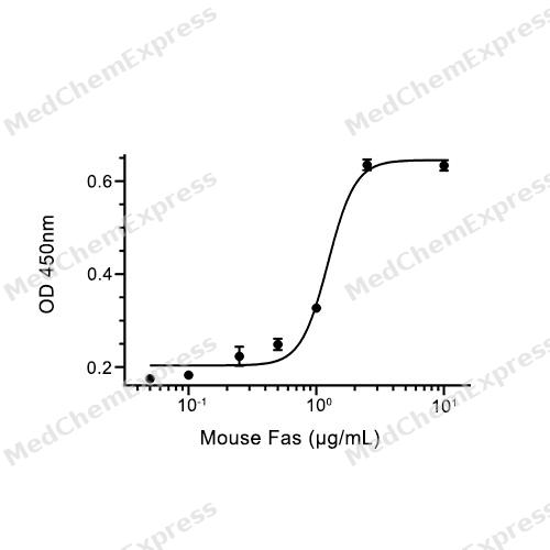
Bioactivity - Cell-Based Assay


Technical Parameters
-
Species Mouse
-
Source HEK293
-
Tag C-hFc
-
Accession
P25446 (Q22-R169)
-
Molecular Construction
-
N-term
-
Fas (Q22-R169)
Accession # P25446 -
hFc
-
C-term
-
-
Protein Length
Extracellular Domain
-
Synonyms
FAS; Fas (TNF Receptor Superfamily, Member 6); Prev. TNFRSF6; Apoptosis Signaling Receptor FAS; Prev. APT1; Apo-1 Antigen; Prev. FAS1; CD95 Antigen; Tumor Necrosis Factor Receptor Superfamily Member 6; Mutant Tumor Necrosis Receptor Superfamily Member 6;
-
AA Sequence
QGTNSISESLKLRRRVRETDKNCSEGLYQGGPFCCQPCQPGKKKVEDCKMNGGTPTCAPCTEGKEYMDKNHYADKCRRCTLCDEEHGLEVETNCTLTQNTKCKCKPDFYCDSPGCEHCVRCASCEHGTLEPCTATSNTNCRKQSPRNR
-
Predicted Molecular Mass
43.3 kDa
-
Molecular Weight
Approximately 60-65 kDa, based on SDS-PAGE under reducing conditions, due to the glycosylation.
-
Glycosylation
Yes
-
Purity
≥ 95%, as determined by Bis-Tris PAGE.


Product Properties
Lyophilized powder.
Lyophilized from a 0.22 μm filtered solution of PBS, pH 7.4, 8% trehalose.
<1 EU/μg, determined by LAL method.
It is not recommended to reconstitute to a concentration less than 100 μg/mL in ddH2O.
Stored at -20°C for 2 years from date of receipt. After reconstitution, it is stable at 4°C for 1 week or -20°C for longer (with carrier protein). It is recommended to freeze aliquots at -20°C or -80°C for extended storage.
Room temperature in continental US; may vary elsewhere.


Documentation
-
Data Sheet (264 KB)
-
SDS (252 KB)
- English - EN (252 KB)
- Français - FR (252 KB)
- Deutsch - DE (252 KB)
- Norwegian - NO (252 KB)
- Español - ES (252 KB)
- Swedish - SV (252 KB)
- Italian - IT (252 KB)
- Korean - KR (252 KB)
- Portuguese - PT (252 KB)
-
Handling Instructions (2659 KB)


References
[1]. Liu C, et al. Differential expression of human Fas mRNA species upon peripheral blood mononuclear cell activation. Biochem J. 1995 Sep 15;310 ( Pt 3)(Pt 3):957-63. [Content Brief]
[2]. Screaton RA, et al. Fas-associated death domain protein interacts with methyl-CpG binding domain protein 4: a potential link between genome surveillance and apoptosis. Proc Natl Acad Sci U S A. 2003 Apr 29;100(9):5211-6. [Content Brief]
[3]. Brenner B, et al. Fas/CD95/Apo-I activates the acidic sphingomyelinase via caspases. Cell Death Differ. 1998 Jan;5(1):29-37. [Content Brief]
[4]. Lee SM, et al. Stimulation of Fas (CD95) induces production of pro-inflammatory mediators through ERK/JNK-dependent activation of NF-κB in THP-1 cells. Cell Immunol. 2011;271(1):157-62. [Content Brief]
[5]. Peng SL, et al. A tumor-suppressor function for Fas (CD95) revealed in T cell-deficient mice. J Exp Med. 1996 Sep 1;184(3):1149-54. [Content Brief]
[6]. Mogil RJ, et al. Fas (CD95) participates in peripheral T cell deletion and associated apoptosis in vivo. Int Immunol. 1995 Sep;7(9):1451-8. [Content Brief]

Calculators
Concentration (start) × Volume (start) = Concentration (final) × Volume (final)


 Powered by Bioz

