



Vitamin D Receptor/VDR Protein, Mouse (sf9, His)

Based on 1 Customer Validation
Vitamin D receptor (VDR) protein is the nuclear receptor of calcitriol, which translocates to the nucleus after binding vitamin D3, thereby coordinating cellular activities.VDR forms a heterodimer with RXR, binds to DNA response elements, and initiates the transcription of vitamin D3-responsive genes.Vitamin D Receptor/VDR Protein, Mouse (sf9, His) is the recombinant mouse-derived Vitamin D Receptor/VDR protein, expressed by Sf9 insect cells , with C-His labeled tag.
- Species: Mouse
- Source: Sf9 insect cells
-
Storage:Stored at -20°C for 2 years from date of receipt. After reconstitution, it is stable at 4°C for 1 week or -20°C for longer (with carrier protein). It is recommended to freeze aliquots at -20°C or -80°C for extended storage.


To place orders, for customer services and technical support, please contact: MedChemExpress USA
Tel: 609-228-6898 E-mail: [email protected] [email protected]


-
 Biological Activity
-
Technical Parameters
-
Product Properties
- Documentation
- Help & FAQs




Biological Activity
Description
Vitamin D receptor (VDR) protein is the nuclear receptor of calcitriol, which translocates to the nucleus after binding vitamin D3, thereby coordinating cellular activities.VDR forms a heterodimer with RXR, binds to DNA response elements, and initiates the transcription of vitamin D3-responsive genes.Vitamin D Receptor/VDR Protein, Mouse (sf9, His) is the recombinant mouse-derived Vitamin D Receptor/VDR protein, expressed by Sf9 insect cells , with C-His labeled tag.
Background
Vitamin D Receptor (VDR) Protein serves as a nuclear receptor for calcitriol, the active form of vitamin D3, mediating its cellular actions. Upon binding vitamin D3, VDR translocates into the nucleus, forming heterodimers with the retinoid X receptor (RXR). The VDR-RXR heterodimers then bind to specific response elements on DNA, initiating the transcription of vitamin D3-responsive target genes. Beyond its central role in calcium homeostasis, VDR also functions as a receptor for the secondary bile acid lithocholic acid (LCA) and its metabolites. In the absence of bound vitamin D3, VDR forms homodimers, while vitamin D3 binding prompts heterodimerization with RXRA. VDR interacts with various coactivators, including MED1, NCOA1, NCOA2, NCOA3, and NCOA6, leading to enhanced transcription of target genes. It also interacts with the corepressor NCOR1, SNW1, IRX4, and exhibits ligand-dependent interactions with CRY1 and CRY2. These molecular interactions highlight the complex regulatory network that governs the transcriptional activity of VDR in response to vitamin D3 and other ligands.
Verified Bioactivity
Measured by its ability to inhibit calcitriol-mediated cytotoxicity using C2C12 cells treated with VDR. The ED50 for this effect is 0.497 μg/mL, corresponding to a specific activity is 2010.454 Unit/mg.
MCE Validation Data
-
Purity - SDS-PAGE
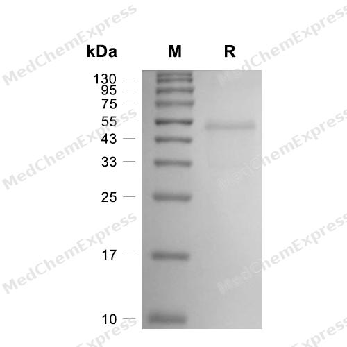
Purity - SDS-PAGE
-
Bioactivity - Cell-Based Assay
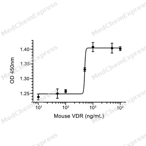
Bioactivity - Cell-Based Assay


Technical Parameters
-
Species Mouse
-
Source Sf9 insect cells
-
Tag C-10*His
-
Accession
P48281 (M1-S422)
-
Molecular Construction
-
N-term
-
VDR (M1-S422)
Accession # P48281 -
His
-
C-term
-
-
Protein Length
Full Length
-
Synonyms
VDR; Vitamin D Receptor; NR1I1; Nuclear Receptor Subfamily 1 Group I Member 1; 1,25-Dihydroxyvitamin D3 Receptor; Vitamin D3 Receptor; PPP1R163; Vitamin D (1,25-Dihydroxyvitamin D3) Receptor; Protein Phosphatase 1, Regulatory Subunit 163; 1,25-Dihydroxyvi
-
AA Sequence
MEAMAASTSLPDPGDFDRNVPRICGVCGDRATGFHFNAMTCEGCKGFFRRSMKRKALFTCPFNGDCRITKDNRRHCQACRLKRCVDIGMMKEFILTDEEVQRKREMIMKRKEEEALKDSLRPKLSEEQQHIIAILLDAHHKTYDPTYADFRDFRPPIRADVSTGSYSPRPTLSFSGDSSSNSDLYTPSLDMMEPASFSTMDLNEEGSDDPSVTLDLSPLSMLPHLADLVSYSIQKVIGFAKMIPGFRDLTSDDQIVLLKSSAIEVIMLRSNQSFTLDDMSWDCGSQDYKYDITDVSRAGHTLELIEPLIKFQVGLKKLNLHEEEHVLLMAICIVSPDRPGVQDAKLVEAIQDRLSNTLQTYIRCRHPPPGSHQLYAKMIQKLADLRSLNEEHSKQYRSLSFQPENSMKLTPLVLEVFGNEIS
-
Predicted Molecular Mass
47.8 kDa
-
Molecular Weight
Approximately 55 kDa, based on SDS-PAGE under reducing conditions.
-
Purity
≥ 90%, as determined by reducing SDS-PAGE.


Product Properties
Lyophilized powder.
Lyophilized from a 0.22 μm filtered solution of PBS, pH 7.4, 0.01% SKL, 5% trehalose.
<1 EU/μg, determined by LAL method.
It is not recommended to reconstitute to a concentration less than 100 μg/mL in ddH2O.
Stored at -20°C for 2 years from date of receipt. After reconstitution, it is stable at 4°C for 1 week or -20°C for longer (with carrier protein). It is recommended to freeze aliquots at -20°C or -80°C for extended storage.
Room temperature in continental US; may vary elsewhere.


Documentation
-
Data Sheet (238 KB)
-
SDS (252 KB)
- English - EN (252 KB)
- Français - FR (252 KB)
- Deutsch - DE (252 KB)
- Norwegian - NO (252 KB)
- Español - ES (252 KB)
- Swedish - SV (252 KB)
- Italian - IT (252 KB)
- Korean - KR (252 KB)
- Portuguese - PT (252 KB)
-
Handling Instructions (2659 KB)

Calculators
Concentration (start) × Volume (start) = Concentration (final) × Volume (final)


 Powered by Bioz

