



ROS-generating agent 1

Based on 1 Customer Validation
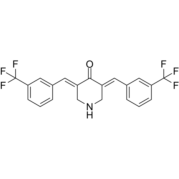
ROS-generating agent 1 is a selective ROS-generating agent. ROS-generating agent 1 inhibits TrxR activity and expression in cancer cells. ROS-generating agent 1 induces ROS-dependent apoptosis and ferroptosis in cancer cells. ROS-generating agent 1 selectively kills lung cancer cells and inhibits the growth of cancer cell xenograft tumors in nude mice. ROS-generating agent 1 can be used for the research of non-small cell lung cancer.
For research use only. We do not sell to patients.
- Purity : 99.11%
- CAS No.: 2369030-41-9
- Formula: C21H15F6NO
- Molecular Weight:411.34
-
Storage:Powder -20°C, 3 years , 4°C, 2 years ; In solvent -80°C, 6 months , -20°C, 1 month

To place orders, for customer services and technical support, please contact: MedChemExpress USA
Tel: 609-228-6898 E-mail: [email protected] [email protected]


-
 Biological Activity
-
Chemical Information
-
Solvent & Solubility
- Protocols
- Purity & Documentation
- References
-
 Help & FAQs


All Caspase Isoforms
More

Biological Activity
Description
Cellular Effect
|
Cell Line

|
Type | Value | Description | References |
|---|---|---|---|---|
| A549 | IC50 |
5.8 μM
Compound: 2c
|
Cytotoxicity against human A549 cells assessed as reduction in cell viability incubated for 48 hrs by MTT assay
Cytotoxicity against human A549 cells assessed as reduction in cell viability incubated for 48 hrs by MTT assay
|
[PMID: 36758308] |
| HepG2 | IC50 |
0.67 μM
Compound: 2c
|
Cytotoxicity against human HepG2 cells assessed as reduction in cell viability incubated for 48 hrs by MTT assay
Cytotoxicity against human HepG2 cells assessed as reduction in cell viability incubated for 48 hrs by MTT assay
|
[PMID: 36758308] |
| HT-1080 | IC50 |
0.93 μM
Compound: 2c
|
Cytotoxicity against human HT-1080 cells assessed as reduction in cell viability incubated for 48 hrs by MTT assay
Cytotoxicity against human HT-1080 cells assessed as reduction in cell viability incubated for 48 hrs by MTT assay
|
[PMID: 36758308] |
| MRC5 | IC50 |
14.09 μM
Compound: 2c
|
Cytotoxicity against human MRC5 cells assessed as reduction in cell viability incubated for 48 hrs by MTT assay
Cytotoxicity against human MRC5 cells assessed as reduction in cell viability incubated for 48 hrs by MTT assay
|
[PMID: 36758308] |
| NCI-H460 | IC50 |
0.44 μM
Compound: 2c
|
Cytotoxicity against human NCI-H460 cells assessed as reduction in cell viability incubated for 48 hrs by MTT assay
Cytotoxicity against human NCI-H460 cells assessed as reduction in cell viability incubated for 48 hrs by MTT assay
|
[PMID: 36758308] |
In Vitro
ROS-generating agent 1 (compound 2c) (48 h) potently and selectively inhibits the viability of NCI-H460 cells (IC50 = 0.44 μM) over normal MRC-5 cells, with a selectivity index of 32.0, and also shows strong cytotoxicity against A549, HepG2, and HT-1080 cancer cells[1].
ROS-generating agent 1 (0.5-2 μM; 1-12 h) selectively induces time- and concentration-dependent ROS accumulation in NCI-H460 cells but not in normal MRC-5 cells[1].
ROS-generating agent 1 (9 h) selectively depletes intracellular GSH levels in NCI-H460 cells in a dose-dependent manner, while having no significant effect on MRC-5 cells[1].
ROS-generating agent 1 (0.5-2 μM; 6 h) dose-dependently inhibits TrxR activity in NCI-H460 cells but has negligible effect on TrxR activity in normal MRC-5 cells[1].
ROS-generating agent 1 (0.5-2 μM; 6 h) potently and dose-dependently inhibits TrxR activity in NCI-H460 cells, with 2 μM causing 59% inhibition[1].
ROS-generating agent 1 (0.5-2 μM; 6 h) dose-dependently reduces intracellular TrxR protein levels in NCI-H460 cells, with 2 μM causing 69% suppression[1].
ROS-generating agent 1 covalently binds to the Sec-498 residue of human TrxR1 with a docking affinity score of −5.494 kcal/mol, forming hydrogen bonds and a cation-π interaction with key active site residues[1].
ROS-generating agent 1 (0.5-1 μM; 48 h) kills NCI-H460 cells via induction of apoptosis and ferroptosis[1].
ROS-generating agent 1 (0.5-2 μM; 12-36 h) concentration- and time-dependently reduces mitochondrial membrane potential in NCI-H460 cells[1].
ROS-generating agent 1 (1-2 μM; 48 h) induces ROS-dependent mitochondrial-mediated apoptosis in NCI-H460 cells, as shown by altered expression of Bcl-2, Bax, caspase-9, and caspase-3[1].
ROS-generating agent 1 (0.25-0.5 μM; 40 h) selectively induces ferroptosis in a dose-dependent manner in NCI-H460 cells[1].
ROS-generating agent 1 (0.5-2 μM; 40 h) dose-dependently increases MDA levels in NCI-H460 cells[1].
ROS-generating agent 1 (0.5-2 μM; 24 h) dose-dependently reduces GPX4 protein levels in NCI-H460 cells[1].
MedChemExpress (MCE) has not independently confirmed the accuracy of these methods. They are for reference only.
-
Cell Line:NCI-H460 cells
-
Concentration:0.5; 1; 2 μM
-
Incubation Time:6 h
-
Result:Inhibited intracellular TrxR protein expression in a dose-dependent manner, with 2 μM suppressing 69% of TrxR expression.
-
Cell Line:NCI-H460 cells
-
Concentration:0.5; 1 μM
-
Incubation Time:48 h
-
Result:Dose-dependently reduced cell viability of NCI-H460 cells.
-
Cell Line:NCI-H460 cells
-
Concentration:1; 2 μM
-
Incubation Time:48 h
-
Result:Triggered down-regulation of anti-apoptotic Bcl-2, up-regulation of pro-apoptotic Bax, and activation of caspase-9 and caspase-3.
These changes were effectively reversed by pretreatment with NAC.
-
Cell Line:NCI-H460 cells
-
Concentration:0.5; 1; 2 μM
-
Incubation Time:24 h
-
Result:Reduced GPX4 protein expression in a concentration-dependent manner.
In Vivo
MedChemExpress (MCE) has not independently confirmed the accuracy of these methods. They are for reference only.
-
Animal Model:BALB/c nude mice (male, 5-6 weeks old) subcutaneously inoculated with NCI-H460 cells[1]
-
Dosage:5; 15 mg/kg
-
Administration:i.p.; every 2 days; 4 weeks
-
Result:Significantly and dose-dependently inhibited NCI-H460 xenograft tumor growth.
Showed no significant changes in body weight.
Exhibited no abnormal histology in liver or kidney tissues via H&E staining.
Increased tumor cell apoptosis.
Down-regulated GPX4 expression in tumor tissues compared to controls.
- RNA extraction experimental
- Detection of Apoptosis
- TUNEL staining for apoptotic DNA fragmentation
- Annexin V plus membrane-impermeant dye apoptosis staining
- ROS/oxidative-stress fluorescent staining
- Patient-Derived Xenograft (PDX)
- Apoptosis Solutions
- Splenic/Portal-Vein Liver Metastasis Xenograft
- Ferroptosis Solutions
- Subcutaneous Cell-Line-Derived Xenograft
- Orthotopic Cell-Line Xenograft
- Intraperitoneal/Peritoneal Dissemination Xenograft


Chemical Information
-
CAS No. 2369030-41-9
-
Appearance Solid
-
Molecular Weight 411.34
-
Formula C21H15F6NO
-
Color Light yellow to yellow
-
SMILES
O=C1/C(CNC/C1=C\C2=CC(C(F)(F)F)=CC=C2)=C/C3=CC=CC(C(F)(F)F)=C3
-
Shipping
Room temperature in continental US; may vary elsewhere.
-
Storage
Powder -20°C 3 years 4°C 2 years In solvent -80°C 6 months -20°C 1 month


Solvent & Solubility
In Vitro:
DMSO : 33.33 mg/mL (81.03 mM; Need ultrasonic; Hygroscopic DMSO has a significant impact on the solubility of product, please use newly opened DMSO)
Please refer to the solubility information to select the appropriate solvent. Once prepared, please aliquot and store the solution to prevent product inactivation from repeated freeze-thaw cycles.
Storage method and period of stock solution: -80°C, 6 months; -20°C, 1 month. When stored at -80°C, please use it within 6 months. When stored at -20°C, please use it within 1 month.
Please refer to the solubility information to select the appropriate solvent. Once prepared, please aliquot and store the solution to prevent product inactivation from repeated freeze-thaw cycles.
Storage method and period of stock solution: -80°C, 6 months; -20°C, 1 month. When stored at -80°C, please use it within 6 months. When stored at -20°C, please use it within 1 month.
Concentration (start) × Volume (start) = Concentration (final) × Volume (final)


Purity & Documentation
-
Data Sheet (275 KB)
-
SDS (393 KB)
- English - EN (393 KB)
- Français - FR (393 KB)
- Deutsch - DE (393 KB)
- Norwegian - NO (393 KB)
- Español - ES (393 KB)
- Swedish - SV (393 KB)
- Italian - IT (393 KB)
- Korean - KR (393 KB)
- Portuguese - PT (393 KB)
-
Handling Instructions (2659 KB)
References


Complete Stock Solution Preparation Table
Please refer to the solubility information to select the appropriate solvent. Once prepared, please aliquot and store the solution to prevent product inactivation from repeated freeze-thaw cycles.
Storage method and period of stock solution: -80°C, 6 months; -20°C, 1 month. When stored at -80°C, please use it within 6 months. When stored at -20°C, please use it within 1 month.
| Optional Solvent | Concentration Solvent Mass | 1 mg | 5 mg | 10 mg | 25 mg |
|---|---|---|---|---|---|
| DMSO | 1 mM | 2.4311 mL | 12.1554 mL | 24.3108 mL | 60.7770 mL |
| 5 mM | 0.4862 mL | 2.4311 mL | 4.8622 mL | 12.1554 mL | |
| 10 mM | 0.2431 mL | 1.2155 mL | 2.4311 mL | 6.0777 mL | |
| 15 mM | 0.1621 mL | 0.8104 mL | 1.6207 mL | 4.0518 mL | |
| 20 mM | 0.1216 mL | 0.6078 mL | 1.2155 mL | 3.0388 mL | |
| 25 mM | 0.0972 mL | 0.4862 mL | 0.9724 mL | 2.4311 mL | |
| 30 mM | 0.0810 mL | 0.4052 mL | 0.8104 mL | 2.0259 mL | |
| 40 mM | 0.0608 mL | 0.3039 mL | 0.6078 mL | 1.5194 mL | |
| 50 mM | 0.0486 mL | 0.2431 mL | 0.4862 mL | 1.2155 mL | |
| 60 mM | 0.0405 mL | 0.2026 mL | 0.4052 mL | 1.0129 mL | |
| 80 mM | 0.0304 mL | 0.1519 mL | 0.3039 mL | 0.7597 mL |


ROS-generating agent 1 Related Classifications
HY-149265 Related Classifications
 Powered by Bioz