



RSH-7

Based on 1 Customer Validation
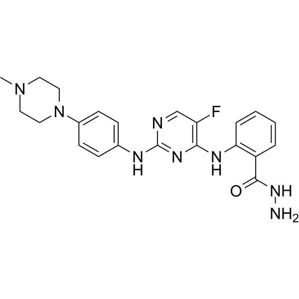
RSH-7 is a potent BTK and FLT3 inhibitor with IC50s of 47, 12 nM, respectively. RSH-7 induces apoptosis and shows antiproliferative activities. RSH-7 inhibits BTK and FLT3 signaling and shows anti-tumor activity.
For research use only. We do not sell to patients.
- Purity : 98.74%
- CAS No.: 2764609-97-2
- Formula: C22H25FN8O
- Molecular Weight:436.49
-
Storage:Powder -20°C, 3 years , 4°C, 2 years ; In solvent -80°C, 6 months , -20°C, 1 month

To place orders, for customer services and technical support, please contact: MedChemExpress USA
Tel: 609-228-6898 E-mail: [email protected] [email protected]


-
 Biological Activity
-
Chemical Information
-
Solvent & Solubility
- Protocols
- Purity & Documentation
- References
-
 Help & FAQs


-
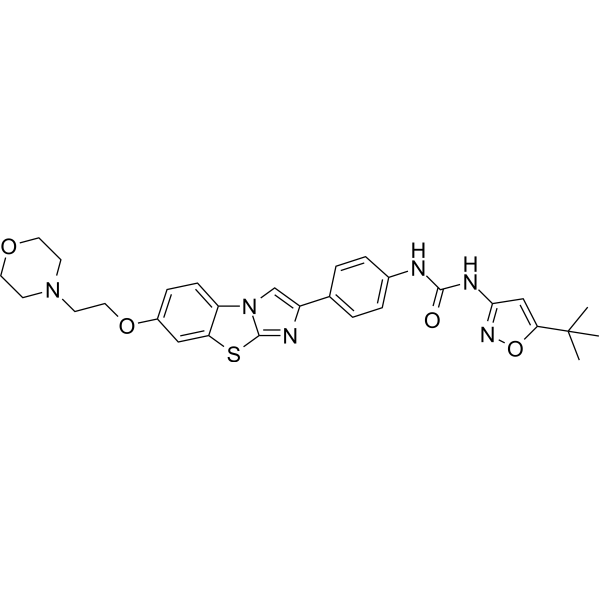
HY-13001
QuizartinibFLT3 Inhibitor -
HY-12333
DenfivontinibFLT3 Inhibitor -
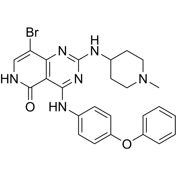
HY-15002
AST 487c-Kit Inhibitor -
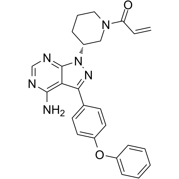
HY-10997
IbrutinibBtk Inhibitor -
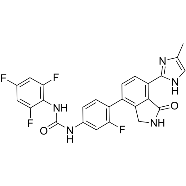
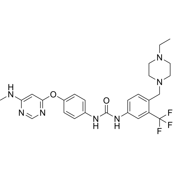
HY-139535
LuxeptinibPan-FLT3/Pan-BTK Inhibitor -
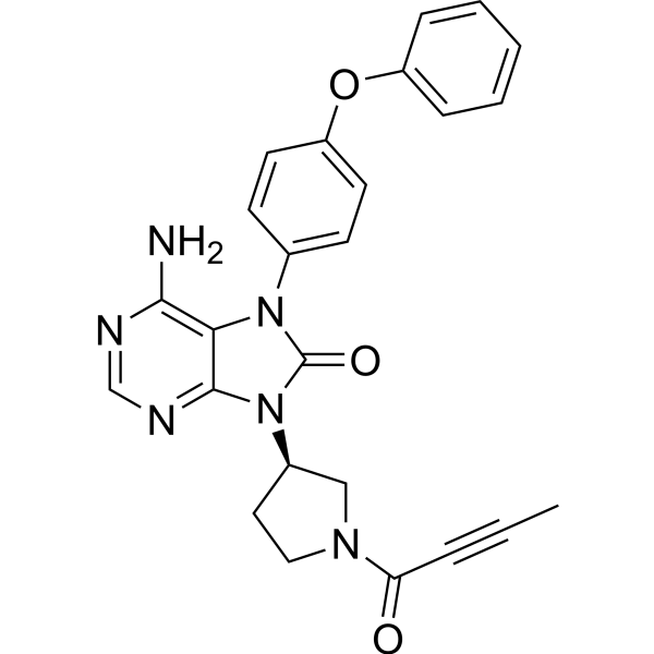
HY-15771
TirabrutinibBTK Inhibitor -
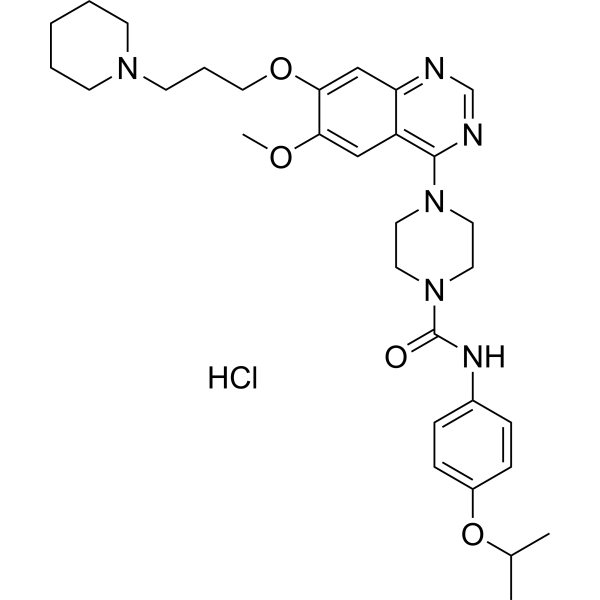
HY-10202A
Tandutinib hydrochlorideFLT3 Inhibitor -
HY-18012
Spebrutinib -

HY-10997R
Ibrutinib (Standard)Btk Inhibitor -
HY-80003
QL47Antiviral Agent/BTK Inhibitor -
HY-15771A
Tirabrutinib hydrochlorideBTK Inhibitor -

HY-13001R
Quizartinib (Standard)FLT3 Inhibitor


Biological Activity
Description
IC50 & Target
IC50: 47 nM (BTK); 12 nM (FLT3)[1]
Cellular Effect
|
Cell Line

|
Type | Value | Description | References |
|---|---|---|---|---|
| JeKo-1 | IC50 |
17 nM
Compound: RSH-7
|
Antiproliferative activity against human JeKo-1 cells assessed as inhibition of cell growth incubated for 72 hrs by CCK8 assay
Antiproliferative activity against human JeKo-1 cells assessed as inhibition of cell growth incubated for 72 hrs by CCK8 assay
|
[PMID: 36399923] |
| K562 | IC50 |
930 nM
Compound: RSH-7
|
Antiproliferative activity against human K562 cells assessed as inhibition of cell growth incubated for 72 hrs by CCK8 assay
Antiproliferative activity against human K562 cells assessed as inhibition of cell growth incubated for 72 hrs by CCK8 assay
|
[PMID: 36399923] |
| MOLT-4 | IC50 |
11 nM
Compound: RSH-7
|
Antiproliferative activity against human MOLT-4 cells assessed as inhibition of cell growth incubated for 72 hrs by CCK8 assay
Antiproliferative activity against human MOLT-4 cells assessed as inhibition of cell growth incubated for 72 hrs by CCK8 assay
|
[PMID: 36399923] |
| MV4-11 | IC50 |
3 nM
Compound: RSH-7
|
Antiproliferative activity against human MV4-11 cells assessed as inhibition of cell growth incubated for 72 hrs by CCK8 assay
Antiproliferative activity against human MV4-11 cells assessed as inhibition of cell growth incubated for 72 hrs by CCK8 assay
|
[PMID: 36399923] |
In Vitro
RSH-7 (1-1000 nM; 72 h) shows antiproliferative activities with IC50s of 17, 3, 11, 930 nM for Jeko-1, MV-4-11, Molt4, K562 cells, respectively[1].
RSH-7 (30, 150, 750 nM; 72 h) decreases the expression of p-BTK (TYR223), p-PLCγ(Tyr1217), p-FLT3 (Tyr589), p-STAT5 (TYR694) in a dose-dependent manner[1].
RSH-7 (30, 150, 750 nM; 72 h) induces apoptosis and increases the expression of BAX, p53, cleaved caspase 3 in a dose dependen manner in jeko-1 cells[1].
MedChemExpress (MCE) has not independently confirmed the accuracy of these methods. They are for reference only.
-
Cell Line:Jeko-1, MV-4-11, Molt4, K562 cells
-
Concentration:1-1000 nM
-
Incubation Time:72 h
-
Result:Showed antiproliferative activities with IC50s of 17, 3, 11, 930 nM for Jeko-1, MV-4-11, Molt4, K562 cells, respectively.
-
Cell Line:jeko-1 cells
-
Concentration:30, 150, 750 nM
-
Incubation Time:72 h
-
Result:Reduced both BTK, PLCγ2, FLT3 and STAT5 phosphorylation in a dose-dependent manner.
-
Cell Line:jeko-1 cells
-
Concentration:30, 150, 750 nM
-
Incubation Time:72 h
-
Result:Dose-dependently induced cell apoptosis and upregulated the expression of pro-apoptotic protein BAX, p53, cleaved caspase 3.
In Vivo
MedChemExpress (MCE) has not independently confirmed the accuracy of these methods. They are for reference only.
-
Animal Model:Female NOD/SCID mice (jeko-1 cell-inoculated xenograft NOD/SCID mice models)[1]
-
Dosage:25, 50 mg/kg
-
Administration:I.p.; daily for 16 days
-
Result:Suppressed tumor growth in a dose-dependent manner, with tumor growth inhibition (TGI) values of 66.95% and 79.78% at doses of 25 and 50 mg/kg,
-
Animal Model:Female NOD/SCID mice (MV4-11 cell-inoculated xenograft NOD/SCID mice models)[1]
-
Dosage:10, 20 mg/kg
-
Administration:I.p.; daily for 21 days
-
Result:Significantly and dose-dependently suppressed the tumor growth with the TGI rates of 74.23% and 94.84% at the dosage of 10 and 20 mg/kg, respectively.


Chemical Information
-
CAS No. 2764609-97-2
-
Appearance Solid
-
Molecular Weight 436.49
-
Formula C22H25FN8O
-
Color Gray to dark gray
-
SMILES
CN1CCN(CC1)C2=CC=C(C=C2)NC3=NC(NC4=C(C=CC=C4)C(NN)=O)=C(C=N3)F
-
Shipping
Room temperature in continental US; may vary elsewhere.
-
Storage
Powder -20°C 3 years 4°C 2 years In solvent -80°C 6 months -20°C 1 month


Solvent & Solubility
In Vitro:
DMSO : 83.33 mg/mL (190.91 mM; Need ultrasonic; Hygroscopic DMSO has a significant impact on the solubility of product, please use newly opened DMSO)
Please refer to the solubility information to select the appropriate solvent. Once prepared, please aliquot and store the solution to prevent product inactivation from repeated freeze-thaw cycles.
Storage method and period of stock solution: -80°C, 6 months; -20°C, 1 month. When stored at -80°C, please use it within 6 months. When stored at -20°C, please use it within 1 month.
Please refer to the solubility information to select the appropriate solvent. Once prepared, please aliquot and store the solution to prevent product inactivation from repeated freeze-thaw cycles.
Storage method and period of stock solution: -80°C, 6 months; -20°C, 1 month. When stored at -80°C, please use it within 6 months. When stored at -20°C, please use it within 1 month.
Concentration (start) × Volume (start) = Concentration (final) × Volume (final)


Purity & Documentation
-
Data Sheet (275 KB)
-
SDS (254 KB)
- English - EN (254 KB)
- Français - FR (254 KB)
- Deutsch - DE (254 KB)
- Norwegian - NO (254 KB)
- Español - ES (254 KB)
- Swedish - SV (254 KB)
- Italian - IT (254 KB)
- Korean - KR (254 KB)
- Portuguese - PT (254 KB)
-
Handling Instructions (2659 KB)
References


Complete Stock Solution Preparation Table
Please refer to the solubility information to select the appropriate solvent. Once prepared, please aliquot and store the solution to prevent product inactivation from repeated freeze-thaw cycles.
Storage method and period of stock solution: -80°C, 6 months; -20°C, 1 month. When stored at -80°C, please use it within 6 months. When stored at -20°C, please use it within 1 month.
| Optional Solvent | Concentration Solvent Mass | 1 mg | 5 mg | 10 mg | 25 mg |
|---|---|---|---|---|---|
| DMSO | 1 mM | 2.2910 mL | 11.4550 mL | 22.9100 mL | 57.2751 mL |
| 5 mM | 0.4582 mL | 2.2910 mL | 4.5820 mL | 11.4550 mL | |
| 10 mM | 0.2291 mL | 1.1455 mL | 2.2910 mL | 5.7275 mL | |
| 15 mM | 0.1527 mL | 0.7637 mL | 1.5273 mL | 3.8183 mL | |
| 20 mM | 0.1146 mL | 0.5728 mL | 1.1455 mL | 2.8638 mL | |
| 25 mM | 0.0916 mL | 0.4582 mL | 0.9164 mL | 2.2910 mL | |
| 30 mM | 0.0764 mL | 0.3818 mL | 0.7637 mL | 1.9092 mL | |
| 40 mM | 0.0573 mL | 0.2864 mL | 0.5728 mL | 1.4319 mL | |
| 50 mM | 0.0458 mL | 0.2291 mL | 0.4582 mL | 1.1455 mL | |
| 60 mM | 0.0382 mL | 0.1909 mL | 0.3818 mL | 0.9546 mL | |
| 80 mM | 0.0286 mL | 0.1432 mL | 0.2864 mL | 0.7159 mL | |
| 100 mM | 0.0229 mL | 0.1146 mL | 0.2291 mL | 0.5728 mL |


RSH-7 Related Classifications
HY-151961 Related Classifications
 Powered by Bioz