



TT01001

Based on 1 Customer Validation
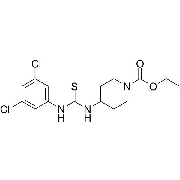
TT01001 is a selective and orally active mitoNEET agonist and a monoamine oxidase B (MAO-B) inhibitor (IC50 = 8.84 μM). TT01001 does not activate PPARγ but interacts with MitoNEET. TT01001 attenuates oxidative stress and neuronal apoptosis by preventing mitoNEET-mediated mitochondrial dysfunction. TT01001 improves type II diabetes and ameliorates mitochondrial function of mice. TT01001 can used for the studies of type II diabetes and neurological disorders .
For research use only. We do not sell to patients.
- Purity : 99.93%
- CAS No.: 1022367-69-6
- Formula: C15H19Cl2N3O2S
- Molecular Weight:376.30
-
Storage:
4°C, protect from light
* In solvent : -80°C, 6 months; -20°C, 1 month (protect from light)

To place orders, for customer services and technical support, please contact: MedChemExpress USA
Tel: 609-228-6898 E-mail: [email protected] [email protected]


-
 Biological Activity
-
Chemical Information
-
Solvent & Solubility
- Protocols
- Purity & Documentation
- References
-
 Help & FAQs




Biological Activity
Description
IC50 & Target
[3]|
MAO-B 8.84 μM (IC50) |
In Vivo
TT01001 (1-9 mg/kg, i.p., single dose) attenuates oxidative stress and neuronal apoptosis by preventing mitoNEET-mediated mitochondrial dysfunction after subarachnoid hemorrhage (SAH) in rats[2].
MedChemExpress (MCE) has not independently confirmed the accuracy of these methods. They are for reference only.
-
Animal Model:Type II diabetes model established in male, 5-week-old mice of C57BL/6J and BKS.Cg-+ Leprdb/+Leprdb (db/db)[1]
-
Dosage:100 mg/kg
-
Administration:Oral administration (p.o.), once daily for 28 days
-
Result:Improved diabetes in db/db Mice without causing weight gain. Was no significant effect on the mtDNA level of the mouse skeletal muscle. Significantly decreased elevated complex II + III activity and no change in complex I + III or IV activity.
-
Animal Model:SAH model established in adult male Sprague-Dawley rats weighting 280-320 g[2]
-
Dosage:Intraperitoneal injection (i.p.), single dose 1 h after SAH
-
Administration:Oral administration (p.o.), once daily for 28 days
-
Result:Improved neurological functions at 24 h after subarachnoid hemorrhage. Reduced the oxidative stress injury at 24 h after subarachnoid hemorrhage. Reduced neuronal apoptosis at 24 h after subarachnoid hemorrhage.
- Detection of Apoptosis
- TUNEL staining for apoptotic DNA fragmentation
- Mitochondrial membrane-potential fluorescent assay
- Annexin V plus membrane-impermeant dye apoptosis staining
- ROS/oxidative-stress fluorescent staining
- Mitochondrial membrane-potential and mitochondrial mass staining
- Apoptosis Solutions
- Research Protocol for Metabolic Diseases


Chemical Information
-
CAS No. 1022367-69-6
-
Appearance Solid
-
Molecular Weight 376.30
-
Formula C15H19Cl2N3O2S
-
Color White to off-white
-
SMILES
O=C(OCC)N1CCC(CC1)NC(NC2=CC(Cl)=CC(Cl)=C2)=S
-
Shipping
Room temperature in continental US; may vary elsewhere.
-
Storage
4°C, protect from light
* In solvent : -80°C, 6 months; -20°C, 1 month (protect from light)


Solvent & Solubility
In Vitro:
DMSO : 125 mg/mL (332.18 mM; Need ultrasonic; Hygroscopic DMSO has a significant impact on the solubility of product, please use newly opened DMSO)
Please refer to the solubility information to select the appropriate solvent. Once prepared, please aliquot and store the solution to prevent product inactivation from repeated freeze-thaw cycles.
Storage method and period of stock solution: -80°C, 6 months; -20°C, 1 month (protect from light). When stored at -80°C, please use it within 6 months. When stored at -20°C, please use it within 1 month.
Please refer to the solubility information to select the appropriate solvent. Once prepared, please aliquot and store the solution to prevent product inactivation from repeated freeze-thaw cycles.
Storage method and period of stock solution: -80°C, 6 months; -20°C, 1 month (protect from light). When stored at -80°C, please use it within 6 months. When stored at -20°C, please use it within 1 month.
Concentration (start) × Volume (start) = Concentration (final) × Volume (final)


Purity & Documentation
-
Data Sheet (272 KB)
-
SDS (394 KB)
- English - EN (394 KB)
- Français - FR (394 KB)
- Deutsch - DE (394 KB)
- Norwegian - NO (394 KB)
- Español - ES (394 KB)
- Swedish - SV (394 KB)
- Italian - IT (394 KB)
- Korean - KR (394 KB)
- Portuguese - PT (394 KB)
-
Handling Instructions (2659 KB)
References
[1]. Takehiro Takahashi, et al. A novel MitoNEET ligand, TT01001, improves diabetes and ameliorates mitochondrial function in db/db mice. J Pharmacol Exp Ther. 2015, 352, 2. [Content Brief]
[2]. Shi G, et al. TT01001 attenuates oxidative stress and neuronal apoptosis by preventing mitoNEET-mediated mitochondrial dysfunction after subarachnoid hemorrhage in rats. Neuroreport. 2020;31(11):845-850. [Content Brief]
[3]. Barbosa Belchior M, et al. Synthesis and evaluation of enzyme inhibition by novel TT01001 derivatives as monoamine oxidase B inhibitors. Bioorg Med Chem Lett. 2025 Mar 1;117:130045. [Content Brief]


Complete Stock Solution Preparation Table
Please refer to the solubility information to select the appropriate solvent. Once prepared, please aliquot and store the solution to prevent product inactivation from repeated freeze-thaw cycles.
Storage method and period of stock solution: -80°C, 6 months; -20°C, 1 month (protect from light). When stored at -80°C, please use it within 6 months. When stored at -20°C, please use it within 1 month.
| Optional Solvent | Concentration Solvent Mass | 1 mg | 5 mg | 10 mg | 25 mg |
|---|---|---|---|---|---|
| DMSO | 1 mM | 2.6575 mL | 13.2873 mL | 26.5745 mL | 66.4364 mL |
| 5 mM | 0.5315 mL | 2.6575 mL | 5.3149 mL | 13.2873 mL | |
| 10 mM | 0.2657 mL | 1.3287 mL | 2.6575 mL | 6.6436 mL | |
| 15 mM | 0.1772 mL | 0.8858 mL | 1.7716 mL | 4.4291 mL | |
| 20 mM | 0.1329 mL | 0.6644 mL | 1.3287 mL | 3.3218 mL | |
| 25 mM | 0.1063 mL | 0.5315 mL | 1.0630 mL | 2.6575 mL | |
| 30 mM | 0.0886 mL | 0.4429 mL | 0.8858 mL | 2.2145 mL | |
| 40 mM | 0.0664 mL | 0.3322 mL | 0.6644 mL | 1.6609 mL | |
| 50 mM | 0.0531 mL | 0.2657 mL | 0.5315 mL | 1.3287 mL | |
| 60 mM | 0.0443 mL | 0.2215 mL | 0.4429 mL | 1.1073 mL | |
| 80 mM | 0.0332 mL | 0.1661 mL | 0.3322 mL | 0.8305 mL | |
| 100 mM | 0.0266 mL | 0.1329 mL | 0.2657 mL | 0.6644 mL |


TT01001 Related Classifications
HY-114520 Related Classifications
 Powered by Bioz