



YL-109
Based on 2 publication(s) in Google Scholar
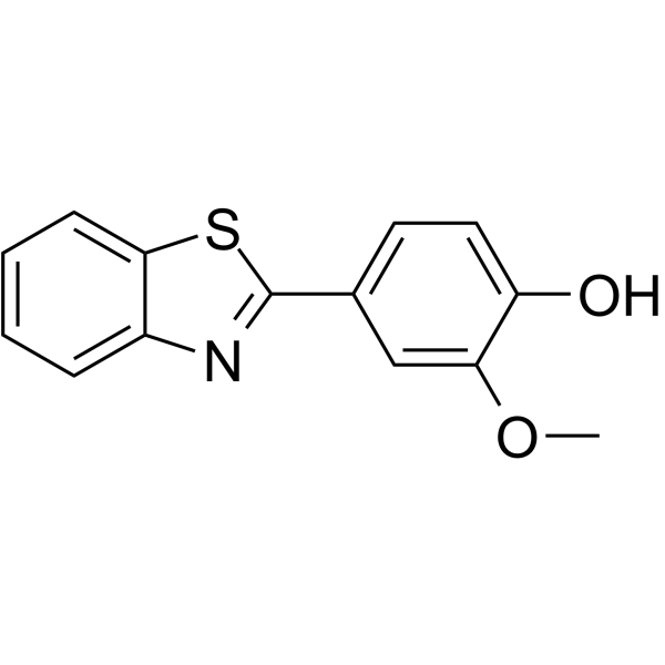
YL-109 is an antitumor agent that can induce carboxyl terminus of Hsp70-interacting protein (CHIP) expression through aryl hydrocarbon receptor (AhR) signaling. YL-109 has ability to inhibit breast cancer cell growth and invasiveness.
For research use only. We do not sell to patients.
- Purity : 98.91%
- CAS No.: 36341-25-0
- Formula: C14H11NO2S
- Molecular Weight:257.31
-
Storage:Powder -20°C, 3 years , 4°C, 2 years ; In solvent -80°C, 2 years , -20°C, 1 year

To place orders, for customer services and technical support, please contact: MedChemExpress USA
Tel: 609-228-6898 E-mail: [email protected] [email protected]


-
 Biological Activity
-
Chemical Information
-
Solvent & Solubility
- Protocols
- Purity & Documentation
- References
-
 Help & FAQs


-
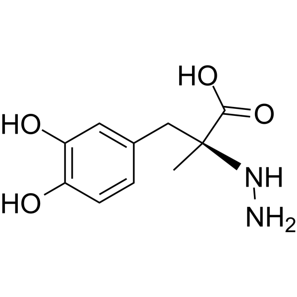
HY-B0311
CarbidopaDecarboxylase Inhibitor -
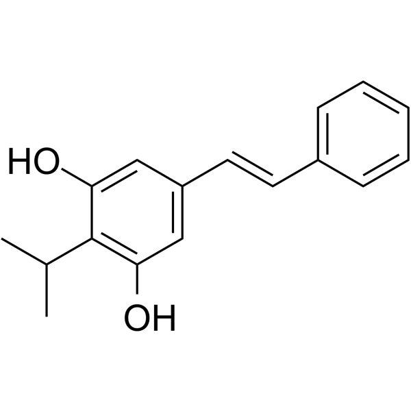
HY-109044
TapinarofAhR Agonist -
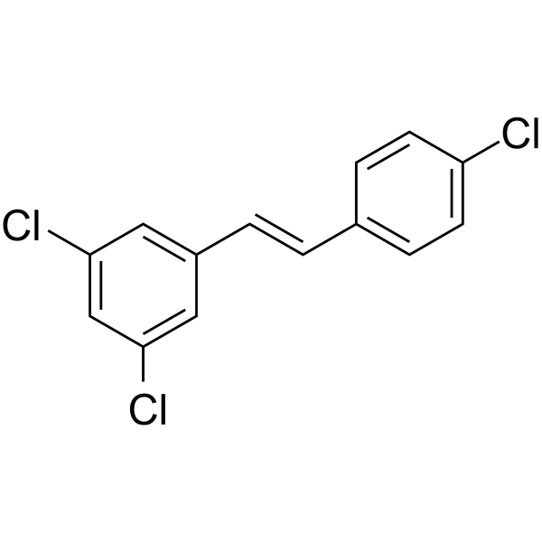
HY-112629
PDM2Aryl Hydrocarbon Receptor Antagonist -
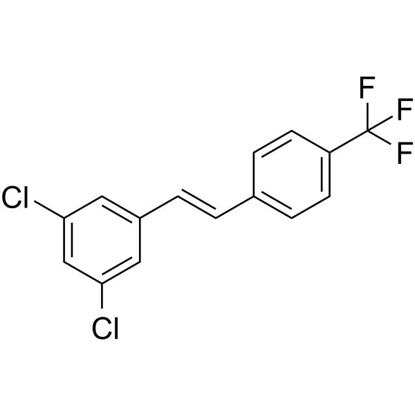
HY-112627
CAY 10465Aryl Hydrocarbon Receptor Agonist -
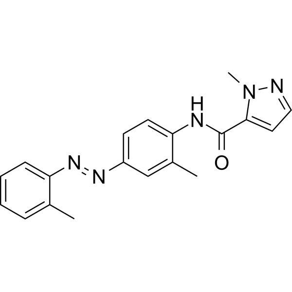
HY-12684
CH-223191AhR Antagonist -

HY-B0311R
Carbidopa (Standard)Decarboxylase Inhibitor -
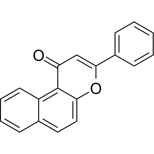
HY-114740
β-NaphthoflavoneAhR agonist -

HY-109044R
Tapinarof (Standard)AhR Agonist -

HY-114740R
β-Naphthoflavone (Standard)AhR agonist
Publications Citing Use of MedChemExpress (MCE) YL-109
More

Biological Activity
Description
In Vitro
YL-109 (0.001-10 μM; 96 h or 24 h) inhibits cell proliferation, motility, and invasiveness in breast cancer cells[1].
YL-109 (1 μM) increases both CHIP mRNA and protein levels in MDA-MB-231 cells[1].
MedChemExpress (MCE) has not independently confirmed the accuracy of these methods. They are for reference only. Further protocols information, click here.
-
Cell Line:MCF-7 and MDA-MB-231 cells
-
Concentration:0.001, 0.01, 0.1, 1, 10 μM
-
Incubation Time:96 hours
-
Result:Strongly inhibited cell proliferation of MCF-7 and MDA-MB-231 cells in a dose-dependent manner (IC50=85.8 nM and 4.02 μM, respectively).
In Vivo
MedChemExpress (MCE) has not independently confirmed the accuracy of these methods. They are for reference only.
-
Animal Model:BALB/cAjcl-nu/nu female mice (4-5 weeks) inoculated with MCF-7 or MDA-MB-231 cells[1]
-
Dosage:15 mg/kg
-
Administration:S.c. every 2 days for 63 days
-
Result:Suppressed tumor growth in mice injected with MCF-7 and MDA-MB-231 cells.


Chemical Information
-
CAS No. 36341-25-0
-
Appearance Solid
-
Molecular Weight 257.31
-
Formula C14H11NO2S
-
Color Off-white to light yellow
-
SMILES
OC1=CC=C(C2=NC3=CC=CC=C3S2)C=C1OC
-
Shipping
Room temperature in continental US; may vary elsewhere.
-
Storage
Powder -20°C 3 years 4°C 2 years In solvent -80°C 2 years -20°C 1 year


Publications (2)
-
Journal Impact Factor
-
Most Recent
-
Biomed Pharmacother
YL-109 attenuates sepsis-associated multiple organ injury through inhibiting the ERK/AP-1 axis and pyroptosis by upregulating CHIP. [Abstract]2024 Jun:175:116633. PMID: 38670049 -


Solvent & Solubility
In Vitro:
DMSO : 100 mg/mL (388.64 mM; Need ultrasonic; Hygroscopic DMSO has a significant impact on the solubility of product, please use newly opened DMSO)
Please refer to the solubility information to select the appropriate solvent. Once prepared, please aliquot and store the solution to prevent product inactivation from repeated freeze-thaw cycles.
Storage method and period of stock solution: -80°C, 2 years; -20°C, 1 year. When stored at -80°C, please use it within 2 years. When stored at -20°C, please use it within 1 year.
Please refer to the solubility information to select the appropriate solvent. Once prepared, please aliquot and store the solution to prevent product inactivation from repeated freeze-thaw cycles.
Storage method and period of stock solution: -80°C, 2 years; -20°C, 1 year. When stored at -80°C, please use it within 2 years. When stored at -20°C, please use it within 1 year.
Concentration (start) × Volume (start) = Concentration (final) × Volume (final)
In Vivo:
Select the appropriate dissolution method based on your experimental animal and administration route.
- For the following dissolution methods, please ensure to first prepare a clear stock solution using an In Vitro approach and then sequentially add co-solvents:
- To ensure reliable experimental results, the clarified stock solution can be appropriately stored based on storage conditions. As for the working solution for In Vivo experiments, it is recommended to prepare freshly and use it on the same day.
- The percentages shown for the solvents indicate their volumetric ratio in the final prepared solution. If precipitation or phase separation occurs during preparation, heat and/or sonication can be used to aid dissolution.
Add each solvent one by one: 10% DMSO 40% PEG300 5% Tween-80 45% Saline
Solubility: ≥ 3 mg/mL (11.66 mM); Clear solution
This protocol yields a clear solution of ≥ 3 mg/mL (saturation unknown).
Taking 1 mL working solution as an example, add 100 μL DMSO stock solution (30.0 mg/mL) to 400 μL PEG300, and mix evenly; then add 50 μL Tween-80 and mix evenly; then add 450 μL Saline to adjust the volume to 1 mL.
Preparation of Saline: Dissolve 0.9 g sodium chloride in ddH₂O and dilute to 100 mL to obtain a clear Saline solution.
Add each solvent one by one: 10% DMSO 90% (20% SBE-β-CD in Saline)
Solubility: 3 mg/mL (11.66 mM); Clear solution; Need ultrasonic
This protocol yields a clear solution of 3 mg/mL.
Taking 1 mL working solution as an example, add 100 μL DMSO stock solution (30.0 mg/mL) to 900 μL 20% SBE-β-CD in Saline, and mix evenly.
Preparation of 20% SBE-β-CD in Saline (4°C, storage for one week): 2 g SBE-β-CD powder is dissolved in 10 mL Saline, completely dissolve until clear.
In Vivo Dissolution Calculator
Please enter the basic information of animal experiments:
-
-
-
-
Recommended: Prepare an additional quantity of animals to account for potential losses during experiments.
Please enter your animal formula composition:
-
%DMSO +
Recommended: Keep the proportion of DMSO in working solution below 2% if your animal is weak.
-
%+
-
+%Tween-80 + +
-
%Saline +
The co-solvents required include: DMSO, . All of co-solvents are available by MedChemExpress (MCE). , Tween 80. All of co-solvents are available by MedChemExpress (MCE).
Working solution concentration: 0.22 mg/mL
Method for preparing stock solution: mg drug dissolved in μL DMSO. Stock solution concentration: mg/mL.
1. Take μL DMSO stock solution;
2. Add μL .
μL , mix evenly;
3. Then add μL Tween 80, mix evenly;
4. Then add μL
Please ensure that the stock solution in the first step is dissolved to a clear state, and add co-solvents in sequence. You can use ultrasonic heating (ultrasonic cleaner, recommended frequency 20-40 kHz), vortexing, etc. to assist dissolution.

Protocols
-
RNA extraction experimental
By lysing cells, releasing RNA, and removing impurities such as proteins and DNA, high-purity RNA products are finally obtained. The commonly used traditional method is the guanidine isothiocyanate/phenol/chloroform method (Trizol), which is suitable for a variety of animal materials including animal tissues, microorganisms, cultured cells, etc., and most plant materials.
-
Protocol for Cell Counting and Cell Density Analysis
Cell counting and cell-density analysis estimate the number of cells in a known volume or field area. Manual hemocytometer counting uses a chamber of defined geometry to convert counted cells into cells/mL, while automated counters and image-analysis workflows detect cell objects from optical, brightfield, fluorescence, impedance, or digital-image features. Trypan blue viability counting is based on dye exclusion: viable cells with intact membranes exclude dye, while non-viable cells with compromised membranes stain blue. The readout is total cell density, viable-cell density, dead-cell density, and percent viability. Cell density can also be estimated from microscopy images by counting objects per image area, from flow cytometry using calibrated volume or reference particles, or from in situ microscopy in bioreactors after calibration against reference methods such as hemocytometer or flow cytometry.
-
Chromatin Immunoprecipitation
Chromatin Immunoprecipitation (ChIP) is a molecular biology technique used to study the interaction between proteins and chromatin.
-
Somatic Cell Culture
A method of simulating the in vivo environment in vitro to maintain the cell growth, differentation and main functions.
-
CCK-8/WST-8 Cell Proliferation Assay
The CCK-8/WST-8 assay is based on the reduction of the water-soluble tetrazolium salt WST-8 to a water-soluble formazan product by cellular dehydrogenases in metabolically active cells, where the generated formazan amount is proportional to the number of living cells and is quantified by measuring absorbance in the visible range, providing a colorimetric readout for cell viability and proliferation assessment. This class of tetrazolium-based assays improves upon earlier MTT-based systems by producing a water-soluble formazan, eliminating the need for organic solubilization steps and enabling direct spectrophotometric measurement in culture medium.
-
Cell Counting-Based Growth Curve Assay
Cell counting-based growth curve assays quantify cell proliferation by directly measuring changes in viable cell number over time using manual or automated counting methods such as hemocytometer-based counting or instrument-assisted cell enumeration, enabling construction of growth curves that reflect population expansion dynamics in response to culture conditions. A widely used approach is trypan blue exclusion with hemocytometer counting, where membrane-compromised (non-viable) cells take up the dye, allowing discrimination between viable and non-viable cells while simultaneously enabling total cell number quantification. Repeated sampling across time points allows estimation of proliferation rate, growth phases, and comparative growth kinetics between experimental conditions.
-
MTT Cell Proliferation Assay
The MTT assay is a colorimetric endpoint assay for estimating viable cell number, cell growth, cytotoxicity, or cell activation in cultured mammalian cells. Living cells reduce the yellow tetrazolium salt MTT into purple/blue formazan, while dead cells do not generate the same signal; the resulting color can be quantified with a multiwell spectrophotometer. MTT reduction is commonly interpreted as a readout of metabolic activity that often correlates with viable cell number, but it should not be treated as a direct cell-counting method unless the assay is optimized for the cell type and experimental condition. Studies show that MTT reduction can involve mitochondrial and non-mitochondrial reducing systems, and formazan may accumulate in intracellular lipid droplets rather than simply marking mitochondria.
-
Breast Cancer Modeling
Breast cancer is a heterogeneous cancer, and it has been distinguished into four subtypes: luminal A, luminal B, HER2-positive and basal-like. Molecular mutations, epigenetic alterations, hormone exposure and immune microenvironment are related to the progression of breast cancer.
-
Cell Viability Determination by MTT Colorimetric Assay
The following protocol uses the MTT colorimetric assay as a classic literature-established method for assessing cell viability/metabolic activity in cultured mammalian cells. MTT[3-(4,5-dimethylthiazol-2-yl)-2,5-diphenyltetrazolium bromide] is reduced by metabolically active cells to a colored formazan product; the amount of formazan is quantified spectrophotometrically and provides an indirect measure of metabolically active viable cells. Importantly, MTT reduction reflects cellular oxidoreductase/metabolic activity rather than an absolute direct count of living cells, so changes in cellular metabolism can alter the signal independently of cell number.


Purity & Documentation
-
Data Sheet (274 KB)
-
SDS (643 KB)
- English - EN (643 KB)
- Français - FR (643 KB)
- Deutsch - DE (643 KB)
- Norwegian - NO (643 KB)
- Español - ES (643 KB)
- Swedish - SV (643 KB)
- Italian - IT (643 KB)
- Korean - KR (643 KB)
- Portuguese - PT (643 KB)
-
Handling Instructions (2659 KB)
References


Complete Stock Solution Preparation Table
Please refer to the solubility information to select the appropriate solvent. Once prepared, please aliquot and store the solution to prevent product inactivation from repeated freeze-thaw cycles.
Storage method and period of stock solution: -80°C, 2 years; -20°C, 1 year. When stored at -80°C, please use it within 2 years. When stored at -20°C, please use it within 1 year.
| Optional Solvent | Concentration Solvent Mass | 1 mg | 5 mg | 10 mg | 25 mg |
|---|---|---|---|---|---|
| DMSO | 1 mM | 3.8864 mL | 19.4318 mL | 38.8636 mL | 97.1591 mL |
| 5 mM | 0.7773 mL | 3.8864 mL | 7.7727 mL | 19.4318 mL | |
| 10 mM | 0.3886 mL | 1.9432 mL | 3.8864 mL | 9.7159 mL | |
| 15 mM | 0.2591 mL | 1.2955 mL | 2.5909 mL | 6.4773 mL | |
| 20 mM | 0.1943 mL | 0.9716 mL | 1.9432 mL | 4.8580 mL | |
| 25 mM | 0.1555 mL | 0.7773 mL | 1.5545 mL | 3.8864 mL | |
| 30 mM | 0.1295 mL | 0.6477 mL | 1.2955 mL | 3.2386 mL | |
| 40 mM | 0.0972 mL | 0.4858 mL | 0.9716 mL | 2.4290 mL | |
| 50 mM | 0.0777 mL | 0.3886 mL | 0.7773 mL | 1.9432 mL | |
| 60 mM | 0.0648 mL | 0.3239 mL | 0.6477 mL | 1.6193 mL | |
| 80 mM | 0.0486 mL | 0.2429 mL | 0.4858 mL | 1.2145 mL | |
| 100 mM | 0.0389 mL | 0.1943 mL | 0.3886 mL | 0.9716 mL |


YL-109 Related Classifications
HY-18619 Related Classifications
 Powered by Bioz