



XTT sodium
Based on 4 publication(s) in Google Scholar
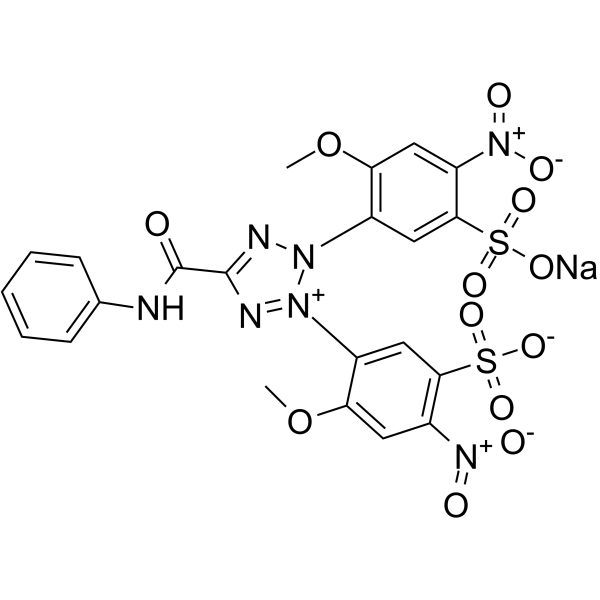
XTT (sodium) is used to assess cell viability as a function of redox potential. Actively respiring cells convert the water-soluble XTT to a water-soluble, orange colored formazan product.
For research use only. We do not sell to patients.
- Purity : 99.85%
- CAS No.: 111072-31-2
- Formula: C22H16N7NaO13S2
- Molecular Weight:673.52
-
Storage:
-20°C, sealed storage, away from moisture
* In solvent : -80°C, 6 months; -20°C, 1 month (sealed storage, away from moisture)

To place orders, for customer services and technical support, please contact: MedChemExpress USA
Tel: 609-228-6898 E-mail: [email protected] [email protected]


-
 Biological Activity
-
Chemical Information
-
Solvent & Solubility
- Protocols
- Purity & Documentation
- References
-
 Help & FAQs


-
HY-W004520
Phenazine methylsulfateApoptosis Iducer -
HY-D0831
WST-8Tetrazolium Dye -
HY-15142
Doxorubicin hydrochlorideTopoisomerase Inhibitor -
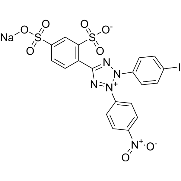
HY-136976
WST-1Cell Proliferation Assay Reagent -
HY-K0010
Protease Inhibitor Cocktail (EDTA-Free, 100× in DMSO) -

HY-K0301
Cell Counting Kit-8 -
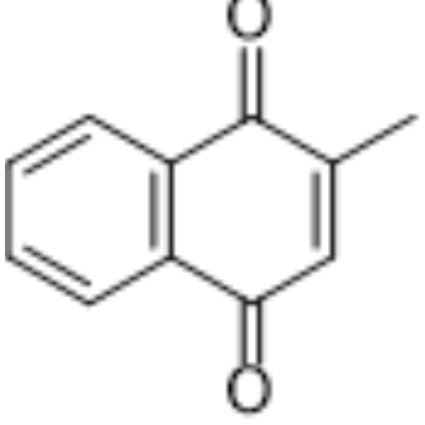
HY-B0332
MenadioneEndogenous Metabolite -
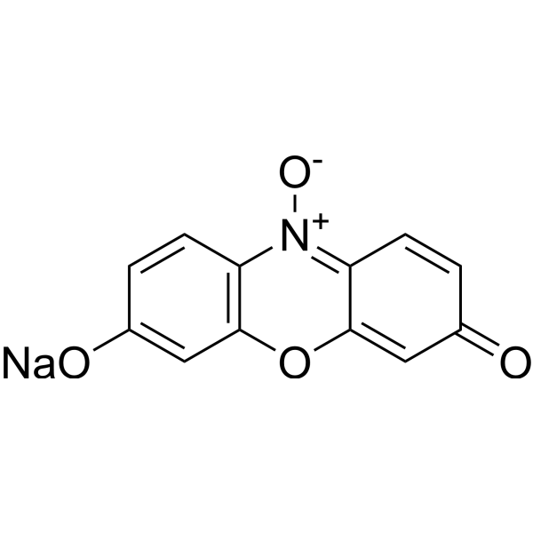
HY-111391
Resazurin sodiumBacterial Inhibitor -

HY-10219
RapamycinmTOR Inhibitor -

HY-B0015
PaclitaxelAntitumor Agent -
HY-D1462
CellTracker Blue CMACFluorescent Dye -
HY-131490
Tetrazolium violetBiochemical Assay Reagent
Publications Citing Use of MedChemExpress (MCE) XTT sodium
More Customer Validation & Images

-
Bio/Physico-chemical Assay
-
Bio/Physico-chemical Assay


Biological Activity
Description
In Vitro
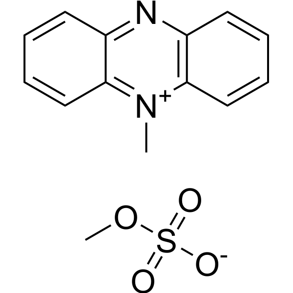
Determining Cell Viability Using XTT (sodium)[2] 1. Solution Preparation (1) XTT (sodium) solution: Dissolve XTT in warm culture medium at a concentration of 1 mg/mL. (2) PMS solution: Prepare PMS solution at a concentration of 100 mM, dissolved in phosphate-buffered saline (PBS), and store at 4°C. (3) Mixing for use: When ready to use, add PMS solution to the XTT solution, resulting in a final PMS concentration of 25 μM. 2. Experimental Procedure (1) Cell culture: Seed the cells to be tested (e.g., HT-2 T cell line) in a 96-well plate, 100 μL medium per well, and incubate at 37°C for 40 hours. (2) Add XTT/PMS solution: Add 25 μL of the XTT/PMS solution to each well and incubate for 4-8 hours. (3) Measure absorbance: At the end of the incubation, measure the absorbance at 450 nm using a microplate reader, with 650 nm as the reference wavelength, and subtract the absorbance of the blank control wells. 3. Washing and Storage Wash the experimental equipment with Milli-Q purified water. Store PMS and XTT solutions protected from light. 4. Notes (1) XTT solution should be freshly prepared, and dissolving it in a 60°C water bath will be easier. (2) The incubation time may vary depending on the experiment, and over-incubation can increase background noise. (3) PMS solution has a short storage time and should be used within one month.
MedChemExpress (MCE) has not independently confirmed the accuracy of these methods. They are for reference only.


Chemical Information
-
CAS No. 111072-31-2
-
Appearance Solid
-
Molecular Weight 673.52
-
Formula C22H16N7NaO13S2
-
Color Light yellow to yellow
-
SMILES
COC1=C(N2N=C(N=[N+]2C3=CC(S([O-])(=O)=O)=C(C=C3OC)[N+]([O-])=O)C(NC4=CC=CC=C4)=O)C=C(S(=O)(O[Na])=O)C([N+]([O-])=O)=C1
-
Shipping
Room temperature in continental US; may vary elsewhere.
-
Storage
-20°C, sealed storage, away from moisture
* In solvent : -80°C, 6 months; -20°C, 1 month (sealed storage, away from moisture)


Publications (4)
-
Journal Impact Factor
-
Most Recent
-
J Nanobiotechnology
A novel tetrahedral framework nucleic acid-based antibiotic delivery system: overcoming biofilm barriers to combat chronic infections. [Abstract]2025 Jul 1;23(1):465. PMID: 40597263
XTT sodium purchased from MedChemExpress. Usage Cited in: J Nanobiotechnology. 2025 Jul 1;23(1):465. [Abstract]
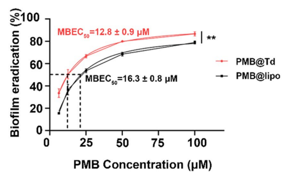
Biofilm eradication rates of PMB@Td and PMB@Lipo at different concentrations was determined by XTT sodium (100 µL; 1 mg/mL) assays.
-
J Med Chem
Activity and Safety Optimization of Mesoricin: A Dual-Domain Antifungal Peptide from Mesorhizobium sp. [Abstract]2025 Apr 8. PMID: 40198836
XTT sodium purchased from MedChemExpress. Usage Cited in: J Med Chem. 2025 Apr 8. [Abstract]
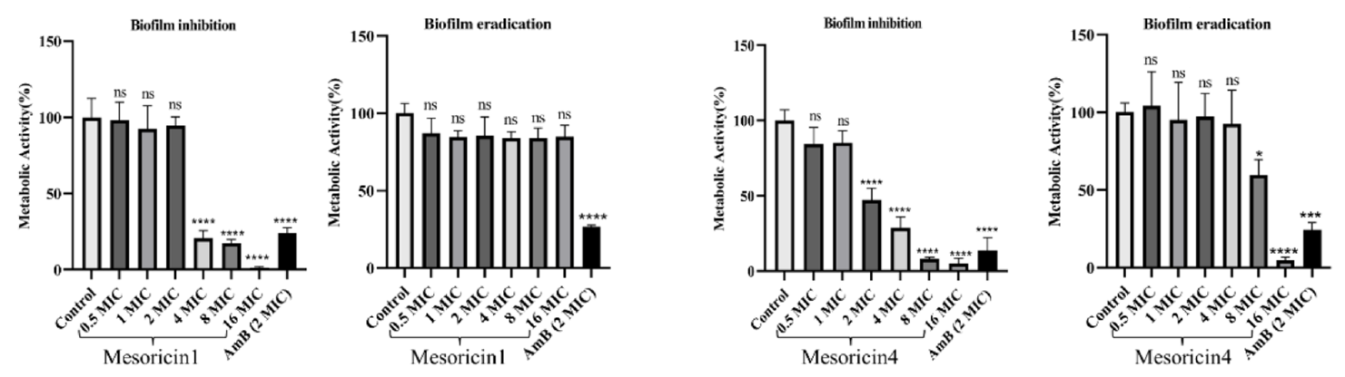
XTT sodium (150 μL; 0.5 mg/mL; 3 h; 37 ℃) was used to assess biofilm inhibition and eradication of C. neoformans H99 by mesoricin1 and mesoricin4 at different concentrations.
-
Molecules
Chemical Characterization, Evaluation of Antimicrobial Potential, and Cytotoxic Activity of Thuja occidentalis L. and Myrtus communis L. Essential Oils for Topical Applications. [Abstract]2026 Apr 7;31(7):1225. PMID: 41976265 -
Curr Microbiol
Berberine Hydrochloride Enhances the Fluconazole Susceptibility of Candida albicans through Multiple Targets. [Abstract]2026 Mar 27;83(5):262. PMID: 41896451


Solvent & Solubility
In Vitro:
DMSO : 40 mg/mL (59.39 mM; ultrasonic and warming and heat to 60°C; Hygroscopic DMSO has a significant impact on the solubility of product, please use newly opened DMSO)
H2O : 10 mg/mL (14.85 mM; ultrasonic and warming and heat to 60°C)
Please refer to the solubility information to select the appropriate solvent. Once prepared, please aliquot and store the solution to prevent product inactivation from repeated freeze-thaw cycles.
Storage method and period of stock solution: -80°C, 6 months; -20°C, 1 month (sealed storage, away from moisture). When stored at -80°C, please use it within 6 months. When stored at -20°C, please use it within 1 month.
* Note: If you choose water as the stock solution, please dilute it to the working solution, then filter and sterilize it with a 0.22 μm filter before use.
Please refer to the solubility information to select the appropriate solvent. Once prepared, please aliquot and store the solution to prevent product inactivation from repeated freeze-thaw cycles.
Storage method and period of stock solution: -80°C, 6 months; -20°C, 1 month (sealed storage, away from moisture). When stored at -80°C, please use it within 6 months. When stored at -20°C, please use it within 1 month.
* Note: If you choose water as the stock solution, please dilute it to the working solution, then filter and sterilize it with a 0.22 μm filter before use.
Concentration (start) × Volume (start) = Concentration (final) × Volume (final)
In Vivo:
Select the appropriate dissolution method based on your experimental animal and administration route.
- For the following dissolution methods, please ensure to first prepare a clear stock solution using an In Vitro approach and then sequentially add co-solvents:
- To ensure reliable experimental results, the clarified stock solution can be appropriately stored based on storage conditions. As for the working solution for In Vivo experiments, it is recommended to prepare freshly and use it on the same day.
- The percentages shown for the solvents indicate their volumetric ratio in the final prepared solution. If precipitation or phase separation occurs during preparation, heat and/or sonication can be used to aid dissolution.
Add each solvent one by one: 10% DMSO 40% PEG300 5% Tween-80 45% Saline
Solubility: ≥ 2.5 mg/mL (3.71 mM); Clear solution
This protocol yields a clear solution of ≥ 2.5 mg/mL (saturation unknown).
Taking 1 mL working solution as an example, add 100 μL DMSO stock solution (25.0 mg/mL) to 400 μL PEG300, and mix evenly; then add 50 μL Tween-80 and mix evenly; then add 450 μL Saline to adjust the volume to 1 mL.
Preparation of Saline: Dissolve 0.9 g sodium chloride in ddH₂O and dilute to 100 mL to obtain a clear Saline solution.
In Vivo Dissolution Calculator
Please enter the basic information of animal experiments:
-
-
-
-
Recommended: Prepare an additional quantity of animals to account for potential losses during experiments.
Please enter your animal formula composition:
-
%DMSO +
Recommended: Keep the proportion of DMSO in working solution below 2% if your animal is weak.
-
%+
-
+%Tween-80 + +
-
%Saline +
The co-solvents required include: DMSO, . All of co-solvents are available by MedChemExpress (MCE). , Tween 80. All of co-solvents are available by MedChemExpress (MCE).
Working solution concentration: 0.22 mg/mL
Method for preparing stock solution: mg drug dissolved in μL DMSO. Stock solution concentration: mg/mL. * In solvent : -80°C, 6 months; -20°C, 1 month (sealed storage, away from moisture)
1. Take μL DMSO stock solution;
2. Add μL .
μL , mix evenly;
3. Then add μL Tween 80, mix evenly;
4. Then add μL
Please ensure that the stock solution in the first step is dissolved to a clear state, and add co-solvents in sequence. You can use ultrasonic heating (ultrasonic cleaner, recommended frequency 20-40 kHz), vortexing, etc. to assist dissolution.


Purity & Documentation
-
Data Sheet (283 KB)
-
SDS (394 KB)
- English - EN (394 KB)
- Français - FR (394 KB)
- Deutsch - DE (394 KB)
- Norwegian - NO (394 KB)
- Español - ES (394 KB)
- Swedish - SV (394 KB)
- Italian - IT (394 KB)
- Korean - KR (394 KB)
- Portuguese - PT (394 KB)
-
Handling Instructions (2659 KB)
References
[1]. Zhao Q, Ernst JT, Hamilton AD, Debnath AK, Jiang S. XTT formazan widely used to detect cell viability inhibits HIV type 1 infection in vitro by targeting gp41. AIDS Res Hum Retroviruses. 2002;18(14):989-997. [Content Brief]
[2]. Roehm NW, et al. An improved colorimetric assay for cell proliferation and viability utilizing the tetrazolium salt XTT. J Immunol Methods. 1991 Sep 13;142(2):257-65. [Content Brief]


Complete Stock Solution Preparation Table
Please refer to the solubility information to select the appropriate solvent. Once prepared, please aliquot and store the solution to prevent product inactivation from repeated freeze-thaw cycles.
Storage method and period of stock solution: -80°C, 6 months; -20°C, 1 month (sealed storage, away from moisture). When stored at -80°C, please use it within 6 months. When stored at -20°C, please use it within 1 month.
| Optional Solvent | Concentration Solvent Mass | 1 mg | 5 mg | 10 mg | 25 mg |
|---|---|---|---|---|---|
| H2O / DMSO | 1 mM | 1.4847 mL | 7.4237 mL | 14.8474 mL | 37.1184 mL |
| 5 mM | 0.2969 mL | 1.4847 mL | 2.9695 mL | 7.4237 mL | |
| 10 mM | 0.1485 mL | 0.7424 mL | 1.4847 mL | 3.7118 mL | |
| DMSO | 15 mM | 0.0990 mL | 0.4949 mL | 0.9898 mL | 2.4746 mL |
| 20 mM | 0.0742 mL | 0.3712 mL | 0.7424 mL | 1.8559 mL | |
| 25 mM | 0.0594 mL | 0.2969 mL | 0.5939 mL | 1.4847 mL | |
| 30 mM | 0.0495 mL | 0.2475 mL | 0.4949 mL | 1.2373 mL | |
| 40 mM | 0.0371 mL | 0.1856 mL | 0.3712 mL | 0.9280 mL | |
| 50 mM | 0.0297 mL | 0.1485 mL | 0.2969 mL | 0.7424 mL |
* Note: If you choose water as the stock solution, please dilute it to the working solution, then filter and sterilize it with a 0.22 μm filter before use.


XTT sodium Related Classifications
HY-122131 Related Classifications
 Powered by Bioz