



MRT68921
Based on 30 publication(s) in Google Scholar
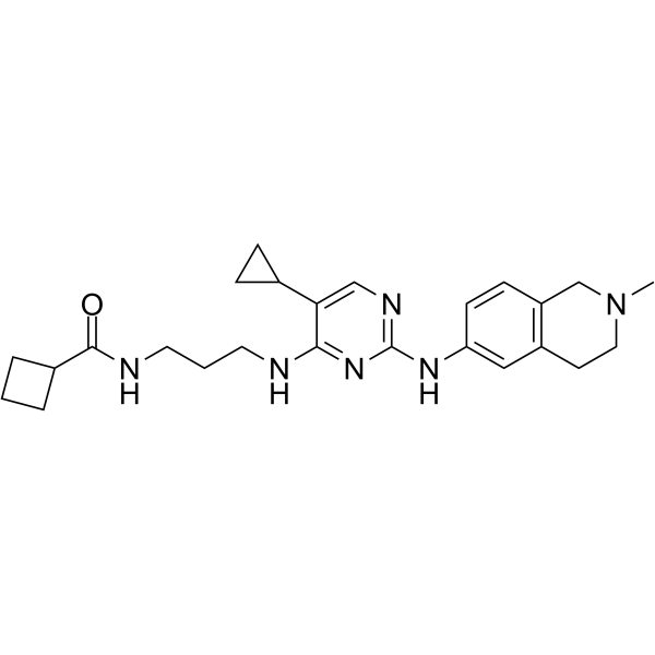
MRT68921 is a potent NUAK1/ULK1 dual inhibitor. MRT68921 inhibits ULK1 and ULK2 with IC50 values of 2.9 nM and 1.1 nM, respectively. MRT68921 can block cells autophagy and kill tumor cells by breaking the balance of oxidative stress signals. MRT68921 can inhibit cell proliferation and induce ROS production and apoptosis. MRT68921 can be used for the research of cancer, such as breast cancer.
Nur für Forschungszwecke. Wir verkaufen nicht an Patienten.
- Reinheit : 98.09%
- CAS. Nr.: 1190379-70-4
- Formel: C25H34N6O
- Molecular Weight:434.58
-
Speicherung:Powder -20°C, 3 years , 4°C, 2 years ; In solvent -80°C, 6 months , -20°C, 1 month

To place orders, for customer services and technical support, please contact: MedChemExpress USA
Tel: 609-228-6898 E-mail: [email protected] [email protected]


-
 Biologische Aktivität
-
Chemical Information
-
Lösungsmittel & Löslichkeit
- Protokoll
- Reinheit & Dokumentation
- Verweise
-
 Help & FAQs


-
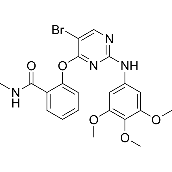
HY-16966
SBI-0206965ULK1/AMPK Inhibitor -
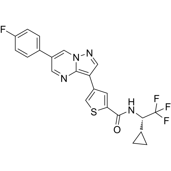
HY-114490
ULK-101ULK1 Inhibitor -
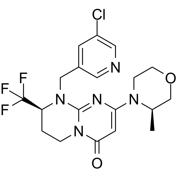
HY-12481
SAR405Vps34 Inhibitor -
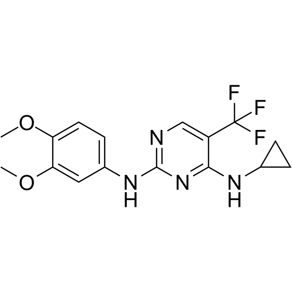
HY-137742
SBP-7455ULK1/ULK2 Autophagy Inhibitor -

HY-100558
Bafilomycin A1V-ATPase Inhibitor -

HY-10219
RapamycinmTOR Inhibitor -
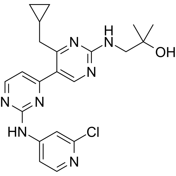
HY-12795
Vps34-IN-1Vps34 Inhibitor -
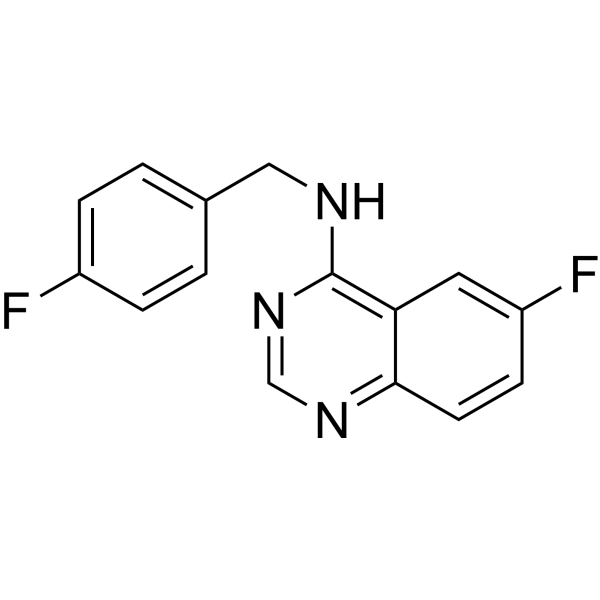
HY-12990
Spautin-1Autophagy Inhibitor -
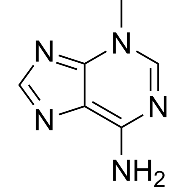
HY-19312
3-MethyladenineAutophagy/PI3K Inhibitor -
HY-100006A
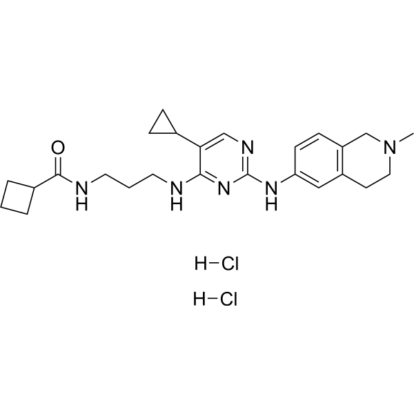
MRT68921 dihydrochlorideNUAK1/ULK1 Inhibitor -
HY-W031727
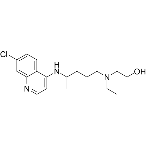
HydroxychloroquineAntimalarial Agent -
HY-160699
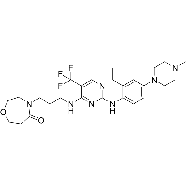
DCC-3116ULK1/2 Inhibitor
Publications Citing Use of MedChemExpress (MCE) MRT68921
More- Nature. 2022 Oct;610(7931):366-372. [Abstract]
- Cancer Cell. 2021 May 10;39(5):678-693.e11. [Abstract]
- Nat Cancer. 2021 May;2(5):503-514. [Abstract]
- Nat Cell Biol. 2026 Apr;28(4):812-827. [Abstract]
- Nat Commun. 2025 Nov 19;16(1):10181. [Abstract]
- ACS Nano. 2024 Jul 2;18(26):16790-16807. [Abstract]
- J Exp Clin Cancer Res. 2026 Apr 23;45(1):104. [Abstract]
- Cell Death Dis. 2026 Jun 16. [Abstract]
- Dev Cell. 2025 Jun 27:S1534-5807(25)00372-7. [Abstract]
- Dev Cell. 2024 Jan 22;59(2):228-243.e7. [Abstract]
- Mol Ther Oncolytics. 2021 Aug 28:23:107-123. [Abstract]
- J Ethnopharmacol. 2024 Dec 5:335:118658. [Abstract]
- Bioorg Chem. 2024 Jun:147:107412. [Abstract]
- Mol Med Rep. 2024 Apr;29(4):67. [Abstract]
- PLoS Pathog. 2024 Aug 13;20(8):e1012461. [Abstract]
- Sci Rep. 2023 Jul 3;13(1):10752. [Abstract]
- Med Oncol. 2026 Mar 3;43(4):163. [Abstract]
- FASEB J. 2026 Jan 31;40(2):e71454. [Abstract]
- mSphere. 2025 Feb 25;10(2):e0053724. [Abstract]
- J Cell Physiol. 2021 Dec;236(12):8110-8121. [Abstract]
- Biochem J. 2019 Mar 12;476(5):875-887. [Abstract]
- Vet Microbiol. 2026 May:316:110993. [Abstract]
- bioRxiv. 2026 Apr 5.
- SSRN. 2025 Jul 25.
- bioRxiv. 2025 Jun 5:2025.05.29.656904. [Abstract]
- Res Sq. 2025 May 07.
- Medical University of South Carolina. 2023 Aug 12.
- Research Square Preprint. 2023 May 22.
- Patent. US20230068698A1.
- Research Square Preprint. 2021 Oct.
 Customer Validation & Images

-
IF
-
WB
-
WB
-
IF
-
Bio/Physico-chemical Assay


Biologische Aktivität
Beschreibung
|
ULK2 1.1 nM (IC50) |
ULK1 2.9 nM (IC50) |
GSK-3β |
Bcl-2 |
In Vitro
MRT68921 (1 μM, 1 h) reduces basal LC3 puncta and blocks basal autophagy in mouse embryonic fibroblasts (MEFs)[1].
MRT68921 (1 μM) reduces LC3-II levels in TBK1 knock-out and matched wild-type MEFs[1].
MRT68921 (24 h) exhibits cytotoxic activity in cancer cells with IC50 values ranging from 1.76-8.91 μM[2].
MRT68921 (0-10 μM, 8-24 h) induces ROS production and apoptosis in NCI-H460 and MNK45 cells[2].
MRT68921 (0-5 μM, 8h) suppresses the NUAK1/MYPT1/Gsk3β andautophagy associated signaling pathway in U251 and MNK45 cells[2].
MedChemExpress (MCE) has not independently confirmed the accuracy of these methods. They are for reference only.
-
Cell Line:U251 and MNK45 cells
-
Concentration:0, 0.5, 1, 2, 3 and 5 μM
-
Incubation Time:8 h
-
Result:Increased cleaved PARP1 expression.
Downregulated phosphorylation of MYPT1 and Gsk3β.
Increased puncta LC3.
In Vivo
MRT68921 (20 mg/kg, s.c., every 2 days for 7 times) inhibits tumor growth in MNK45 tumor mice models[2].
MRT68921 (20 mg/kg, i.p., daily for 7 times) reduces the number of lung metastatic nodules in 4T1 tumor mice models[2].
MedChemExpress (MCE) has not independently confirmed the accuracy of these methods. They are for reference only.
-
Animal Model:NCI-H460 tumor mice models[2]
-
Dosage:10, 20, or 40 mg/kg
-
Administration:Subcutaneously injection, daily for 7 times
-
Result:Reduced tumor volume.
Increased Bax levels and decreased Bcl-2 levels.
-
Animal Model:MNK45 tumor mice models[2]
-
Dosage:20 mg/kg
-
Administration:Subcutaneously injection, every 2 days for 7 times
-
Result:Reduced tumor volume.
-
Animal Model:4T1 tumor mice models[2]
-
Dosage:20 mg/kg
-
Administration:Intraperitoneally injection, daily for 7 times
-
Result:Reduced the number of lung metastatic nodules.
Had no abnormal structures of the heart, kidney, liver, and spleen.
- Annexin V plus membrane-impermeant dye apoptosis staining
- ROS/oxidative-stress fluorescent staining
- BrdU Incorporation Assay
- Somatic Cell Culture
- CFSE Dye Dilution Proliferation Assay
- Dye-dilution cell tracking and proliferation staining
- CCK-8/WST-8 Cell Proliferation Assay
- Protocol for Cell Cycle
- Cell Counting-Based Growth Curve Assay
- Colony Formation (Clonogenic) Assay
- Breast Cancer Modeling
- Cell Viability Determination by MTT Colorimetric Assay
- EdU Incorporation Assay (Click Chemistry-Based DNA Synthesis Measurement)
- Ki-67 Immunostaining Proliferation Assay
- Detection of Apoptosis
- TUNEL staining for apoptotic DNA fragmentation
- PCNA Immunodetection Proliferation Assay
- Protocol for Cell Counting and Cell Density Analysis
- Autophagy
- Apoptosis Solutions
- Lysosome and acidic-vesicle live-cell staining
- MTT Cell Proliferation Assay
- Macroautophagy Solutions


Chemical Information
-
CAS. Nr. 1190379-70-4
-
Appearance Solid
-
Molecular Weight 434.58
-
Formel C25H34N6O
-
Color Light yellow to yellow
-
SMILES
O=C(C1CCC1)NCCCNC2=NC(NC3=CC4=C(CN(C)CC4)C=C3)=NC=C2C5CC5
-
Versand
Room temperature in continental US; may vary elsewhere.
-
Speicherung
Powder -20°C 3 years 4°C 2 years In solvent -80°C 6 months -20°C 1 month


Publications (30)
-
Journal Impact Factor
-
Most Recent
-
Nature
2022 Oct;610(7931):366-372. PMID: 36198801
MRT68921 purchased from MedChemExpress. Usage Cited in: Nature. 2022 Oct;610(7931):366-372. [Abstract]
KPC cells plated on wild-type or R/R ECM or plastic were incubated in CM or LG medium with or without EIPA, MBQ-167 (MBQ), MRT68921 dihydrochloride (MRT, 600 nM), EIPA + MRT or MBQ + MRT for 24 h.
-
Cancer Cell
2021 May 10;39(5):678-693.e11. PMID: 33740421
MRT68921 purchased from MedChemExpress. Usage Cited in: Cancer Cell. 2021 May 10;39(5):678-693.e11. [Abstract]
Autophagosomes and macropinosomes in MIA PaCa-2 cells treated +/− MRT68921 dihydrochloride (600 nM). Scale bar, 10 μm.

MRT68921 purchased from MedChemExpress. Usage Cited in: Cancer Cell. 2021 May 10;39(5):678-693.e11. [Abstract]
KC6141 cells were incubated with vehicle, the ULK1/2 inhibitor MRT68921 dihydrochloride (MRT, 600 nM), EIPA or MRT + EIPA and total viable cells were measured.

MRT68921 purchased from MedChemExpress. Usage Cited in: Cancer Cell. 2021 May 10;39(5):678-693.e11. [Abstract]
C57BL/6 mice bearing KC6141 tumors or Nude mice bearing 1334 and 1444 allografts were treated with vehicle, MRT68921 dihydrochloride, EIPA, or MRT68921 dihydrochloride (10 mg/kg)+ EIPA for 3 weeks or 15 days. Whereas MRT68921 dihydrochloride or EIPA alone had a small effect on KC6141 or 1444 or 1334 tumor growth, the two agents together strongly inhibited tumors.
-
Nat Cancer
ULK1 inhibition overcomes compromised antigen presentation and restores antitumor immunity in LKB1 mutant lung cancer. [Abstract]2021 May;2(5):503-514. PMID: 34142094
MRT68921 purchased from MedChemExpress. Usage Cited in: Nat Cancer. 2021 May;2(5):503-514. [Abstract]
Cell growth inhibition in KL and KP cells in response to autophagy inhibitors chloroquine (CQ) or ULK1 inhibitor MRT68921 dihydrochloride (MRT). Data shown represents one of three independent experiments. n=3 cell cultures for each cell line with each treatment condition.
-
Nat Cell Biol
TOLLIP targets GSDME-NT-carrying endocytic vesicles for autophagy to regulate pyroptosis and chemotherapy efficacy. [Abstract]2026 Apr;28(4):812-827. PMID: 41803502 -
Nat Commun
PRDX1 promotes testosterone synthesis and attenuates aging via redox regulation of ATG4B to modulate lipophagy. [Abstract]2025 Nov 19;16(1):10181. PMID: 41261096 -
ACS Nano
Realistic Nanoplastics Induced Pulmonary Damage via the Crosstalk of Ferritinophagy and Mitochondrial Dysfunction. [Abstract]2024 Jul 2;18(26):16790-16807. PMID: 38869479
MRT68921 purchased from MedChemExpress. Usage Cited in: ACS Nano. 2024 Jul 2;18(26):16790-16807. [Abstract]
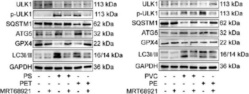
TC-1 cells were pretreated with 1 μM MRT68921 for 1 h, followed by NP treatment for 24 h. MRT68921 inhibited the upregulation of NP-induced p-ULK1, ATG5, and LC3-II and the downregulation of SQSTM1 and GPX4.
-
J Exp Clin Cancer Res
Ferrodoxin 1 (FDX1) drives paclitaxel resistance in ovarian cancer via copper metabolism and ULK1/ATG13-mediated autophagy: overcome by pH/ROS-responsive PPD/PDP@si-FDX1 nanomicelles. [Abstract]2026 Apr 23;45(1):104. PMID: 42026685 -
Cell Death Dis
Pictilisib and nutrient stress synergize to induce methuosis via PI(4,5)P2-dependent macropinocytic dysregulation in cancer cells. [Abstract]2026 Jun 16. PMID: 42303628 -
Dev Cell
2025 Jun 27:S1534-5807(25)00372-7. PMID: 40609542
MRT68921 purchased from MedChemExpress. Usage Cited in: Dev Cell. 2025 Jun 27:S1534-5807(25)00372-7. [Abstract]
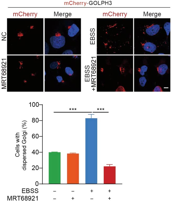
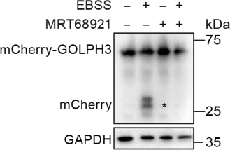
Confocal images of mCherry-GOLPH3-expressing U2OS cells. Cells were either untreated or treated with EBSS for 12 hours, in the presence or absence of MRT68921 (250 μM). Right: Quantification of the percentage of cells with dispersed mCherry-GOLPH3. A total of 300 cells for each sample were quantified. n = 3. Scale bar: 10 µm.
MRT68921 purchased from MedChemExpress. Usage Cited in: Dev Cell. 2025 Jun 27:S1534-5807(25)00372-7. [Abstract]
Inhibition of EBSS-induced mCherry-GOLPH3 cleavage by ULK1 inhibitor MRT68921 (250 μM). U2OS cells transfected with the mCherry-GOLPH3 plasmid were left untreated or treated with EBSS, in the presence or absence of MRT68921 for 8 hours.
-
Dev Cell
Autophagy supports PDGFRA-dependent brain tumor development by enhancing oncogenic signaling. [Abstract]2024 Jan 22;59(2):228-243.e7. PMID: 38113891 -
Mol Ther Oncolytics
Oleanolic acid blocks the purine salvage pathway for cancer therapy by inactivating SOD1 and stimulating lysosomal proteolysis. [Abstract]2021 Aug 28:23:107-123. PMID: 34703880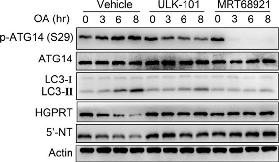
MRT68921 purchased from MedChemExpress. Usage Cited in: Mol Ther Oncolytics. 2021 Aug 28:23:107-123. [Abstract]
The effects of two ULK1 inhibitors, ULK-101 (1 μM) and MRT68921 (1 μM), on levels of phospho-ATG14 (Ser29), ATG14, LC3-I/II, HGPRT, and 5′-NT in OA-treated A549 cells. The results showed that both ULK1 inhibitors rapidly downregulated phosphorylated ATG14 at Ser29, a well-established substrate of ULK1, reduced LC3-II isoform expression, and stabilized HGPRT and 5′-N.
-
J Ethnopharmacol
2024 Dec 5:335:118658. PMID: 39103023
MRT68921 purchased from MedChemExpress. Usage Cited in: J Ethnopharmacol. 2024 Dec 5:335:118658. [Abstract]
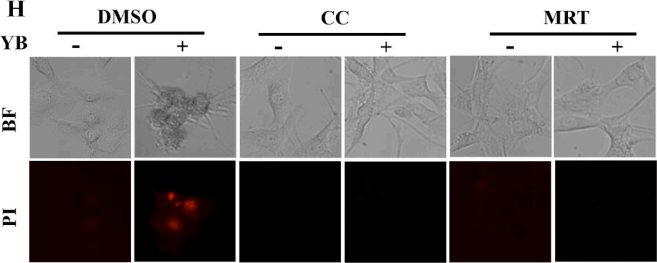
MRT68921 (MRT, 5 μM; 2 h) attenuates the accumulation of LC3-II, thereby reducing YB treatment-induced necroptosis.
-
Bioorg Chem
Identification of FTY720 and COH29 as novel topoisomerase I catalytic inhibitors by experimental and computational studies. [Abstract]2024 Jun:147:107412. PMID: 38696845 -
Mol Med Rep
2024 Apr;29(4):67. PMID: 38456519 -
PLoS Pathog
A small protein encoded by PCBP1-AS1 is identified as a key regulator of influenza virus replication via enhancing autophagy. [Abstract]2024 Aug 13;20(8):e1012461. PMID: 39137200 -
Sci Rep
2023 Jul 3;13(1):10752. PMID: 37400460 -
Med Oncol
ULK1-driven autophagy modulation alters tumor-promoting pathways in triple-negative breast cancer. [Abstract]2026 Mar 3;43(4):163. PMID: 41774346 -
FASEB J
EM2, a Novel Elephantopus mollis H.B.K. Monomer, Enhances Radiosensitivity in Cervical Cancer Through Dual Inhibition of AKT and Autophagy. [Abstract]2026 Jan 31;40(2):e71454. PMID: 41527777 -
mSphere
Inhibition of Unc-51-like-kinase is mitoprotective during Pseudomonas aeruginosa infection in corneal epithelial cells. [Abstract]2025 Feb 25;10(2):e0053724. PMID: 39791872 -
J Cell Physiol
2021 Dec;236(12):8110-8121. PMID: 34101831 -
Biochem J
Conservation of structure, function and inhibitor binding in UNC-51-like kinase 1 and 2 (ULK1/2). [Abstract]2019 Mar 12;476(5):875-887. PMID: 30782972 -
Vet Microbiol
Porcine reproductive and respiratory syndrome virus nsp2 protects viral RNA-dependent RNA polymerase from autophagic degradation. [Abstract]2026 May:316:110993. PMID: 41861694 -
-
-
bioRxiv
2025 Jun 5:2025.05.29.656904. PMID: 40501952 -
-
-
-
-


Lösungsmittel & Löslichkeit
In Vitro:
DMSO : 100 mg/mL (230.11 mM; Need ultrasonic; Hygroscopic DMSO has a significant impact on the solubility of product, please use newly opened DMSO)
Please refer to the solubility information to select the appropriate solvent. Once prepared, please aliquot and store the solution to prevent product inactivation from repeated freeze-thaw cycles.
Storage method and period of stock solution: -80°C, 6 months; -20°C, 1 month. When stored at -80°C, please use it within 6 months. When stored at -20°C, please use it within 1 month.
Please refer to the solubility information to select the appropriate solvent. Once prepared, please aliquot and store the solution to prevent product inactivation from repeated freeze-thaw cycles.
Storage method and period of stock solution: -80°C, 6 months; -20°C, 1 month. When stored at -80°C, please use it within 6 months. When stored at -20°C, please use it within 1 month.
Konzentration (Stammlösung) × Volumen (Stammlösung) = Konzentration (Ziellösung) × Volumen (Ziellösung)
In Vivo:
Select the appropriate dissolution method based on your experimental animal and administration route.
- For the following dissolution methods, please ensure to first prepare a clear stock solution using an In Vitro approach and then sequentially add co-solvents:
- To ensure reliable experimental results, the clarified stock solution can be appropriately stored based on storage conditions. As for the working solution for In Vivo experiments, it is recommended to prepare freshly and use it on the same day.
- The percentages shown for the solvents indicate their volumetric ratio in the final prepared solution. If precipitation or phase separation occurs during preparation, heat and/or sonication can be used to aid dissolution.
Add each solvent one by one: 10% DMSO 40% PEG300 5% Tween-80 45% Saline
Solubility: 2.5 mg/mL (5.75 mM); Clear solution; Need ultrasonic
This protocol yields a clear solution of 2.5 mg/mL.
Taking 1 mL working solution as an example, add 100 μL DMSO stock solution (25.0 mg/mL) to 400 μL PEG300, and mix evenly; then add 50 μL Tween-80 and mix evenly; then add 450 μL Saline to adjust the volume to 1 mL.
Preparation of Saline: Dissolve 0.9 g sodium chloride in ddH₂O and dilute to 100 mL to obtain a clear Saline solution.
Add each solvent one by one: 10% DMSO 90% (20% SBE-β-CD in Saline)
Solubility: 2.5 mg/mL (5.75 mM); Clear solution; Need ultrasonic
This protocol yields a clear solution of 2.5 mg/mL.
Taking 1 mL working solution as an example, add 100 μL DMSO stock solution (25.0 mg/mL) to 900 μL 20% SBE-β-CD in Saline, and mix evenly.
Preparation of 20% SBE-β-CD in Saline (4°C, storage for one week): 2 g SBE-β-CD powder is dissolved in 10 mL Saline, completely dissolve until clear.
In Vivo Dissolution Calculator
Please enter the basic information of animal experiments:
-
-
-
-
Recommended: Prepare an additional quantity of animals to account for potential losses during experiments.
Please enter your animal formula composition:
-
%DMSO +
Recommended: Keep the proportion of DMSO in working solution below 2% if your animal is weak.
-
%+
-
+%Tween-80 + +
-
%Saline +
The co-solvents required include: DMSO, . All of co-solvents are available by MedChemExpress (MCE). , Tween 80. All of co-solvents are available by MedChemExpress (MCE).
Working solution concentration: 0.22 mg/mL
Method for preparing stock solution: mg drug dissolved in μL DMSO. Stock solution concentration: mg/mL.
1. Take μL DMSO stock solution;
2. Add μL .
μL , mix evenly;
3. Then add μL Tween 80, mix evenly;
4. Then add μL
Please ensure that the stock solution in the first step is dissolved to a clear state, and add co-solvents in sequence. You can use ultrasonic heating (ultrasonic cleaner, recommended frequency 20-40 kHz), vortexing, etc. to assist dissolution.


Reinheit & Dokumentation
-
Data Sheet (276 KB)
-
SDS (393 KB)
- English - EN (393 KB)
- Français - FR (393 KB)
- Deutsch - DE (393 KB)
- Norwegian - NO (393 KB)
- Español - ES (393 KB)
- Swedish - SV (393 KB)
- Italian - IT (393 KB)
- Korean - KR (393 KB)
- Portuguese - PT (393 KB)
-
Handling Instructions (2659 KB)
Verweise
[1]. Petherick KJ, et al. Pharmacological inhibition of ULK1 kinase blocks mammalian target of rapamycin (mTOR)-dependent autophagy. J Biol Chem. 2015 May 1;290(18):11376-83. [Content Brief]
[2]. Chen Y, et al. Dual targeting of NUAK1 and ULK1 using the multitargeted inhibitor MRT68921 exerts potent antitumor activities. Cell Death Dis. 2020 Sep 1;11(8):712. [Content Brief]


Complete Stock Solution Preparation Table
Please refer to the solubility information to select the appropriate solvent. Once prepared, please aliquot and store the solution to prevent product inactivation from repeated freeze-thaw cycles.
Storage method and period of stock solution: -80°C, 6 months; -20°C, 1 month. When stored at -80°C, please use it within 6 months. When stored at -20°C, please use it within 1 month.
| Optional Solvent | Concentration Solvent Mass | 1 mg | 5 mg | 10 mg | 25 mg |
|---|---|---|---|---|---|
| DMSO | 1 mM | 2.3011 mL | 11.5054 mL | 23.0107 mL | 57.5268 mL |
| 5 mM | 0.4602 mL | 2.3011 mL | 4.6021 mL | 11.5054 mL | |
| 10 mM | 0.2301 mL | 1.1505 mL | 2.3011 mL | 5.7527 mL | |
| 15 mM | 0.1534 mL | 0.7670 mL | 1.5340 mL | 3.8351 mL | |
| 20 mM | 0.1151 mL | 0.5753 mL | 1.1505 mL | 2.8763 mL | |
| 25 mM | 0.0920 mL | 0.4602 mL | 0.9204 mL | 2.3011 mL | |
| 30 mM | 0.0767 mL | 0.3835 mL | 0.7670 mL | 1.9176 mL | |
| 40 mM | 0.0575 mL | 0.2876 mL | 0.5753 mL | 1.4382 mL | |
| 50 mM | 0.0460 mL | 0.2301 mL | 0.4602 mL | 1.1505 mL | |
| 60 mM | 0.0384 mL | 0.1918 mL | 0.3835 mL | 0.9588 mL | |
| 80 mM | 0.0288 mL | 0.1438 mL | 0.2876 mL | 0.7191 mL | |
| 100 mM | 0.0230 mL | 0.1151 mL | 0.2301 mL | 0.5753 mL |


MRT68921 Related Classifications
HY-100006 Related Classifications
 Powered by Bioz