



C5b-9 Antibody
(Synonyms: C5; C6; C7; C8; C9; Complement component 5; Complement component 6; Complement component 7; Complement component 8; Complement component 9; MAC; Membrane attack complex; TCC; Terminal complement complex.)Based on 1 publication(s) in Google Scholar
C5b-9 Antibody is a Rabbit-derived and non-conjugated IgG polyclonal antibody, targeting to C5b-9.
-
Host:
Rabbit
-
Isotype:
IgG
-
Application:
WB, IHC-P, IHC-F, ICC/IF
-
Reactivity :
Human, Rat
-
Formulation:
Supplied in 0.01M TBS(pH7.4) with 1% BSA, 0.03% Proclin300 and 50% Glycerol or 0.01M TBS (pH7.4) with 1% BSA, 0.02% Proclin300 and 50% Glycerol.
-
Conjugation:
Non-conjugated


To place orders, for customer services and technical support, please contact: MedChemExpress USA
Tel: 609-228-6898 E-mail: [email protected] [email protected]



Publications Citing Use of MedChemExpress (MCE) C5b-9 Antibody
More
 1 Publications Citing Use of MCE C5b-9 Antibody (1)
-
IHC


Applications
| Application |
WB

WB: Western Blot
|
IHC-P
IHC-P: Immunohistochemistry-Paraffin
|
IHC-F
IHC-F: Immunohistochemistry-Frozen
|
ICC/IF
ICC/IF: Immunocytochemistry/
Immunofluorescence |
|---|---|---|---|---|
| Dilution Ratio | 1:500-2000 | 1:100-500 | 1:100-500 | 1:100-500 |


Product Details
C5b-9 Antibody is a Rabbit-derived and non-conjugated IgG polyclonal antibody, targeting to C5b-9.
-
Host Rabbit

-
Clonality Polyclonal

-
Species ReactivityHuman, Rat Predicted Reactivity: Mouse
Note: The predicted reactivity is for reference only and should not be considered a guarantee of product performance.
-
Observed Molecular WeightObserved band size: 72 kDa
Note: Due to possible protein modifications or aggregation, the molecular weight should be confirmed by actual measurement, and the predicted value is for reference only.

-
Calculated Molecular Weight Predicted band size: 72 kDa

Entrez Gene: 727 Human ; 15139 Mouse ;
SwissProt: P01031 Human ; P02748 Human ; P07357 Human ; P10643 Human ; P13671 Human ; P06684 Mouse ; P08650 Rat
OMIM: 609536 Human
Purified Human SC5b-9 Complex
Endogenous
affinity purified
Non-conjugated
Unmodified
IgG


Product Properties
-
Appearance
Solution
-
Formulation
Supplied in 0.01M TBS(pH7.4) with 1% BSA, 0.03% Proclin300 and 50% Glycerol or 0.01M TBS (pH7.4) with 1% BSA, 0.02% Proclin300 and 50% Glycerol.
-
Concentration
Batch-dependent, Please check the COA for the concentration of each lot. Check Lot Concentration
-
Storage & Stability
Stored at -20°C for 1 year. Avoid repeated freeze / thaw cycles.
-
Shipping
Shipping with blue ice.


Publications (1)
-
Journal Impact Factor
-
Most Recent
-
Exp Cell Res
Membrane attack complex impairs hepatocyte autophagy in alcohol-related liver disease by regulating the SIRT1-FOXO3 signaling. [Abstract]2025 Aug 15;451(2):114713. PMID: 40834969
C5b-9 Antibody purchased from MedChemExpress. Usage Cited in: Exp Cell Res. 2025 Aug 15;451(2):114713. [Abstract]
C5b-9 levels in the liver were determined by IHC.


Verification Images
-
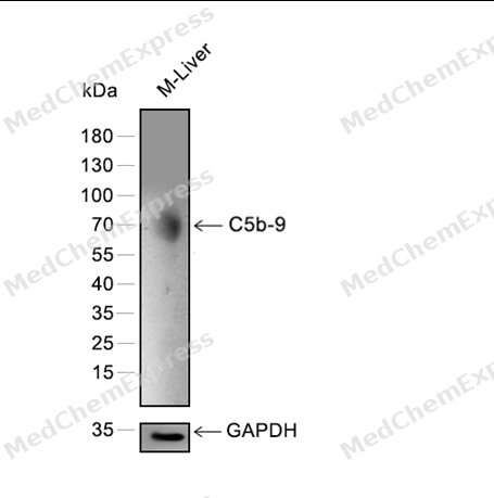 Western blot analysis of extracts from M-Liver using C5b-9 antibody. Proteins were transferred to a PVDF membrane and blocked with 5% nonfat powdered milk in PBST for 2 hour at room temperature. The primary antibody (1/2000) and Loading control antibody (GAPDH, 1/3000) was diluted with 5% nonfat powdered milk in PBST at 4°C overnight. Goat Anti-Rabbit IgG-HRP Secondary Antibody (1/8,000) was incubated for 45min at room temperature.
 -
 Immunohistochemical analysis of paraffin-embedded human oral cancer tissue using C5b-9 Antibody (HY-P81185, 1/500). The section was pretreated using heat mediated antigen retrieval with sodium citrate buffer (pH 6.0) for 8 minutes. The tissues were blocked with quick block buffer for 0.5 hours at room temperature, washed with PBS and PBST, and then incubated with the primary antibody overnight at 4℃. The detection was performed using an HRP conjugated compact polymer system. DAB was used as the chromogen. Tissues were counterstained with hematoxylin and mounted with DPX.
 -
 Immunohistochemical analysis of paraffin-embedded human liver cancer tissue using C5b-9 Antibody (HY-P81185, 1/500). The section was pretreated using heat mediated antigen retrieval with sodium citrate buffer (pH 6.0) for 8 minutes. The tissues were blocked with quick block buffer for 0.5 hours at room temperature, washed with PBS and PBST, and then incubated with the primary antibody overnight at 4℃. The detection was performed using an HRP conjugated compact polymer system. DAB was used as the chromogen. Tissues were counterstained with hematoxylin and mounted with DPX.
 -
 Immunohistochemical analysis of paraffin-embedded human thyroid cancer tissue using C5b-9 Antibody (HY-P81185, 1/500). The section was pretreated using heat mediated antigen retrieval with sodium citrate buffer (pH 6.0) for 8 minutes. The tissues were blocked with quick block buffer for 0.5 hours at room temperature, washed with PBS and PBST, and then incubated with the primary antibody overnight at 4℃. The detection was performed using an HRP conjugated compact polymer system. DAB was used as the chromogen. Tissues were counterstained with hematoxylin and mounted with DPX.
 -
 Immunohistochemical analysis of paraffin-embedded human urothelial cancer tissue using C5b-9 Antibody (HY-P81185, 1/500). The section was pretreated using heat mediated antigen retrieval with sodium citrate buffer (pH 6.0) for 8 minutes. The tissues were blocked with quick block buffer for 0.5 hours at room temperature, washed with PBS and PBST, and then incubated with the primary antibody overnight at 4℃. The detection was performed using an HRP conjugated compact polymer system. DAB was used as the chromogen. Tissues were counterstained with hematoxylin and mounted with DPX.
 -
 Immunohistochemical analysis of paraffin-embedded human colon cancer tissue using C5b-9 Antibody (HY-P81185, 1/500). The section was pretreated using heat mediated antigen retrieval with sodium citrate buffer (pH 6.0) for 8 minutes. The tissues were blocked with quick block buffer for 0.5 hours at room temperature, washed with PBS and PBST, and then incubated with the primary antibody overnight at 4℃. The detection was performed using an HRP conjugated compact polymer system. DAB was used as the chromogen. Tissues were counterstained with hematoxylin and mounted with DPX.
 -
 Immunohistochemical analysis of paraffin-embedded human rectum cancer tissue using C5b-9 Antibody (HY-P81185, 1/500). The section was pretreated using heat mediated antigen retrieval with sodium citrate buffer (pH 6.0) for 8 minutes. The tissues were blocked with quick block buffer for 0.5 hours at room temperature, washed with PBS and PBST, and then incubated with the primary antibody overnight at 4℃. The detection was performed using an HRP conjugated compact polymer system. DAB was used as the chromogen. Tissues were counterstained with hematoxylin and mounted with DPX.
 -
 Immunocytochemistry analysis of Hela cells labeling C5b-9 with C5b-9 Antibody (HY-P81185) at 1/100 dilution. Cells were fixed in 4% paraformaldehyde for 15 minutes at room temperature, permeabilized with 0.1% Triton X-100 in PBS for 15 minutes at room temperature, then blocked with quick block buffer for 10 minutes at room temperature. Cells were then incubated with C5b-9 Antibody (HY-P81185) at 1/100 dilution in quick block buffer overnight at 4 ℃. AF488-conjugated Goat Anti-Rabbit IgG H&L (HY-P8002, Green) was used as the secondary antibody at 1/1,000 dilution. PBS instead of the primary antibody was used as the secondary antibody only control. The Nuclear counterstain was DAPI (Blue).
 -
 Immunocytochemistry analysis of Hela cells labeling C5b-9 with C5b-9 Antibody (HY-P81185) at 1/300 dilution. Cells were fixed in 4% paraformaldehyde for 15 minutes at room temperature, permeabilized with 0.1% Triton X-100 in PBS for 15 minutes at room temperature, then blocked with quick block buffer for 10 minutes at room temperature. Cells were then incubated with C5b-9 Antibody (HY-P81185) at 1/300 dilution in quick block buffer overnight at 4 ℃. AF488-conjugated Goat Anti-Rabbit IgG H&L (HY-P8002, Green) was used as the secondary antibody at 1/1,000 dilution. PBS instead of the primary antibody was used as the secondary antibody only control. The Nuclear counterstain was DAPI (Blue).


Background
-
Function
C5b-9 is a Pore-forming component of the membrane attack complex (MAC), a multiprotein complex activated by the complement cascade, which inserts into a target cell membrane and forms a pore, leading to target cell membrane rupture and cell lysis. The MAC is initiated by proteolytic cleavage of C5 into complement C5b in response to the classical, alternative, lectin and GZMK complement pathways. The complement pathways consist in a cascade of proteins that leads to phagocytosis and breakdown of pathogens and signaling that strengthens the adaptive immune system. Constitutes the pore-forming subunit of the MAC complex: during MAC assembly, C9 associates with the C5b8 intermediate complex, and polymerizes to complete the pore[1][2][3][4][5][6][7][8][9][10][11].
-
Subcellular Localization
Secreted; Target cell membrane; Multi-pass membrane protein
-
Expression
Tissue_specificity:Plasma (at protein level) -
Subunit
Homooligomer; about 20 C9 chains oligomerize to give rise to a huge beta-barrel that forms a 100 Angstrom diameter pore in target membranes (PubMed:26841934, PubMed:30111885, PubMed:34752492). Component of the membrane attack complex (MAC), composed of complement C5b, C6, C7, C8A, C8B, C8G and multiple copies of the pore-forming subunit C9 (PubMed:22832194, PubMed:26841837, PubMed:26841934, PubMed:27052168, PubMed:30552328, PubMed:31061395, PubMed:34752492, PubMed:6177822)
-
Synonyms
C5; C6; C7; C8; C9; Complement component 5; Complement component 6; Complement component 7; Complement component 8; Complement component 9; MAC; Membrane attack complex; TCC; Terminal complement complex.
Documentation
-
Data Sheet (262 KB)
-
SDS (252 KB)
- English - EN (252 KB)
- Français - FR (252 KB)
- Deutsch - DE (252 KB)
- Norwegian - NO (252 KB)
- Español - ES (252 KB)
- Swedish - SV (252 KB)
- Italian - IT (252 KB)
- Korean - KR (252 KB)
- Portuguese - PT (252 KB)
-
User Guide for Antibodies (1077 KB)
References
[1]. Hadders MA, et al. Assembly and regulation of the membrane attack complex based on structures of C5b6 and sC5b9. Cell Rep. 2012 Mar 29;1(3):200-7. [Content Brief]
[2]. Serna M, et al. Structural basis of complement membrane attack complex formation. Nat Commun. 2016 Feb 4;7:10587. [Content Brief]
[3]. Dudkina NV, et al. Structure of the poly-C9 component of the complement membrane attack complex. Nat Commun. 2016 Feb 4;7:10588. [Content Brief]
[4]. Sharp TH, et al. Heterogeneous MAC Initiator and Pore Structures in a Lipid Bilayer by Phase-Plate Cryo-electron Tomography. Cell Rep. 2016 Apr 5;15(1):1-8. [Content Brief]
[5]. Menny A, et al. CryoEM reveals how the complement membrane attack complex ruptures lipid bilayers. Nat Commun. 2018 Dec 14;9(1):5316. [Content Brief]
[6]. Podack ER, et al. Molecular organization of C9 within the membrane attack complex of complement. Induction of circular C9 polymerization by the C5b-8 assembly. J Exp Med. 1982 Jul 1;156(1):268-82. [Content Brief]
[7]. Lassiter HA, et al. The administration of complement component C9 enhances the survival of neonatal rats with Escherichia coli sepsis. Pediatr Res. 1997 Jul;42(1):128-36. [Content Brief]
[8]. Witzel-Schlömp K, et al. Heterogeneity in the genetic basis of human complement C9 deficiency. Immunogenetics. 1998 Jul;48(2):144-7. [Content Brief]
[9]. Spicer BA, et al. The first transmembrane region of complement component-9 acts as a brake on its self-assembly. Nat Commun. 2018 Aug 15;9(1):3266. [Content Brief]
[10]. Doorduijn DJ, et al. Polymerization of C9 enhances bacterial cell envelope damage and killing by membrane attack complex pores. PLoS Pathog. 2021 Nov;17(11):e1010051. [Content Brief]
[11]. DiScipio RG, et al. The architecture of complement component C9 and poly(C9). J Biol Chem. 1985 Nov 25;260(27):14802-9. [Content Brief]

 Powered by Bioz
