



Fas Ligand Antibody (YA4042)
(Synonyms: FASLG; APTL; FASL; CD95L; ALPS1B; CD95-L; TNFSF6; TNLG1A; APT1LG1; Fas Ligand Antibody )Based on 1 Customer Validation
Fas Ligand Antibody (YA4042) is a Mouse-derived and non-conjugated IgG1 monoclonal antibody, targeting to Fas Ligand.
-
Host:
Mouse
-
Isotype:
IgG1
-
Application:
ICC/IF, FC, ELISA
-
Reactivity :
Human
-
Formulation:
Supplied in PBS with 0.05% sodium azide
-
Conjugation:
Non-conjugated


To place orders, for customer services and technical support, please contact: MedChemExpress USA
Tel: 609-228-6898 E-mail: [email protected] [email protected]





Applications
| Application |
ICC/IF

ICC/IF: Immunocytochemistry/
Immunofluorescence |
FC
FC: Flow Cytometry
|
ELISA
ELISA: Enzyme Linked Immunosorbent Assay
|
|---|---|---|---|
| Dilution Ratio | 1:50-1:200 | 1:200-1:400 | 1:10000 |


Product Details
Fas Ligand Antibody (YA4042) is a Mouse-derived and non-conjugated IgG1 monoclonal antibody, targeting to Fas Ligand.
-
Host Mouse

-
Clonality Monoclonal

-
Species ReactivityHuman
-
Observed Molecular WeightObserved band size: 31 kDa
Note: Due to possible protein modifications or aggregation, the molecular weight should be confirmed by actual measurement, and the predicted value is for reference only.

-
Calculated Molecular Weight Predicted band size: 31 kDa

Purified recombinant fragment of human CD178 (AA: extra 103-281) expressed in E. Coli.
affinity purified.
Non-conjugated
Unmodified
IgG1


Product Properties
-
Appearance
Solution
-
Formulation
Supplied in PBS with 0.05% sodium azide
-
Concentration
Batch-dependent, Please check the COA for the concentration of each lot. Check Lot Concentration
-
Storage & Stability
Stored at -20°C for 1 year. Avoid repeated freeze / thaw cycles.
-
Versand
Shipping with blue ice.


Verification Images
-
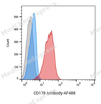 Flow cytometric analysis of 1X10^6 Hela cells labeling CD178 Antibody(red). Cells were fixed with 4% paraformaldehyde and permeabilised with 0.2% Triton X-100. Then stained with the primary antibody at 1/400 dilution overnight at 4℃. AF 488 Goat Anti-mouse IgG H&L was used as the secondary antibody at 1/1,000 dilution for 45 minutes at room temperature. Mouse IgG Isotype Control (blue) was used as the isotype control, cells without incubation with primary antibody were used as the unlabeled control (black).
 -
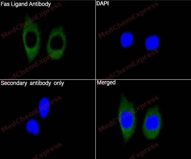 Immunocytochemistry analysis of HeLa cells labeling Fas Ligand with Fas Ligand Antibody (HY-P84345) at 1/100 dilution. Cells were fixed in 4% paraformaldehyde for 15 minutes at room temperature, permeabilized with 0.1% Triton X-100 in PBS for 15 minutes at room temperature, then blocked with quick block buffer for 10 minutes at room temperature. Cells were then incubated with Fas Ligand Antibody (HY-P84345) at 1/100 dilution in quick block buffer overnight at 4 ℃. AF488-conjugated Goat Anti-Mouse IgG H&L(HY-P8005, Green) was used as the secondary antibody at 1/1,000 dilution. PBS instead of the primary antibody was used as the secondary antibody only control. The Nuclear counterstain was DAPI (Blue).
 -
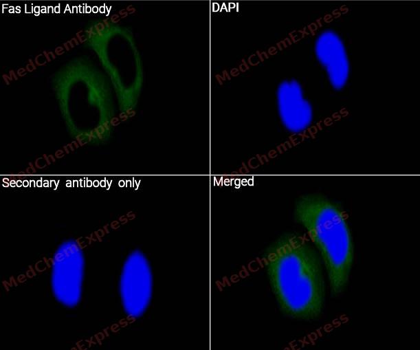 Immunocytochemistry analysis of HePG2 cells labeling Fas Ligand with Fas Ligand Antibody (HY-P84345) at 1/100 dilution. Cells were fixed in 4% paraformaldehyde for 15 minutes at room temperature, permeabilized with 0.1% Triton X-100 in PBS for 15 minutes at room temperature, then blocked with quick block buffer for 10 minutes at room temperature. Cells were then incubated with Fas Ligand Antibody (HY-P84345) at 1/100 dilution in quick block buffer overnight at 4 ℃. AF488-conjugated Goat Anti-Mouse IgG H&L(HY-P8005, Green) was used as the secondary antibody at 1/1,000 dilution. PBS instead of the primary antibody was used as the secondary antibody only control. The Nuclear counterstain was DAPI (Blue).


Background
-
Function
Fas Ligand is a Cytokine that binds to TNFRSF6/FAS, a receptor that transduces the apoptotic signal into cells. Involved in cytotoxic T-cell-mediated apoptosis, natural killer cell-mediated apoptosis and in T-cell development. Initiates fratricidal/suicidal activation-induced cell death (AICD) in antigen-activated T-cells contributing to the termination of immune responses. TNFRSF6/FAS-mediated apoptosis also has a role in the induction of peripheral tolerance. Binds to TNFRSF6B/DcR3, a decoy receptor that blocks apoptosis; Induces FAS-mediated activation of NF-kappa-B, initiating non-apoptotic signaling pathways. Can induce apoptosis but does not appear to be essential for this process; Cytoplasmic form induces gene transcription inhibition[1][2][3][4][5].
-
Subcellular Localization
Cell membrane; Single-pass type II membrane protein; Cytoplasmic vesicle lumen; Lysosome lumen; Secreted; Nucleus
-
Isoforms & Post-Translational Modification
P48023 has 2 isomers: P48023-1: 31485 Da (predicted); P48023-2: 14006 Da (predicted).
The soluble form derives from the membrane form by proteolytic processing. The membrane-bound form undergoes two successive intramembrane proteolytic cleavages. The first one is processed by ADAM10 producing an N-terminal fragment, which lacks the receptor-binding extracellular domain. This ADAM10-processed FasL (FasL APL) remnant form is still membrane anchored and further processed by SPPL2A that liberates the FasL intracellular domain (FasL ICD). FasL shedding by ADAM10 is a prerequisite for subsequent intramembrane cleavage by SPPL2A in T-cells;N-glycosylated (PubMed:9228058). Glycosylation enhances apoptotic activity (PubMed:27806260);Phosphorylated by FGR on tyrosine residues; this is required for ubiquitination and subsequent internalization;Monoubiquitinated -
Subunit
Homotrimer (PubMed:27806260). Interacts with ARHGAP9, BAIAP2L1, BTK, CACNB3, CACNB4, CRK, DLG2, DNMBP, DOCK4, EPS8L3, FGR, FYB1, FYN, HCK, ITK, ITSN2, KALRN, LYN, MACC1, MIA, MPP4, MYO15A, NCF1, NCK1, NCK2, NCKIPSD, OSTF1, PIK3R1, PSTPIP1, RIMBP3C, SAMSN1, SH3GL3, SH3PXD2B, SH3PXD2A, SH3RF2, SKAP2, SNX33, SNX9, SORBS3, SPTA1, SRC, SRGAP1, SRGAP2, SRGAP3, TEC, TJP3 and YES1
-
SwissProt ID
-
Synonyms
FASLG; APTL; FASL; CD95L; ALPS1B; CD95-L; TNFSF6; TNLG1A; APT1LG1; Fas Ligand Antibody


Documentation
-
Data Sheet (234 KB)
-
SDS (252 KB)
- English - EN (252 KB)
- Français - FR (252 KB)
- Deutsch - DE (252 KB)
- Norwegian - NO (252 KB)
- Español - ES (252 KB)
- Swedish - SV (252 KB)
- Italian - IT (252 KB)
- Korean - KR (252 KB)
- Portuguese - PT (252 KB)
-
User Guide for Antibodies (1077 KB)

 Powered by Bioz
