



CEACAM1 Antibody (YA3496)
(Synonyms: BGP-1; BGPI; CD66a; CEACAM1)
Based on 1 Customer Validation
CEACAM1 Antibody (YA3496) is a Rabbit-derived and non-conjugated IgG monoclonal antibody, targeting to CEACAM1.
-
Host:
Rabbit
-
Isotype:
IgG
-
Application:
WB, IHC-P, ICC/IF, IP
-
Reactivity :
Human
-
Formulation:
Supplied in 50 mM Tris-Glycine(pH 7.4), 0.15 M NaCl, 40% Glycerol, 0.01% sodium azide and 0.05% BSA.
-
Conjugation:
Non-conjugated


To place orders, for customer services and technical support, please contact: MedChemExpress USA
Tel: 609-228-6898 E-mail: [email protected] [email protected]





Applications
| Application |
WB

WB: Western Blot
|
IHC-P
IHC-P: Immunohistochemistry-Paraffin
|
ICC/IF
ICC/IF: Immunocytochemistry/
Immunofluorescence |
IP
IP: Immunoprecipitation
|
|---|---|---|---|---|
| Dilution Ratio | 1:1000-1:2000 | 1:100-1:200 | 1:50-1:200 | 1:50 |


Product Details
CEACAM1 Antibody (YA3496) is a Rabbit-derived and non-conjugated IgG monoclonal antibody, targeting to CEACAM1.
-
Host Rabbit

-
Clonality Recombinant,Monoclonal

-
Species ReactivityHuman
-
Observed Molecular WeightObserved band size: 120-150 kDa
Note: Due to possible protein modifications or aggregation, the molecular weight should be confirmed by actual measurement, and the predicted value is for reference only.

-
Calculated Molecular Weight Predicted band size: 58 kDa

A synthesized peptide derived from human CEACAM1 aa480-526/526.
Endogenous
Affinity Chromatography
Non-conjugated
Unmodified
IgG


Product Properties
-
Appearance
Solution
-
Formulation
Supplied in 50 mM Tris-Glycine(pH 7.4), 0.15 M NaCl, 40% Glycerol, 0.01% sodium azide and 0.05% BSA.
-
Concentration
Batch-dependent, Please check the COA for the concentration of each lot. Check Lot Concentration
-
Storage & Stability
Stored at -20°C for 1 year. Avoid repeated freeze / thaw cycles.
-
Shipping
Shipping with blue ice.


Verification Images
-
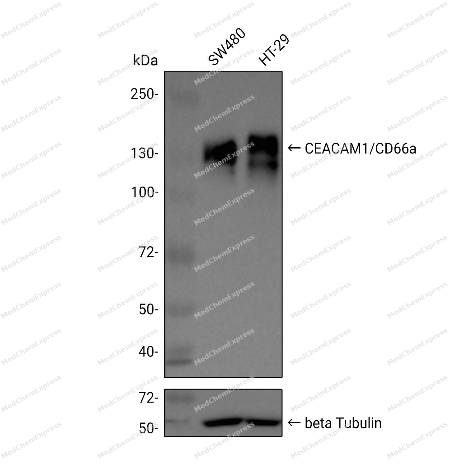 Western blot analysis was performed on extracts from SW480 (lane 1, 15 μg), and HT-29 (lane 2, 15 μg) using CEACAM1/CD66a Rabbit mAb. Proteins were transferred to a PVDF membrane and blocked with 5% non - fat milk in TBST at 4°C overnight. The primary antibody (1:1000 dilution) and the loading control antibody (beta-Tubulin(HRP), HY-P80955A, 1:5000 dilution) was incubated in 5% non-fat milk in TBST for 1 hour at 37°C. Goat Anti - Rabbit IgG - HRP Secondary Antibody (1:20000 dilution) was then applied for 40 minutes at 37°C.
 -
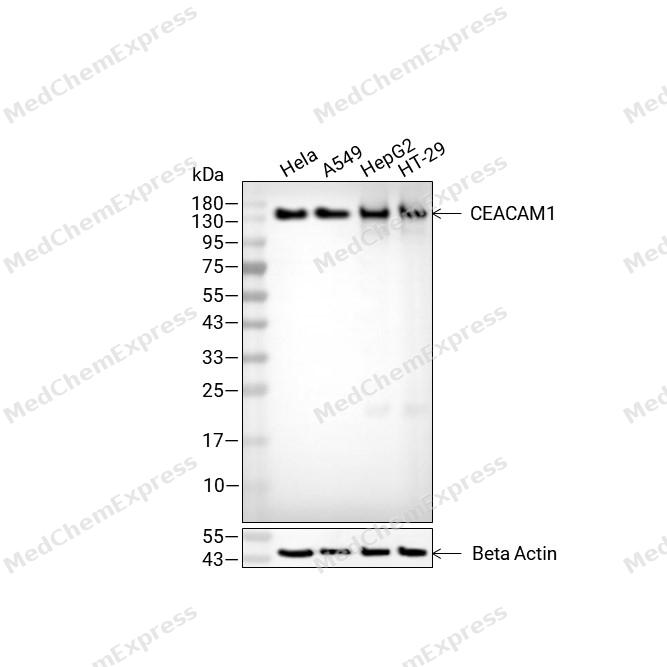 Western blot analysis of extracts from Hela (lane2(20μg), A549 (lane3(20μg), HepG2 (lane4(20μg) and HT-29 (lane5(20μg) using CEACAM1 Antibody (HY-P80985). Proteins were transferred to a PVDF membrane and blocked with 5% non-fat milk in TBST for 2 hour at room temperature. The primary antibody (1/2000) and Loading control antibody (Beta Actin, HY-P80993, 1/10,000) was used in 5% non-fat milk in TBST at 4°C overnight. Goat Anti-Rabbit IgG-HRP Secondary Antibody (HY-P8001 ,1/10,000) was used for 1 hour at room temperature.
 -
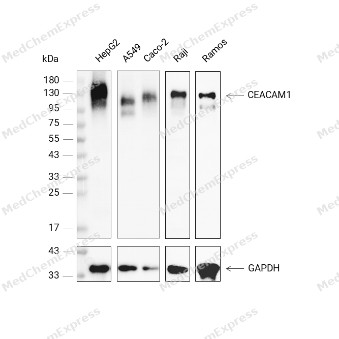 Western blot analysis was performed on protein extracts (40 μg) from HepG2 (lane 2), A549 (lane 3), Caco-2 (lane 4), Raji (lane 5), and Ramos (lane 6) using CEACAM1 antibody. Proteins were transferred onto a 0.45 μm PVDF membrane using the Trans-Blot® Turbo™ system for 13 min. The membrane was then blocked with 5% nonfat milk in TBST (HY-K1025) for 1 h at room temperature. The primary antibody (1:1000) and loading control antibody GAPDH Antibody (HRP) (HY-P80954A) (1:5000) were diluted in 5% nonfat milk in TBST and incubated with the membrane overnight at 4°C. After washing, the membrane of primary antibody was incubated with HRP-conjugated goat anti-rabbit/mouse IgG secondary antibody (HY-P8001/HY-P8004) (1:5000) diluted in 5% nonfat milk in TBST for 1 h at room temperature. Protein bands were visualized using an Ultra High Sensitivity ECL detection kit (HY-K1005).
 -
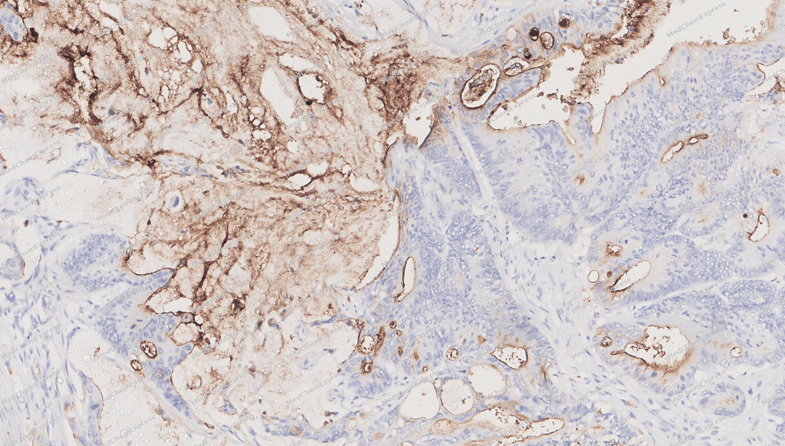 Immunohistochemical analysis of paraffin-embedded human mammary gland tissue using CEACAM1 antibody was performed. The section was pretreated using high-temperature mediated EDTA antigen retrieval buffer (pH 9.0), for 20 minutes. The tissues were incubated with primary antibody (HY-P80985, 1:100 dilution) at room temperature for 20 minutes. Detection was performed using an HRP conjugated compact polymer system. DAB was used as the chromogen. The tissues were counterstained with hematoxylin and mounted with neutral balsam mounting medium.
 -
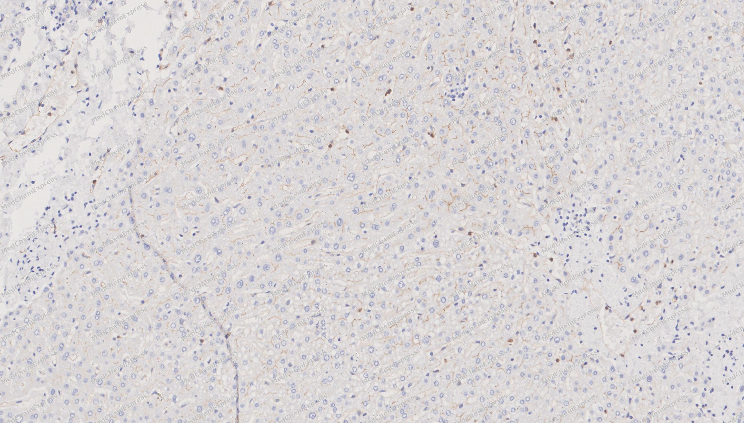 Immunohistochemical analysis of paraffin-embedded human thyroid tissue using CEACAM1 antibody was performed. The section was pretreated using high-temperature mediated EDTA antigen retrieval buffer (pH 9.0), for 20 minutes. The tissues were incubated with primary antibody (HY-P80985, 1:100 dilution) at room temperature for 20 minutes. Detection was performed using an HRP conjugated compact polymer system. DAB was used as the chromogen. The tissues were counterstained with hematoxylin and mounted with neutral balsam mounting medium.
 -
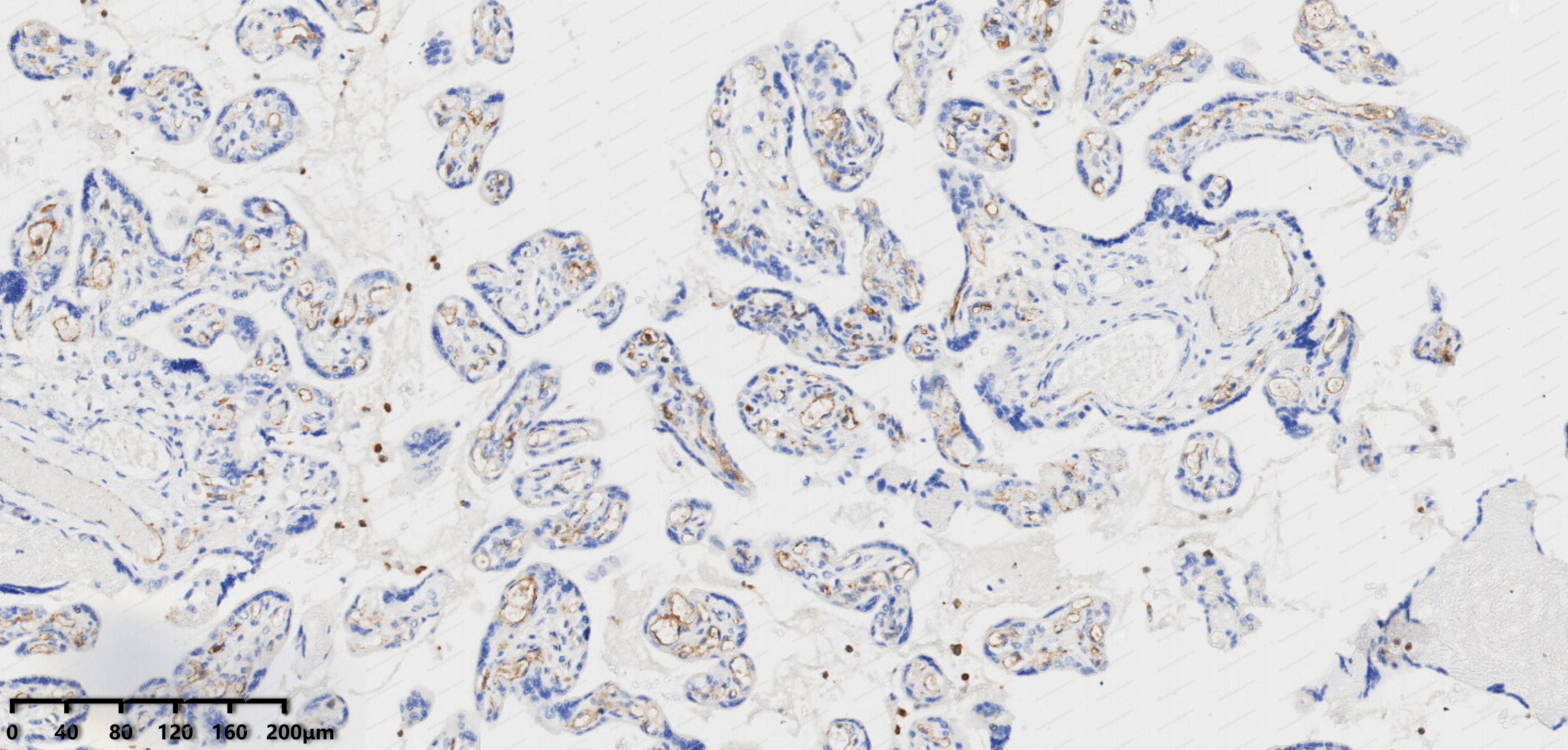 Immunohistochemical analysis of paraffin-embedded human placenta tissue using CEACAM1 antibody was performed. The section was pretreated using high-temperature mediated EDTA antigen retrieval buffer (pH 9.0), for 20 minutes. The tissues were incubated with primary antibody (HY-P80985, 1:100 dilution) at room temperature for 20 minutes. Detection was performed using an HRP conjugated compact polymer system. DAB was used as the chromogen. The tissues were counterstained with hematoxylin and mounted with neutral balsam mounting medium.
 -
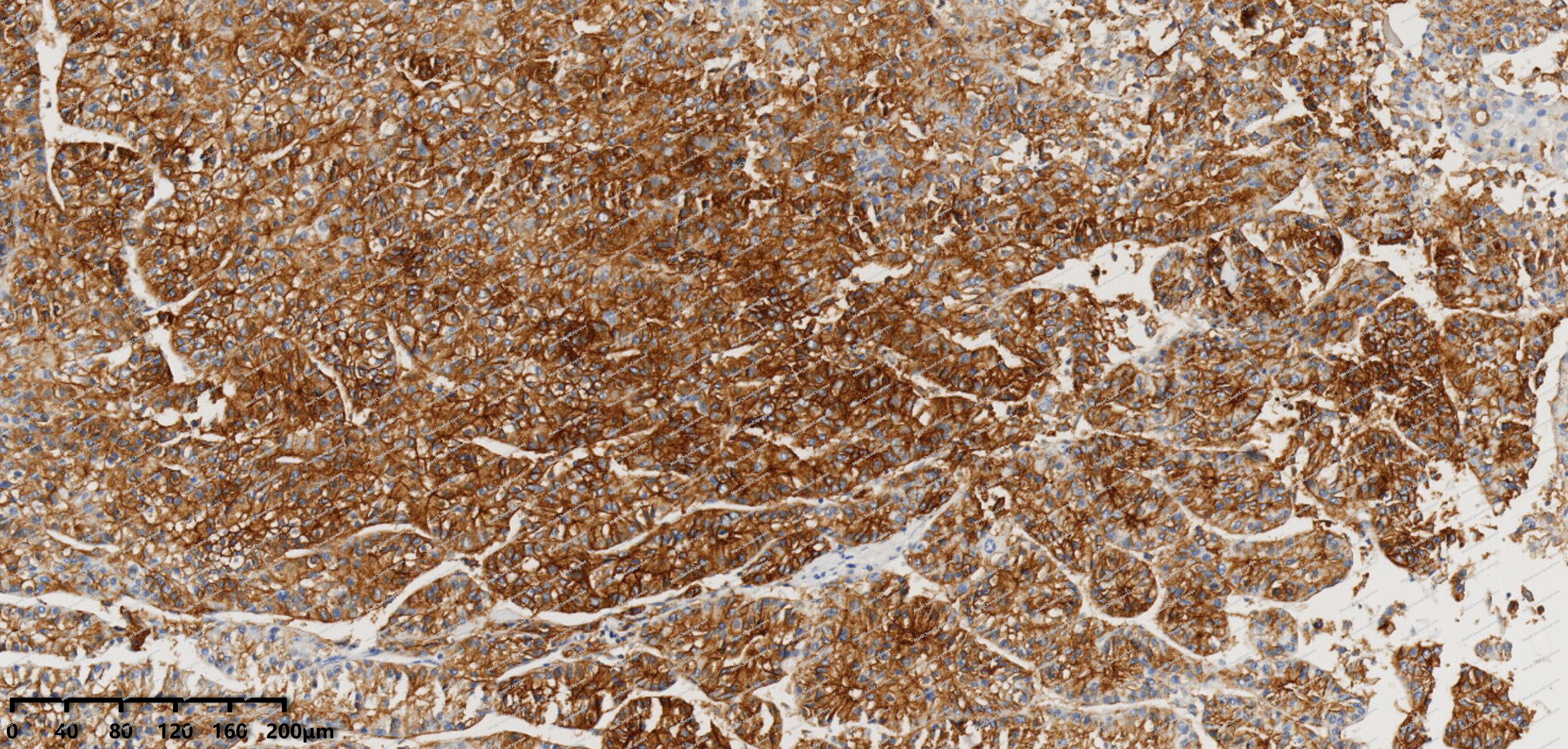 Immunohistochemical analysis of paraffin-embedded human liver cancer tissue using CEACAM1 antibody was performed. The section was pretreated using high-temperature mediated EDTA antigen retrieval buffer (pH 9.0), for 20 minutes. The tissues were incubated with primary antibody (HY-P80985, 1:100 dilution) at room temperature for 20 minutes. Detection was performed using an HRP conjugated compact polymer system. DAB was used as the chromogen. The tissues were counterstained with hematoxylin and mounted with neutral balsam mounting medium.
 -
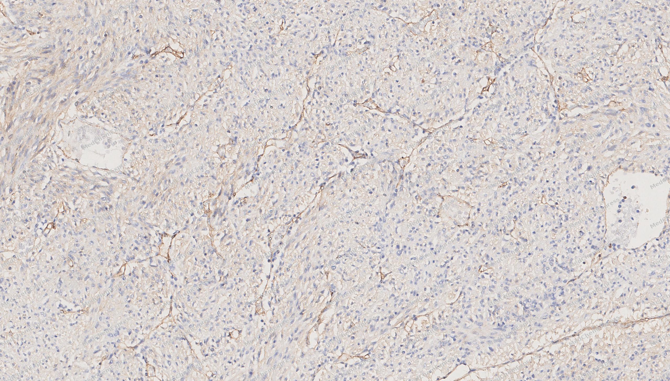 Immunohistochemical analysis of paraffin-embedded human Stromal tumor tissue using CEACAM1 antibody was performed. The section was pretreated using high-temperature mediated EDTA antigen retrieval buffer (pH 9.0), for 20 minutes. The tissues were incubated with primary antibody (HY-P80985, 1:100 dilution) at room temperature for 20 minutes. Detection was performed using an HRP conjugated compact polymer system. DAB was used as the chromogen. The tissues were counterstained with hematoxylin and mounted with neutral balsam mounting medium.
 -
 Immunocytochemistry analysis of HT-29 cells labeling CEACAM1 with CEACAM1 Antibody (HY-P80985) at 1/100 dilution. Cells were fixed in 4% paraformaldehyde for 15 minutes at room temperature, permeabilized with 0.1% Triton X-100 in PBS for 15 minutes at room temperature, then blocked with quick block buffer for 10 minutes at room temperature. Cells were then incubated with CEACAM1 Antibody (HY-P80985) at 1/100 dilution in quick block buffer overnight at 4 ℃. AF488-conjugated Goat Anti-Rabbit IgG H&L(HY-P8002, Green) was used as the secondary antibody at 1/1,000 dilution. PBS instead of the primary antibody was used as the secondary antibody only control. The Nuclear counterstain was DAPI (Blue).
 -
 Immunocytochemistry analysis of HT-29 cells labeling CEACAM1 with CEACAM1 Antibody (HY-P80985) at 1/200 dilution. Cells were fixed in 4% paraformaldehyde for 15 minutes at room temperature, permeabilized with 0.1% Triton X-100 in PBS for 15 minutes at room temperature, then blocked with quick block buffer for 10 minutes at room temperature. Cells were then incubated with CEACAM1 Antibody (HY-P80985) at 1/200 dilution in quick block buffer overnight at 4 ℃. AF488-conjugated Goat Anti-Rabbit IgG H&L(HY-P8002, Green) was used as the secondary antibody at 1/1,000 dilution. PBS instead of the primary antibody was used as the secondary antibody only control. The Nuclear counterstain was DAPI (Blue).


Background
-
Function
Carcinoembryonic antigen-related cell adhesion molecule 1 (CEACAM1), also known as CD66a, BGP, or C-CAM, is an extensively studied, multifunctional transmembrane protein which is the primordial member of the carcinoembryonic antigen cell adhesion molecul
-
Subcellular Localization
Cell membrane; Single-pass type I membrane protein; Lateral cell membrane; Apical cell membrane; Basal cell membrane; Cell junction; Cell junction, adherens junction; Secreted; Secreted; Secreted; Cell membrane; Single-pass type I membrane protein; Cell membrane; Single-pass type I membrane protein; Cell membrane; Single-pass type I membrane protein; Cell membrane; Single-pass type I membrane protein; Cytoplasmic vesicle, secretory vesicle membrane; Lateral cell membrane; Apical cell membrane; Basal cell membrane; Cell junction; Cell junction, adherens junction; Cell projection, microvillus membrane; Single-pass type I membrane protein; Apical cell membrane; Single-pass type I membrane protein
-
Expression
Tissue_specificity:It is expressed in colonic columnar epithelial cells (protein level) (PubMed: 10436421) . The predominant form expressed on T cells is the one containing a long cytoplasmic domain (PubMed: 18424730) . It is also expressed in granulocytes and lymphocytes. Leukocytes express only isoforms 6 and 1 (PubMed: 11994468) .
Induction:Induced in primary T cells by activation with IL-2 -
Isoforms & Post-Translational Modification
P13688 has 11 isomers: P13688-1: 57560 Da (predicted); P13688-2: 45950 Da (predicted); P13688-3: 35302 Da (predicted); P13688-4: 38578 Da (predicted); P13688-5: 50349 Da (predicted); P13688-6: 46910 Da (predicted); P13688-7: 27431 Da (predicted); P13688-8: 50521 Da (predicted); P13688-9: 43062 Da (predicted); P13688-10: 51147 Da (predicted); P13688-11: 39871 Da (predicted).
Phosphorylated on serine and tyrosine (By similarity). Isoform 1 is phosphorylated on tyrosine by Src family kinases like SRC and LCK and by receptor like CSF3R, EGFR and INSR upon stimulation (PubMed:15467833, PubMed:18424730, PubMed:7478590). Phosphorylated at Ser-508; mediates activity. Phosphorylated at Tyr-493; regulates activity (By similarity). Phosphorylated at Tyr-493 by EGFR and INSR upon stimulation; this phosphorylation is Ser-508-phosphorylation-dependent; mediates cellular internalization; increases interaction with downstream proteins like SHC1 and FASN (By similarity). Phosphorylated at Tyr-493 and Tyr-520 by LCK; mediates PTPN6 association and is regulated by homophilic ligation of CEACAM1 in the absence of T cell activation (PubMed:18424730). Phosphorylated at Tyr-520; mediates interaction with PTPN11 (By similarity);Phosphorylated on serine and threonine -
Subunit
Monomer. Oligomer. Heterodimer. Homodimer (PubMed:26483485). Cis-dimer/oligomer (via Ig-like C2-type and/or via cytoplasmic domains); induced by trans-homophilic cell adhesion through an allosteric mechanism transmitted by the Ig-like V-type domain, and is regulated by intracellular calcium and calmodulin. Interacts (via cytoplasmic domain) with calmodulin in a calcium dependent manner; reduces homophilic cell adhesion through dissociation of dimer (By similarity). Isoform 1 interacts (via cytoplasmic domain) with PTPN11 (preferentially) and PTPN6; cis-homodimer form is preferred; this interaction is decreased by formation of Isoform 1 /Isoform 8 cis-heterodimers and is dependent on the monomer/dimer equilibrium; this interaction is phosphorylation-dependent (PubMed:23696226). Isoform 1 interacts with LYN (By similarity). Isoform 1 interacts (via cytoplasmic domain) with SRC (via SH2 domain); this interaction is regulated by trans-homophilic cell adhesion (PubMed:7478590). Isoform 1 interacts (via cytoplasmic domain) with LCK; mediates phosphorylation at Tyr-493 and Tyr-520 resulting in PTPN6 association. Isoform 1 interacts with PTPN6; this interaction is phosphorylation-dependent and causes a profound decrease in TCR stimulation-induced CD247 and ZAP70 phosphorylation. Isoform 1 interacts with TCR/CD3 complex through TCR beta chain and CD3E; colocalizes at the cell surface and upon stimulation of the TCR/CD3 complex recruits PTPN6 in the TCR/CD3 complex, resulting in dephosphorylation of CD247 and ZAP70 (PubMed:18424730). Isoform 1 interacts (via cytoplasmic domain) with SHC1 (via SH2 domain); SHC1 mediates interaction with INSR or EGFR in a Ser-508 phosphorylation-dependent manner (By similarity). Isoform 1 interacts with EGFR; the interaction is indirect (PubMed:15467833). Isoform 1 interacts with CSF3R; down-regulates the CSF3R-STAT3 pathway through recruitment of PTPN6 that dephosphorylates CSF3R (By similarity). Isoform 1 (phosphorylated form) interacts with TLR4 and SYK; recruits PTPN6 that dephosphorylates SYK, reducing the production of reactive oxygen species (ROS) and lysosome disruption, leading to a reduction of the inflammasome activity (By similarity). Isoform 1 interacts with FLNA; inhibits cell migration and cell scattering by interfering with the interaction of FLNA with RALA (PubMed:16291724). Isoform 1 interacts (via cytoplasmic domain) with PXN; the interaction is phosphotyrosyl-dependent (PubMed:11035932). Isoform 1 interacts with KLRK1; recruits PTPN6 that dephosphorylates VAV1 (PubMed:23696226). Isoform 1 interacts with CEACAM8 (PubMed:11994468). Isoform 1 interacts with FASN; this interaction is insulin and phosphorylation-dependent; reduces fatty-acid synthase activity (By similarity). Interacts (via Ig-like V-type) with HAVCR2 (via Ig-like V-type); facilitates the maturation and cell surface expression of HAVCR2 thereby regulating T cell tolerance induction (PubMed:25363763). Isoform 8 interacts (via the cytoplasmic domain) with ANXA2; this interaction is regulated by phosphorylation and appears in the AIIt complex (PubMed:14522961). Interacts (via Lewis X moieties) with CD209 (via C-type lectin domain); this interaction is regulated by the glycosylation pattern of CEACAM1 on cell types and regulates contact between dendritic cells and neutrophils (PubMed:16246332)
-
SwissProt ID
-
Synonyms
BGP-1; BGPI; CD66a; CEACAM1
-
Research Field
Tags & Cell Markers


Documentation
-
Data Sheet (235 KB)
-
SDS (251 KB)
- English - EN (251 KB)
- Français - FR (251 KB)
- Deutsch - DE (251 KB)
- Norwegian - NO (251 KB)
- Español - ES (251 KB)
- Swedish - SV (251 KB)
- Italian - IT (251 KB)
- Korean - KR (251 KB)
- Portuguese - PT (251 KB)
-
User Guide for Antibodies (1077 KB)

 Powered by Bioz
