



E2F-1 Antibody (YA471)
(Synonyms: RBBP3, E2F1, Transcription factor E2F1, E2F-1, PBR3, Retinoblastoma-associated protein 1, Retinoblastoma-binding protein 3, pRB-binding protein E2F-1, RBAP-1, RBBP-3)
Based on 1 Customer Validation
E2F-1 Antibody (YA471) is a Rabbit-derived and non-conjugated IgG monoclonal antibody, targeting to E2F-1.
-
Host:
Rabbit
-
Isotype:
IgG
-
Application:
WB, ICC/IF, IHC-P, IP, FC
-
Reactivity :
Human, Mouse
-
Formulation:
Supplied in 1*TBS (pH7.4), 0.05% BSA and 40% Glycerol. Preservative: 0.05% Sodium Azide.
-
Conjugation:
Non-conjugated


To place orders, for customer services and technical support, please contact: MedChemExpress USA
Tel: 609-228-6898 E-mail: [email protected] [email protected]





Applications
| Application |
WB

WB: Western Blot
|
ICC/IF
ICC/IF: Immunocytochemistry/
Immunofluorescence |
IHC-P
IHC-P: Immunohistochemistry-Paraffin
|
FC
FC: Flow Cytometry
|
IP
IP: Immunoprecipitation
|
|---|---|---|---|---|---|
| Dilution Ratio | 1:1000 | 1:100-1:500 | 1:50-1:200 | 1:50-1:100 | Use at an assay dependent concentration. |


Product Details
E2F-1 Antibody (YA471) is a Rabbit-derived and non-conjugated IgG monoclonal antibody, targeting to E2F-1.
-
Host Rabbit

-
Clonality Recombinant,Monoclonal

-
Species ReactivityHuman, Mouse
-
Observed Molecular WeightObserved band size: 60 kDa
Note: Due to possible protein modifications or aggregation, the molecular weight should be confirmed by actual measurement, and the predicted value is for reference only.

-
Calculated Molecular Weight Predicted band size: 47 kDa

Entrez Gene: 1869 Human ; 13555 Mouse ; 399489 Rat
SwissProt: Q01094 Human ; Q61501 Mouse ; O09139 Rat
OMIM: 189971 Human
Synthetic peptide corresponding to Human E2F1.AA range:371-404.
Endogenous
Protein A affinity purified.
Non-conjugated
Unmodified
IgG


Product Properties
-
Appearance
Solution
-
Formulation
Supplied in 1*TBS (pH7.4), 0.05% BSA and 40% Glycerol. Preservative: 0.05% Sodium Azide.
-
Concentration
Batch-dependent, Please check the COA for the concentration of each lot. Check Lot Concentration
-
Storage & Stability
Stored at -20°C for 1 year. Avoid repeated freeze / thaw cycles.
-
Shipping
Shipping with blue ice.


Verification Images
-
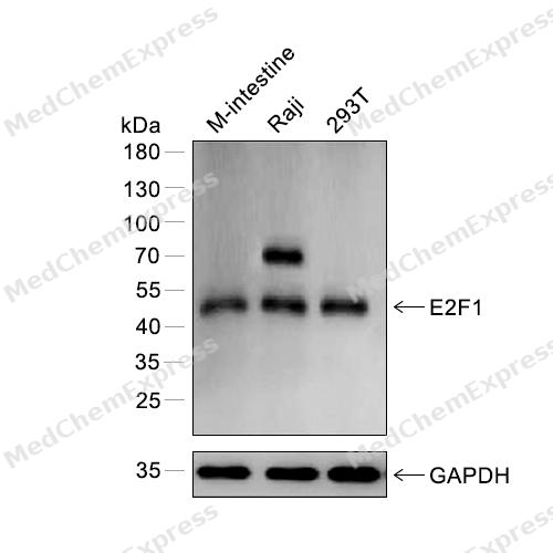 Western blot analysis of extracts from M-intestine (lane 1) and Raji (lane 2) and 293T (lane 3) using E2F1 antibody. Proteins were transferred to a PVDF membrane and blocked with 5% nonfat powdered milk in PBST for 2 hour at room temperature. The primary antibody (1/1000) and loading control antibody (GAPDH, 1/3000) was diluted with 5% nonfat powdered milk in PBST at 4°C overnight. Goat Anti-Rabbit IgG-HRP Secondary Antibody (1/8,000) was incubated for 45min at room temperature.
 -
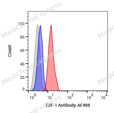 Flow cytometric analysis of 1X10⁶ HepG2 cells labeling E2F-1 Antibody (HY-P80491, red). Cells were fixed with 4% paraformaldehyde and permeabilised with 90% methanol. Then stained with the primary antibody at 1/100 dilution for an hour at 4℃. AF488-conjugated Goat Anti-Rabbit IgG H&L (HY-P8002) was used as the secondary antibody at 1/1,000 dilution for 30 minutes at 4℃. Rabbit IgG Isotype Control (HY-P80879, blue) was used as the isotype control, cells without incubation with primary antibody were used as the unlabeled control (black).
 -
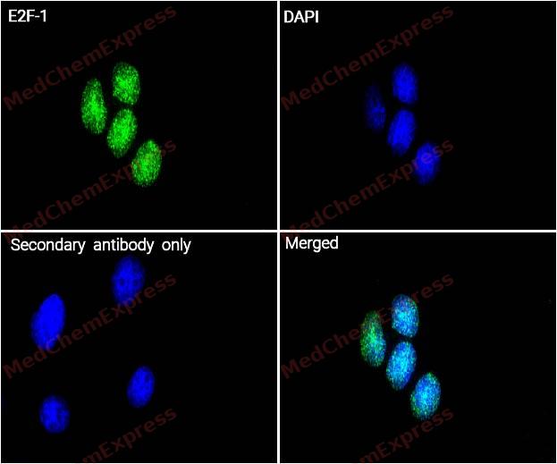 Immunocytochemistry analysis of Hela cells labeling E2F-1 with E2F-1 Antibody (HY-P80491) at 1/100 dilution. Cells were fixed in 4% paraformaldehyde for 15 minutes at room temperature, permeabilized with 0.1% Triton X-100 in PBS for 15 minutes at room temperature, then blocked with quick block buffer for 10 minutes at room temperature. Cells were then incubated with E2F-1 Antibody (HY-P80491) at 1/100 dilution in quick block buffer overnight at 4 ℃. AF488-conjugated Goat Anti-Rabbit IgG H&L(HY-P8002, Green) was used as the secondary antibody at 1/1,000 dilution. PBS instead of the primary antibody was used as the secondary antibody only control. The Nuclear counterstain was DAPI (Blue).
 -
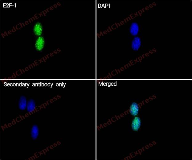 Immunocytochemistry analysis of Hela cells labeling E2F-1 with E2F-1 Antibody (HY-P80491) at 1/200 dilution. Cells were fixed in 4% paraformaldehyde for 15 minutes at room temperature, permeabilized with 0.1% Triton X-100 in PBS for 15 minutes at room temperature, then blocked with quick block buffer for 10 minutes at room temperature. Cells were then incubated with E2F-1 Antibody (HY-P80491) at 1/200 dilution in quick block buffer overnight at 4 ℃. AF488-conjugated Goat Anti-Rabbit IgG H&L(HY-P8002, Green) was used as the secondary antibody at 1/1,000 dilution. PBS instead of the primary antibody was used as the secondary antibody only control. The Nuclear counterstain was DAPI (Blue).


Background
-
Function
E2F-1 transcription activator that binds DNA cooperatively with DP proteins through the E2 recognition site, 5'-TTTC[CG]CGC-3' found in the promoter region of a number of genes whose products are involved in cell cycle regulation or in DNA replication. The DRTF1/E2F complex functions in the control of cell-cycle progression from G1 to S phase. E2F1 binds preferentially RB1 in a cell-cycle dependent manner. It can mediate both cell proliferation and TP53/p53-dependent apoptosis. Blocks adipocyte differentiation by binding to specific promoters repressing CEBPA binding to its target gene promoters. Directly activates transcription of PEG10. Positively regulates transcription of RRP1B[1][2][3][4][5][6][7][8][9].
-
Subcellular Localization
Nucleus
-
Subunit
Component of the DRTF1/E2F transcription factor complex. Forms heterodimers with DP family members. The E2F1 complex binds specifically hypophosphorylated RB1, the interaction represses E2F1-driven transcription (PubMed:8336704). During the cell cycle, RB1 becomes phosphorylated in mid-to-late G1 phase, detaches from the DRTF1/E2F complex, rendering E2F transcriptionally active. Viral oncoproteins, notably E1A, T-antigen and HPV E7, are capable of sequestering RB1, thus releasing the active complex. Interacts with TRRAP, which probably mediates its interaction with histone acetyltransferase complexes, leading to transcription activation. Binds TOPBP1 and EAPP. Interacts with ARID3A. Interacts with TRIM28; the interaction inhibits E2F1 acetylation through recruiting HDAC1 and represses its transcriptional activity. Interaction with KAT2B; the interaction acetylates E2F1 enhancing its DNA-binding and transcriptional activity. Interacts with BIRC2/c-IAP1 (via BIR domains). The complex TFDP1:E2F1 interacts with CEBPA; the interaction prevents CEBPA binding to target genes promoters and represses its transcriptional activity (PubMed:20176812). Interacts with RRP1B (PubMed:20040599). Interacts with HCFC1 (PubMed:23629655). Interacts with KMT2E; the interaction is probably indirect and is mediated via HCFC1 (PubMed:23629655). Interacts with DCAF5 and L3MBTL3; the interaction requires methylation at Lys-185 and is necessary to target E2F1 for ubiquitination by the CRL4-DCAF5 E3 ubiquitin ligase complex (PubMed:29691401)
-
SwissProt ID
-
Synonyms
RBBP3, E2F1, Transcription factor E2F1, E2F-1, PBR3, Retinoblastoma-associated protein 1, Retinoblastoma-binding protein 3, pRB-binding protein E2F-1, RBAP-1, RBBP-3
-
Research Field
Epigenetics and Nuclear Signaling


Documentation
-
Data Sheet (262 KB)
-
SDS (252 KB)
- English - EN (252 KB)
- Français - FR (252 KB)
- Deutsch - DE (252 KB)
- Norwegian - NO (252 KB)
- Español - ES (252 KB)
- Swedish - SV (252 KB)
- Italian - IT (252 KB)
- Korean - KR (252 KB)
- Portuguese - PT (252 KB)
-
User Guide for Antibodies (1077 KB)
References
[1]. Martínez-Balbás MA, et al. Regulation of E2F1 activity by acetylation. EMBO J. 2000 Feb 15;19(4):662-71. [Content Brief]
[2]. Stevens C, et al. Chk2 activates E2F-1 in response to DNA damage. Nat Cell Biol. 2003 May;5(5):401-9. [Content Brief]
[3]. García-Alvarez G, et al. Glycogen synthase kinase-3beta binds to E2F1 and regulates its transcriptional activity. Biochim Biophys Acta. 2007 Mar;1773(3):375-82. [Content Brief]
[4]. Wang C, et al. Regulation of E2F1 function by the nuclear corepressor KAP1. J Biol Chem. 2007 Oct 12;282(41):29902-9. [Content Brief]
[5]. Wang C, et al. PEG10 directly regulated by E2Fs might have a role in the development of hepatocellular carcinoma. FEBS Lett. 2008 Aug 6;582(18):2793-8. [Content Brief]
[6]. Wang D, et al. Phosphorylated E2F1 is stabilized by nuclear USP11 to drive Peg10 gene expression and activate lung epithelial cells. J Mol Cell Biol. 2018 Feb 1;10(1):60-73. [Content Brief]
[7]. Wu X, et al. p53 and E2F-1 cooperate to mediate apoptosis. Proc Natl Acad Sci U S A. 1994 Apr 26;91(9):3602-6. [Content Brief]
[8]. Zaragoza K, et al. Repression of transcriptional activity of C/EBPalpha by E2F-dimerization partner complexes. Mol Cell Biol. 2010 May;30(9):2293-304. [Content Brief]
[9]. Paik JC, et al. Regulation of E2F1-induced apoptosis by the nucleolar protein RRP1B. J Biol Chem. 2010 Feb 26;285(9):6348-63. [Content Brief]

 Powered by Bioz
