



C-erbB-3/HER3 Antibody (YA457)
(Synonyms: ERBB3; HER3; Receptor tyrosine-protein kinase erbB-3; Proto-oncogene-like protein c-ErbB-3; Tyrosine kinase-type cell surface receptor HER3)Based on 1 Customer Validation
C-erbB-3/HER3 Antibody (YA457) is a Rabbit-derived and non-conjugated IgG monoclonal antibody, targeting to C-erbB-3/HER3.
-
Host:
Rabbit
-
Isotype:
IgG
-
Application:
WB, IHC-P, FC
-
Reactivity :
Human, Mouse
-
Formulation:
Supplied in 50 mM Tris-Glycine (pH 7.4), 0.15 M NaCl, 40% Glycerol and 0.05% BSA. Preservative: 0.01% Sodium azide
-
Conjugation:
Non-conjugated


To place orders, for customer services and technical support, please contact: MedChemExpress USA
Tel: 609-228-6898 E-mail: [email protected] [email protected]





Applications
| Application |
WB

WB: Western Blot
|
IHC-P
IHC-P: Immunohistochemistry-Paraffin
|
FC
FC: Flow Cytometry
|
|---|---|---|---|
| Dilution Ratio | 1:500-1:1000 | 1:100-1:200 | 1:20-1:50 |


Product Details
C-erbB-3/HER3 Antibody (YA457) is a Rabbit-derived and non-conjugated IgG monoclonal antibody, targeting to C-erbB-3/HER3.
-
Host Rabbit

-
Clonality Recombinant,Monoclonal

-
Species ReactivityHuman, Mouse
-
Observed Molecular WeightObserved band size: 185 kDa
Note: Due to possible protein modifications or aggregation, the molecular weight should be confirmed by actual measurement, and the predicted value is for reference only.

-
Calculated Molecular Weight Predicted band size: 148 kDa

Synthetic peptide corresponding to Human ErbB3 aa1094-1111.
Endogenous
affinity purified
Non-conjugated
Unmodified
IgG


Product Properties
-
Appearance
Solution
-
Formulation
Supplied in 50 mM Tris-Glycine (pH 7.4), 0.15 M NaCl, 40% Glycerol and 0.05% BSA. Preservative: 0.01% Sodium azide
-
Concentration
Batch-dependent, Please check the COA for the concentration of each lot. Check Lot Concentration
-
Storage & Stability
Stored at -20°C for 1 year. Avoid repeated freeze / thaw cycles.
-
Shipping
Shipping with blue ice.


Verification Images
-
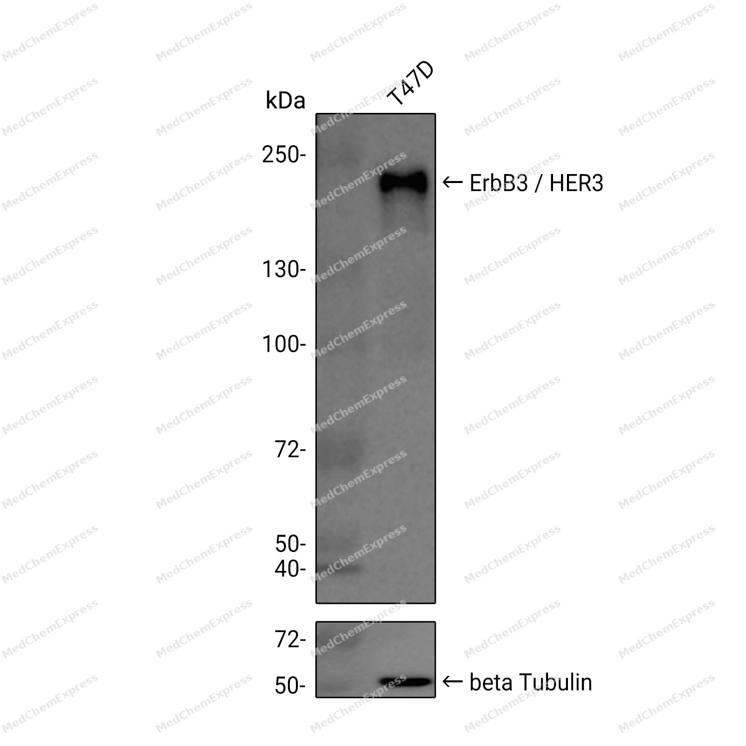 Western blot analysis was performed on extracts from T47D (lane 1, 15 μg) using ErbB3 / HER3 Rabbit mAb. Proteins were transferred to a PVDF membrane and blocked with 5% non - fat milk in TBST at 4°C overnight. The primary antibody (1:1000 dilution) and the loading control antibody (beta-Tubulin(HRP), HY-P80955A, 1:5000 dilution) was incubated in 5% non-fat milk in TBST for 1 hour at 37°C. Goat Anti - Rabbit IgG - HRP Secondary Antibody (1:20000 dilution) was then applied for 40 minutes at 37°C.
 -
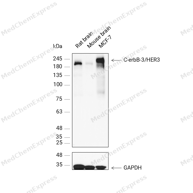 Western blot analysis was performed on protein extracts (30 μg) from Rat brain (lane 1), Mouse brain (lane 2), and MCF-7 (lane 3) using C-erbB-3/HER3 antibody. Proteins were transferred onto a 0.45 μm PVDF membrane using a wet transfer system for 90 min. The membrane was then blocked with 5% nonfat milk in TBST (HY-K1025) for 1 h at room temperature. Thhe primary antibody (1:500) and loading control antibody GAPDH Antibody (HRP) (HY-P80954A) (1:5000) were diluted in 5% nonfat milk in TBST and incubated with the membrane overnight at 4°C. After washing, the membrane of primary antibody was incubated with HRP-conjugated goat anti-rabbit/mouse IgG secondary antibody (HY-P8001/HY-P8004) (1:5000) diluted in 5% nonfat milk in TBST for 1 h at room temperature. Protein bands were visualized using an Ultra High Sensitivity ECL detection kit (HY-K1005).
 -
 Western blot analysis of extracts from MCF-7 (lane 2, 20ug), SK-Br-3 (lane 3, 20ug), Mouse skin (lane 4, 20ug) and K562 (lane 5, 20ug) using C-erbB-3/HER3 Antibody (YA457) (HY-P80659). Proteins were transferred to a PVDF membrane and blocked with 5% non-fat milk in TBST at 4°C overnight. The primary antibody (1/1000) and Loading control antibody (Beta Actin, HY-P80993, 1/10,000) was used in 5% non-fat milk in TBST for 2 hour at room temperature. Goat Anti-Rabbit IgG-HRP Secondary Antibody (HY-P8001, 1/10,000) was used for 1 hour at room temperature.


Background
-
Function
C-erbB-3/HER3 is a Tyrosine-protein kinase that plays an essential role as cell surface receptor for neuregulins. Binds to neuregulin-1 (NRG1) and is activated by it; ligand-binding increases phosphorylation on tyrosine residues and promotes its association with the p85 subunit of phosphatidylinositol 3-kinase. May also be activated by CSPG5. Involved in the regulation of myeloid cell differentiation[1][2][3].
-
Subcellular Localization
Cell membrane; Single-pass type I membrane protein; Secreted
-
Expression
Tissue_specificity:Epithelial tissue and brain -
Isoforms & Post-Translational Modification
P21860 has 5 isomers: P21860-1: 148098 Da (predicted); P21860-2: 20136 Da (predicted); P21860-3: 36490 Da (predicted); P21860-4: 141752 Da (predicted); P21860-5: 77426 Da (predicted).
Autophosphorylated (PubMed:20351256). Ligand-binding increases phosphorylation on tyrosine residues and promotes its association with the p85 subunit of phosphatidylinositol 3-kinase (PubMed:20682778) -
Subunit
Monomer and homodimer. Heterodimer with each of the other ERBB receptors (Potential). Interacts with CSPG5 (PubMed:15358134). Interacts with GRB7 (PubMed:9516479). Interacts with MUC1 (PubMed:12939402). Interacts with MYOC (By similarity). Interacts with isoform 2 of PA2G4 (PubMed:11325528, PubMed:16832058). Found in a ternary complex with NRG1 and ITGAV:ITGB3 or ITGA6:ITGB4 (PubMed:20682778)
-
SwissProt ID
-
Synonyms
ERBB3; HER3; Receptor tyrosine-protein kinase erbB-3; Proto-oncogene-like protein c-ErbB-3; Tyrosine kinase-type cell surface receptor HER3
-
Research Field
Cell Biology


Documentation
-
Data Sheet (262 KB)
-
SDS (252 KB)
- English - EN (252 KB)
- Français - FR (252 KB)
- Deutsch - DE (252 KB)
- Norwegian - NO (252 KB)
- Español - ES (252 KB)
- Swedish - SV (252 KB)
- Italian - IT (252 KB)
- Korean - KR (252 KB)
- Portuguese - PT (252 KB)
-
User Guide for Antibodies (1077 KB)
References
[1]. Ieguchi K, et al. Direct binding of the EGF-like domain of neuregulin-1 to integrins ({alpha}v{beta}3 and {alpha}6{beta}4) is involved in neuregulin-1/ErbB signaling. J Biol Chem. 2010 Oct 8;285(41):31388-98. [Content Brief]
[2]. Kinugasa Y, et al. Neuroglycan C, a novel member of the neuregulin family. Biochem Biophys Res Commun. 2004 Sep 3;321(4):1045-9. [Content Brief]
[3]. Braunstein EM, et al. A germline ERBB3 variant is a candidate for predisposition to erythroid MDS/erythroleukemia. Leukemia. 2016 Nov;30(11):2242-2245. [Content Brief]

 Powered by Bioz
