



Fibronectin Antibody (YA6107)
(Synonyms: FN1; FN; Fibronectin; FN; Cold-insoluble globulin; CIG)
Based on 1 Customer Validation
Fibronectin Antibody (YA6107) is a Rabbit-derived and non-conjugated IgG monoclonal antibody, targeting to Fibronectin.
-
Host:
Rabbit
-
Isotype:
IgG
-
Application:
WB, IHC-P, ICC/IF, ELISA
-
Reactivity :
Human, Mouse, Rat
-
Formulation:
Supplied in PBS, 50% glycerol, 0.05% Proclin 300, 0.05%BSA
-
Conjugation:
Non-conjugated


To place orders, for customer services and technical support, please contact: MedChemExpress USA
Tel: 609-228-6898 E-mail: [email protected] [email protected]





Applications
| Application |
IHC-P

IHC-P: Immunohistochemistry-Paraffin
|
WB
WB: Western Blot
|
ICC/IF
ICC/IF: Immunocytochemistry/
Immunofluorescence |
ELISA
ELISA: Enzyme Linked Immunosorbent Assay
|
|---|---|---|---|---|
| Dilution Ratio | 1:1000-1:5000 | 1:2000-1:10000 | 1:200-1:1000 | 1:5000-1:20000 |


Product Details
Fibronectin Antibody (YA6107) is a Rabbit-derived and non-conjugated IgG monoclonal antibody, targeting to Fibronectin.
-
Host Rabbit

-
Clonality Monoclonal

-
Species ReactivityHuman, Mouse, Rat
-
Observed Molecular WeightObserved band size: 285 kDa
Note: Due to possible protein modifications or aggregation, the molecular weight should be confirmed by actual measurement, and the predicted value is for reference only.

-
Calculated Molecular Weight Predicted band size: 272 kDa

Protein A
Non-conjugated
Unmodified
IgG


Product Properties
-
Appearance
Solution
-
Formulation
Supplied in PBS, 50% glycerol, 0.05% Proclin 300, 0.05%BSA
-
Concentration
Batch-dependent, Please check the COA for the concentration of each lot. Check Lot Concentration
-
Storage & Stability
Stored at -20°C for 1 year. Avoid repeated freeze / thaw cycles.
-
Shipping
Shipping with blue ice.


Verification Images
-
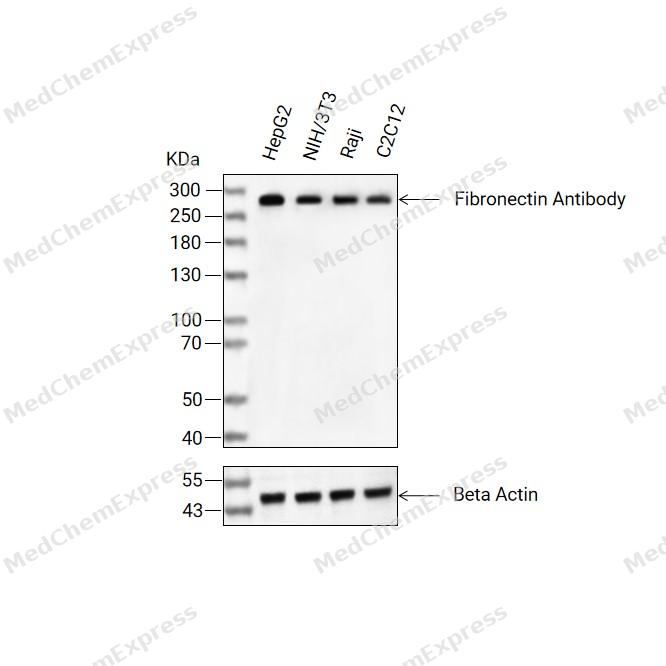 Western blot analysis of extracts from HepG2 (lane 2(20μg), NIH/3T3 (lane 3(20μg), Raji (lane 4(20μg), C2C12 (lane 5(20μg) using Fibronectin Antibody. Proteins were transferred to a PVDF membrane and blocked with 5% BSA in TBST for 2 hour at room temperature. The primary antibody (1/1000) and Loading control antibody (Beta Actin, HY-P80438, 1/3000) was used in 5% BSA in TBST at 4°C overnight. Goat Anti-Mouse/Rabbit IgG-HRP Secondary Antibody (HY-P8004/HY-P8001, 1/10,000) was used for 1 hour at room temperature.
 -
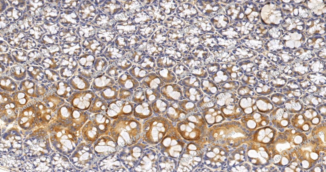 Immunohistochemical analysis of paraffin-embedded mouse colon tissue using Fibronectin Antibody (HY-P86415, 1/4000). The section was pretreated using heat mediated antigen retrieval with sodium citrate buffer (pH 6.0) for 8 minutes. The tissues were blocked with quick block buffer for 0.5 hours at room temperature, washed with PBS and PBST, and then incubated with the primary antibody overnight at 4℃. The detection was performed using an HRP conjugated compact polymer system. DAB was used as the chromogen. Tissues were counterstained with hematoxylin and mounted with DPX.
 -
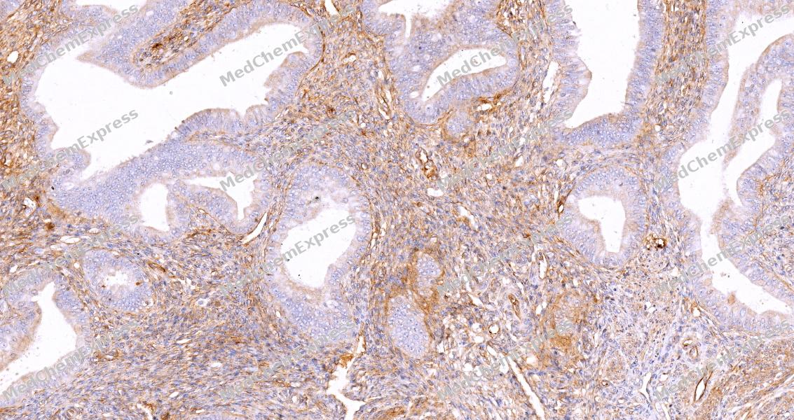 Immunohistochemical analysis of paraffin-embedded human endometrium tissue using Fibronectin Antibody (HY-P86415, 1/4000). The section was pretreated using heat mediated antigen retrieval with sodium citrate buffer (pH 6.0) for 8 minutes. The tissues were blocked with quick block buffer for 0.5 hours at room temperature, washed with PBS and PBST, and then incubated with the primary antibody overnight at 4℃. The detection was performed using an HRP conjugated compact polymer system. DAB was used as the chromogen. Tissues were counterstained with hematoxylin and mounted with DPX.
 -
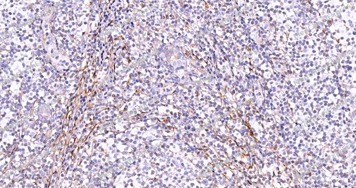 Immunohistochemical analysis of paraffin-embedded human breast tissue using Fibronectin Antibody (HY-P86415, 1/4000). The section was pretreated using heat mediated antigen retrieval with sodium citrate buffer (pH 6.0) for 8 minutes. The tissues were blocked with quick block buffer for 0.5 hours at room temperature, washed with PBS and PBST, and then incubated with the primary antibody overnight at 4℃. The detection was performed using an HRP conjugated compact polymer system. DAB was used as the chromogen. Tissues were counterstained with hematoxylin and mounted with DPX.
 -
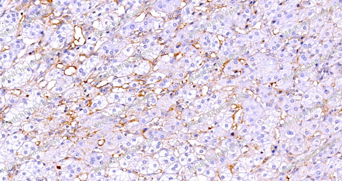 Immunohistochemical analysis of paraffin-embedded human adrenal gland tissue using Fibronectin Antibody (HY-P86415, 1/4000). The section was pretreated using heat mediated antigen retrieval with sodium citrate buffer (pH 6.0) for 8 minutes. The tissues were blocked with quick block buffer for 0.5 hours at room temperature, washed with PBS and PBST, and then incubated with the primary antibody overnight at 4℃. The detection was performed using an HRP conjugated compact polymer system. DAB was used as the chromogen. Tissues were counterstained with hematoxylin and mounted with DPX.
 -
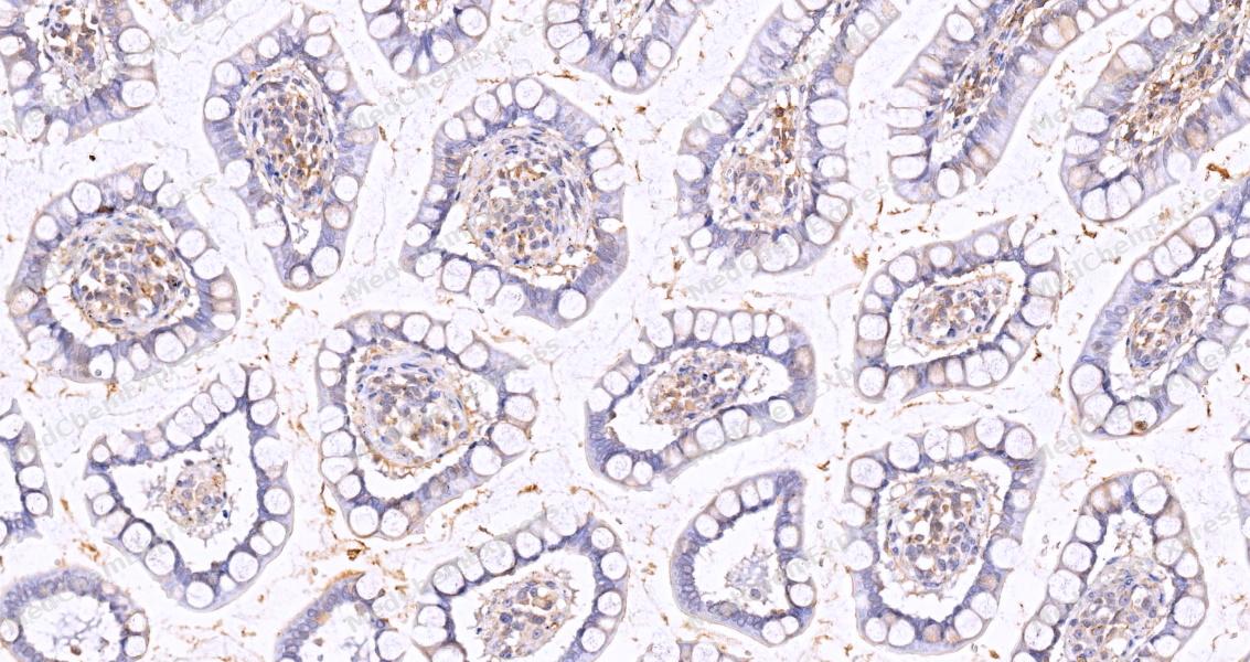 Immunohistochemical analysis of paraffin-embedded human small intestine tissue using Fibronectin Antibody (HY-P86415, 1/4000). The section was pretreated using heat mediated antigen retrieval with sodium citrate buffer (pH 6.0) for 8 minutes. The tissues were blocked with quick block buffer for 0.5 hours at room temperature, washed with PBS and PBST, and then incubated with the primary antibody overnight at 4℃. The detection was performed using an HRP conjugated compact polymer system. DAB was used as the chromogen. Tissues were counterstained with hematoxylin and mounted with DPX.
 -
 Immunocytochemistry analysis of Hela cells labeling Fibronectin with Fibronectin Antibody (HY-P86415) at 1/200 dilution. Cells were fixed in 4% paraformaldehyde for 15 minutes at room temperature, permeabilized with 0.1% Triton X-100 in PBS for 15 minutes at room temperature, then blocked with quick block buffer for 10 minutes at room temperature. Cells were then incubated with Fibronectin Antibody (HY-P86415) at 1/200 dilution in quick block buffer overnight at 4 ℃. AF488-conjugated Goat Anti-Rabbit IgG H&L(HY-P8002, Green) was used as the secondary antibody at 1/1,000 dilution. PBS instead of the primary antibody was used as the secondary antibody only control. The Nuclear counterstain was DAPI (Blue).
 -
 Immunocytochemistry analysis of HepG2 cells labeling Fibronectin with Fibronectin Antibody (HY-P86415) at 1/200 dilution. Cells were fixed in 4% paraformaldehyde for 15 minutes at room temperature, permeabilized with 0.1% Triton X-100 in PBS for 15 minutes at room temperature, then blocked with quick block buffer for 10 minutes at room temperature. Cells were then incubated with Fibronectin Antibody (HY-P86415) at 1/200 dilution in quick block buffer overnight at 4 ℃. AF488-conjugated Goat Anti-Rabbit IgG H&L(HY-P8002, Green) was used as the secondary antibody at 1/1,000 dilution. PBS instead of the primary antibody was used as the secondary antibody only control. The Nuclear counterstain was DAPI (Blue).


Background
-
Function
Fibronectin is a Fibronectins bind cell surfaces and various compounds including collagen, fibrin, heparin, DNA, and actin. Fibronectins are involved in cell adhesion, cell motility, opsonization, wound healing, and maintenance of cell shape. Involved in osteoblast compaction through the fibronectin fibrillogenesis cell-mediated matrix assembly process, essential for osteoblast mineralization. Participates in the regulation of type I collagen deposition by osteoblasts. Acts as a ligand for the LILRB4 receptor, inhibiting FCGR1A/CD64-mediated monocyte activation; Binds fibronectin and induces fibril formation. This fibronectin polymer, named superfibronectin, exhibits enhanced adhesive properties. Both anastellin and superfibronectin inhibit tumor growth, angiogenesis and metastasis. Anastellin activates p38 MAPK and inhibits lysophospholipid signaling; Secreted by contracting muscle, induces liver autophagy, a degradative pathway for nutrient mobilization and damage removal, and systemic insulin sensitization via hepatic ITGA5:ITGB1 integrin receptor signaling[1][2][3][4][5].
-
Subcellular Localization
Secreted, extracellular space, extracellular matrix; Secreted
-
Expression
Tissue_specificity:Fibronectin (protein level) is expressed in the internal limiting membrane of the retina and around blood vessels (PubMed: 29777959) . Plasma fibronectin (soluble dimer form) is secreted by hepatocytes. Cellular fibronectin (dimer or cross-linked polymer form) is produced by fibroblasts, epithelial cells, and other cell types and deposited in the extracellular matrix in fibrous form. Urine contains Ugl-Y1, Ugl-Y2, and Ugl-Y3 (PubMed: 17614963) . -
Isoforms & Post-Translational Modification
P02751 has 17 isomers: P02751-15: 272320 Da (predicted); P02751-1: 262625 Da (predicted); P02751-2: 71971 Da (predicted); P02751-3: 259216 Da (predicted); P02751-4: 222976 Da (predicted); P02751-5: 243334 Da (predicted); P02751-6: 240509 Da (predicted); P02751-7: 268912 Da (predicted); P02751-8: 252811 Da (predicted); P02751-9: 246688 Da (predicted); P02751-10: 239626 Da (predicted); P02751-11: 262406 Da (predicted); P02751-12: 221292 Da (predicted); P02751-13: 249322 Da (predicted); P02751-14: 249402 Da (predicted); P02751-16: 73683 Da (predicted); P02751-17: 256502 Da (predicted).
Sulfated;It is not known whether both or only one of Thr-2155 and Thr-2156 are/is glycosylated;Forms covalent cross-links mediated by a transglutaminase, such as F13A or TGM2, between a glutamine and the epsilon-amino group of a lysine residue, forming homopolymers and heteropolymers (e.g. fibrinogen-fibronectin, collagen-fibronectin heteropolymers);Phosphorylated by FAM20C in the extracellular medium;Proteolytic processing produces the C-terminal NC1 peptide, anastellin;Some lysine residues are oxidized to allysine by LOXL3, promoting fibronectin activation and matrix formation;Serotonylated on Gln residues by TGM2 in response to hypoxia -
Subunit
Mostly heterodimers or multimers of alternatively spliced variants, connected by 2 disulfide bonds near the carboxyl ends; to a lesser extent homodimers. Interacts with FBLN1, AMBP, TNR, LGALS3BP and COL13A1. Interacts with FBLN7 (By similarity). Interacts with COMP (PubMed:12225811). Interacts (via type III repeats 9-14) with TNFAIP6 (via CUB domain); this interaction enhances fibronectin fibril assembly. TNFAIP6 may act as a bridging molecule between FN1 and THBS1 (PubMed:18042364). Interacts with TNR; the interaction inhibits cell adhesion and neurite outgrowth (By similarity). Interacts with FST3 and MYOC. Interacts with SVEP1 (By similarity)
-
SwissProt ID
-
Synonyms
FN1; FN; Fibronectin; FN; Cold-insoluble globulin; CIG


Documentation
-
Data Sheet (234 KB)
-
SDS (251 KB)
- English - EN (251 KB)
- Français - FR (251 KB)
- Deutsch - DE (251 KB)
- Norwegian - NO (251 KB)
- Español - ES (251 KB)
- Swedish - SV (251 KB)
- Italian - IT (251 KB)
- Korean - KR (251 KB)
- Portuguese - PT (251 KB)
-
User Guide for Antibodies (1077 KB)

 Powered by Bioz
