



HDAC2 Antibody (YA393)
(Synonyms: Histone deacetylase 2, HD2, Protein deacylase HDAC2, HDAC2)
Based on 1 Customer Validation
HDAC2 Antibody (YA393) is a Rabbit-derived and non-conjugated IgG monoclonal antibody, targeting to HDAC2.
-
Host:
Rabbit
-
Isotype:
IgG
-
Application:
WB, ICC/IF, IHC-P, IP, FC
-
Reactivity :
Human, Mouse, Rat
-
Formulation:
Supplied in 1*TBS (pH7.4), 0.05% BSA and 40% Glycerol. Preservative: 0.05% Sodium Azide.
-
Conjugation:
Non-conjugated


To place orders, for customer services and technical support, please contact: MedChemExpress USA
Tel: 609-228-6898 E-mail: [email protected] [email protected]





Applications
| Application |
WB

WB: Western Blot
|
ICC/IF
ICC/IF: Immunocytochemistry/
Immunofluorescence |
IHC-P
IHC-P: Immunohistochemistry-Paraffin
|
FC
FC: Flow Cytometry
|
IP
IP: Immunoprecipitation
|
|---|---|---|---|---|---|
| Dilution Ratio | 1:500-1:2000 | 1:50-1:200 | 1:50-1:200 | 1:50-1:100 | Use at an assay dependent concentration. |


Product Details
HDAC2 Antibody (YA393) is a Rabbit-derived and non-conjugated IgG monoclonal antibody, targeting to HDAC2.
-
Host Rabbit

-
Clonality Recombinant,Monoclonal

-
Species ReactivityHuman, Mouse, Rat
-
Observed Molecular WeightObserved band size: 55 kDa
Note: Due to possible protein modifications or aggregation, the molecular weight should be confirmed by actual measurement, and the predicted value is for reference only.

-
Calculated Molecular Weight Predicted band size: 55 kDa

Entrez Gene: 3066 Human ; 15182 Mouse ; 84577 Rat
SwissProt: Q92769 Human ; P70288 Mouse ;
OMIM: 605164 Human
Synthetic peptide corresponding to Human HDAC2.AA range:439-488.
Endogenous
Protein A affinity purified.
Non-conjugated
Unmodified
IgG


Product Properties
-
Appearance
Solution
-
Formulation
Supplied in 1*TBS (pH7.4), 0.05% BSA and 40% Glycerol. Preservative: 0.05% Sodium Azide.
-
Concentration
Batch-dependent, Please check the COA for the concentration of each lot. Check Lot Concentration
-
Storage & Stability
Stored at -20°C for 1 year. Avoid repeated freeze / thaw cycles.
-
Shipping
Shipping with blue ice.


Verification Images
-
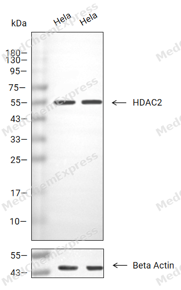 Western blot analysis of extracts from Hela(lane 2(20μg), Hela (lane 3(40μg),using HDAC2 Antibody. Proteins were transferred to a PVDF membrane and blocked with 5% BSA in TBST for 2 hour at room temperature. The primary antibody and Loading control antibody (Beta Actin, HY-P80438, 1/3000) was used in 5% BSA in TBST at 4°C overnight. Goat Anti-Mouse/Rabbit IgG-HRP Secondary Antibody (HY-P8004/HY-P8001, 1/10,000) was used for 1 hour at room temperature.
 -
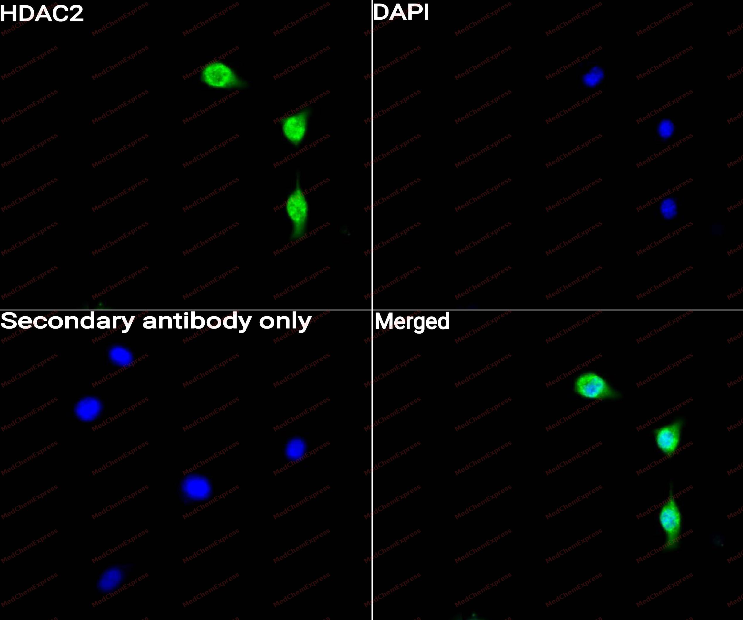 Immunocytochemistry analysis of NIH3T3 cells labeling HDAC2 with HDAC2 Antibody (HY-P80151) at 1/50 dilution. Cells were fixed in 4% paraformaldehyde for 15 minutes at room temperature, permeabilized with 0.1% Triton X-100 for 10 minutes at room temperature, then blocked with QuickBlock™ Blocking Buffer for Immunol Staining for 10 min at room temperature. Cells were then incubated with HDAC2 Antibody (HY-P80151) at 1/50 dilution in QuickBlock™ Blocking Buffer for Immunol Staining at 4 ℃. Alexa Fluor® 488-conjugated AffiniPure Goat Anti-Rabbit IgG H&L(HY-P8002, Green) was used as the secondary antibody at 1/1,000 dilution. PBS instead of the primary antibody was used as the secondary antibody only control. The Nuclear counterstain was DAPI (Blue).
 -
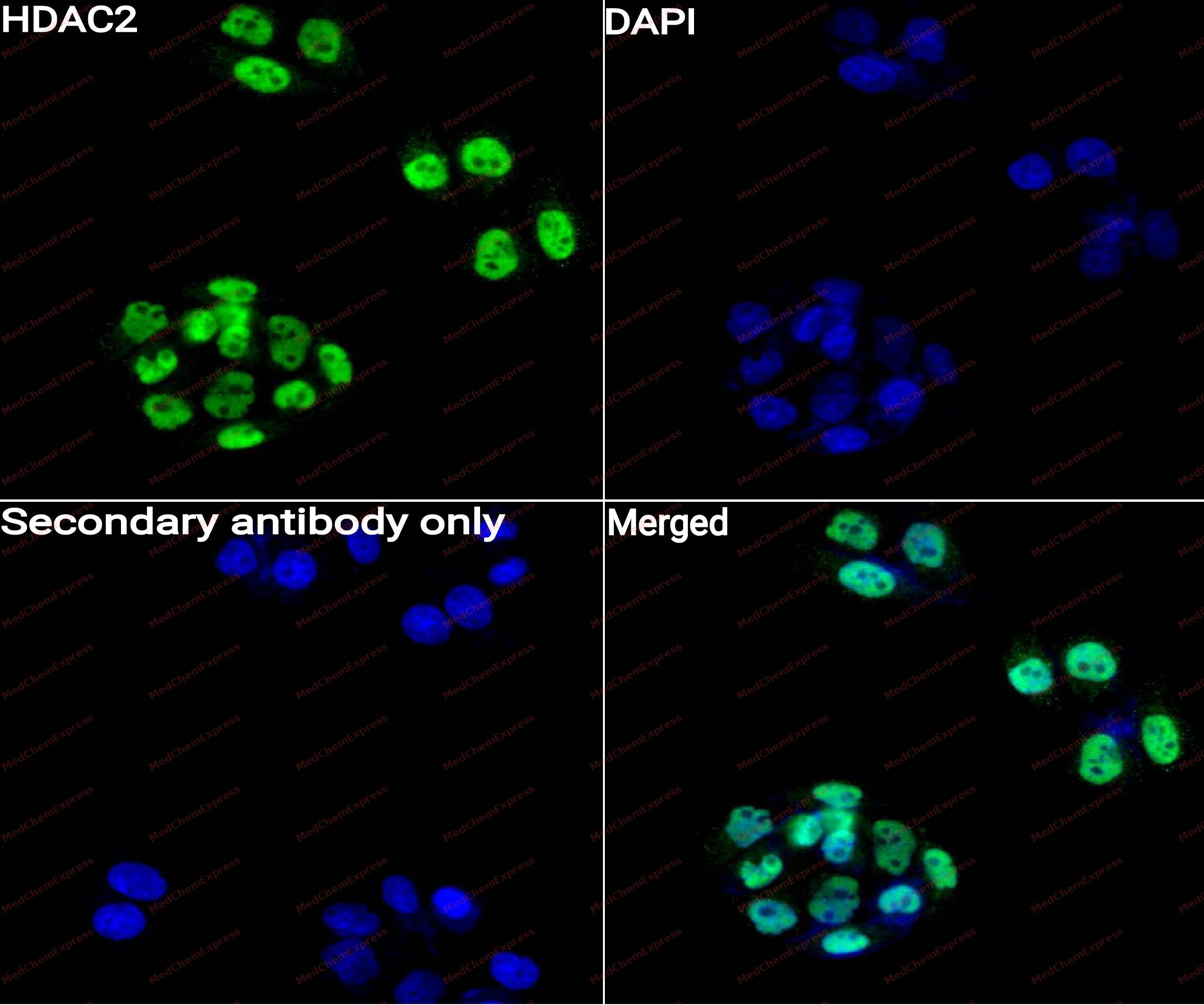 Immunocytochemistry analysis of Hela cells labeling HDAC2 with HDAC2 Antibody (HY-P80151) at 1/50 dilution. Cells were fixed in 4% paraformaldehyde for 15 minutes at room temperature, permeabilized with 0.1% Triton X-100 for 10 minutes at room temperature, then blocked with QuickBlock™ Blocking Buffer for Immunol Staining for 10 min at room temperature. Cells were then incubated with HDAC2 Antibody (HY-P80151)at 1/50 dilution in QuickBlock™ Blocking Buffer for Immunol Staining at 4 ℃. Alexa Fluor® 488-conjugated AffiniPure Goat Anti-Rabbit IgG H&L(HY-P8002,Green) was used as the secondary antibody at 1/1,000 dilution. PBS instead of the primary antibody was used as the secondary antibody only control. The Nuclear counterstain was DAPI (Blue).
 -
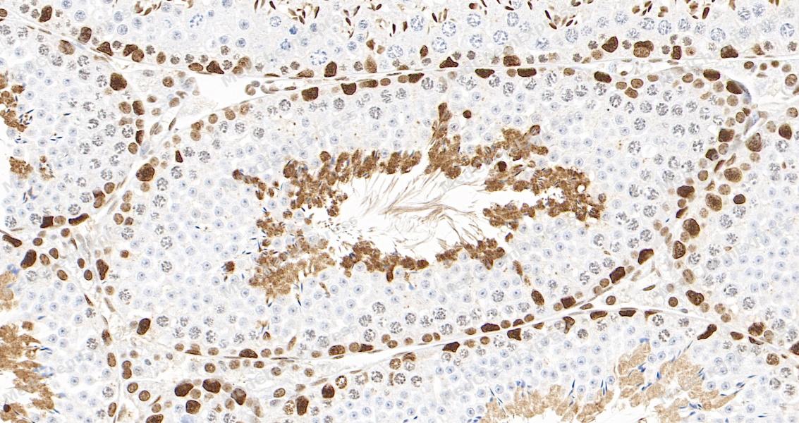 Immunohistochemical analysis of paraffin-embedded mouse testis tissue using HDAC2 Antibody. The section was pre-treated using heat mediated antigen retrieval with Tris-EDTA buffer (pH 9.0) for 8 minutes. The tissues were blocked in QuickBlock for 20 minutes at room temperature, washed with ddH2O and PBS, and then probed with the primary antibody at 1/100 dilution in 4℃ overnight. The detection was performed using an HRP conjugated compact polymer system. DAB was used as the chromogen. Tissues were counterstained with hematoxylin and mounted with DPX.
 -
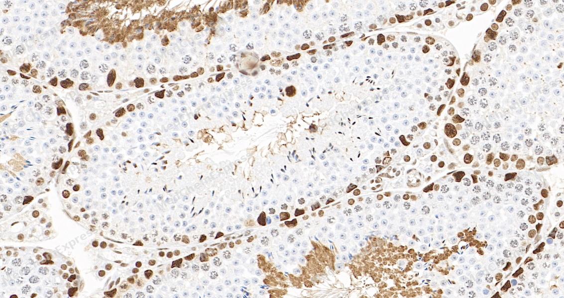 Immunohistochemical analysis of paraffin-embedded mouse testis tissue using HDAC2 Antibody. The section was pre-treated using heat mediated antigen retrieval with Tris-EDTA buffer (pH 9.0) for 8 minutes. The tissues were blocked in QuickBlock for 20 minutes at room temperature, washed with ddH2O and PBS, and then probed with the primary antibody at 1/100 dilution in 4℃ overnight. The detection was performed using an HRP conjugated compact polymer system. DAB was used as the chromogen. Tissues were counterstained with hematoxylin and mounted with DPX.
 -
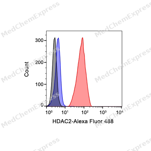 Flow cytometric analysis of 1X10^6 Hela cells labeling HDAC2 Antibody (HY-P80151, red). Cells were fixed with 4% paraformaldehyde and permeabilised with 90% methanol. Then stained with the primary antibody at 1/1000 dilution for an hour at 4℃. Alexa Fluor® 488-conjugated AffiniPure Goat Anti-Rabbit IgG H&L (HY-P8002) was used as the secondary antibody at 1/1,000 dilution for 30 minutes at 4℃. Rabbit IgG Isotype Control (HY-P80879, blue) was used as the isotype control, cells without incubation with primary antibody were used as the unlabeled control (black).


Background
-
Function
HDAC2 (histone deacetylase 2) is a class I histone deacetylase that removes acetyl groups from histone lysine residues and functions within multiprotein transcriptional repressor complexes to regulate gene expression, cell-cycle progression, and developmental processes[1][2]. Mechanistically, HDAC2 promotes chromatin condensation and transcriptional repression through histone deacetylation, thereby influencing cellular homeostasis and epigenetic regulation of gene activity[3][4]. Because histone acetylation and deacetylation dynamically control chromatin accessibility, HDAC2 participates in biological pathways linked to cell growth, cell death, and signal transduction[2][3]. In disease contexts, altered HDAC2 expression has been associated with multiple pathological conditions, particularly cancer and neurodegenerative disorders, where dysregulated epigenetic control contributes to disease progression[2]. Therefore, HDAC2 has emerged as an important experimental target for investigating epigenetic mechanisms underlying tumor biology and neurological dysfunction[2]. Compared with closely related class I isoforms, especially HDAC1, HDAC2 exhibits both overlapping and unique biological functions, with evidence supporting distinct as well as redundant roles in the regulation of proliferation and tumorigenesis[5]. This distinction has increased interest in isoform-selective approaches that can dissect HDAC2-specific functions while minimizing the limitations associated with broad-spectrum HDAC inhibition[2]. For experimental applications, selective HDAC2 inhibitors are widely investigated as chemical tools for studying transcriptional regulation and disease-associated epigenetic networks, and isoform-selective inhibition is considered advantageous because it may provide improved specificity and reduced adverse effects relative to pan-HDAC inhibitors[2].
-
Subcellular Localization
Nucleus; Cytoplasm
-
Expression
Tissue_specificity:Widely expressed; low concentrations in the brain and lungs. -
Subunit
Part of the core histone deacetylase (HDAC) complex composed of HDAC1, HDAC2, RBBP4 and RBBP7, the core complex associates with SIN3, SAP18 and SAP30 to form the SIN3 HDAC complex (PubMed:10904264). Component of the nucleosome remodeling and deacetylase (NuRD) repressor complex, composed of core proteins MTA1, MTA2, MTA3, RBBP4, RBBP7, HDAC1, HDAC2, MBD2, MBD3, and peripherally associated proteins CDK2AP1, CDK2AP2, GATAD2A, GATAD2B, CHD3, CHD4 and CHD5 (PubMed:16428440, PubMed:25593309, PubMed:28977666, PubMed:33283408). The exact stoichiometry of the NuRD complex is unknown, and some subunits such as MBD2 and MBD3, GATAD2A and GATAD2B, and CHD3, CHD4 and CHD5 define mutually exclusive NuRD complexes (PubMed:16428440, PubMed:28977666, PubMed:33283408). Component of a RCOR/GFI/KDM1A/HDAC complex (By similarity). Component of a BHC histone deacetylase complex that contains HDAC1, HDAC2, HMG20B, KDM1A, RCOR1 and PHF21A (PubMed:12493763). The BHC complex may also contain ZMYM2, ZNF217, ZMYM3, GSE1 and GTF2I (PubMed:12493763). Part of a complex containing the core histones H2A, H2B, H3 and H4, DEK and unphosphorylated DAXX (PubMed:12140263). Part of a complex containing ATR and CHD4 (PubMed:10545197). Forms a heterologous complex at least with YY1 (PubMed:8917507). Interacts in the late S-phase of DNA-replication with DNMT1 in the other transcriptional repressor complex composed of DNMT1, DMAP1, PCNA, CAF1 (PubMed:10888872). Component of a mSin3A corepressor complex that contains SIN3A, SAP130, SUDS3, ARID4B, HDAC1 and HDAC2 (PubMed:12724404). Part of a complex composed of TRIM28, HDAC1, HDAC2 and EHMT2 (PubMed:10904264). Part of a complex containing at least CDYL, MIER1, MIER2, HDAC1 and HDAC2 (PubMed:19061646). Component of a histone deacetylase complex containing DNTTIP1, ZNF541, HDAC1 and HDAC2 (PubMed:21573134). Forms a complex comprising APPL1, RUVBL2, APPL2, CTNNB1 and HDAC1 (PubMed:19433865). Interacts directly with GFI1. Interacts directly with GFI1B (By similarity). Interacts with APEX1; the interaction is not dependent on the acetylated status of APEX1 (PubMed:14633989). Interacts with ATR (PubMed:10545197). Interacts with BCL6 (non-acetylated form) (PubMed:12402037, PubMed:18212045). Interacts with BEND3 (PubMed:21914818). Interacts with CBFA2T3 (PubMed:11533236). Interacts with CDK2AP1 (PubMed:20523938). Interacts with CHD4 (PubMed:25593309). Interacts with CHD5 (By similarity). Interacts with CHFR (PubMed:19182791). Interacts with CRY1 (By similarity). Interacts with DNMT1 (PubMed:10888872). Interacts with GATAD2A (PubMed:33283408). Interacts with HCFC1 (PubMed:12670868). Interacts with HDAC7 (By similarity). Interacts with HDAC10 (PubMed:11739383). Interacts with INSM1 (By similarity). Interacts with KDM4A (PubMed:15927959). Interacts with MACROH2A1 (via the non-histone region) (By similarity). Interacts with MBD3L2 (PubMed:15701600). Interacts with MTA1, with a preference for sumoylated MTA1 (PubMed:21965678, PubMed:24970816). Interacts with NACC2 (PubMed:22926524). Interacts with NRIP1 (PubMed:15060175). Interacts with PELP1 (PubMed:15456770). Interacts with PIMREG (PubMed:18757745). Interacts with PRDM6 (By similarity). Interacts with PWWP2B (By similarity). Interacts with SAP30 (By similarity). Interacts with SAP30L (PubMed:16820529). Interacts with SETDB1 (By similarity). Interacts with SIX3 (By similarity). Interacts with SMARCAD1 (PubMed:21549307). Interacts with SNW1 (PubMed:10644367). Interacts with SPHK2 (PubMed:19729656). Interacts with SPEN/MINT (PubMed:11331609). Interacts (CK2 phosphorylated form) with SP3 (PubMed:12176973). Interacts with SUV39H1 (By similarity). Interacts with TSHZ3 (via its N-terminus) (PubMed:19343227). Interacts with ZMYND8 (PubMed:25593309). Interacts with ZNF431 (By similarity). Interacts with ZNF263; recruited to the SIX3 promoter along with other proteins involved in chromatin modification and transcriptional corepression where it contributes to transcriptional repression (PubMed:32051553). Identified in a complex with HDAC1, KCTD19, DNTTIP1 and ZNF541 (By similarity). Component of the SIN3B complex, which includes SIN3B, HDAC2, PHF12 and MORF4L1; interacts directly with all subunits (PubMed:37137925)
-
SwissProt ID
-
Synonyms
Histone deacetylase 2, HD2, Protein deacylase HDAC2, HDAC2
-
Research Field
Epigenetics and Nuclear Signaling


Documentation
-
Data Sheet (263 KB)
-
SDS (251 KB)
- English - EN (251 KB)
- Français - FR (251 KB)
- Deutsch - DE (251 KB)
- Norwegian - NO (251 KB)
- Español - ES (251 KB)
- Swedish - SV (251 KB)
- Italian - IT (251 KB)
- Korean - KR (251 KB)
- Portuguese - PT (251 KB)
-
User Guide for Antibodies (1077 KB)
References
[1]. HDAC2 gene information from NCBI.
[2]. Marinović M, et al. Further investigation of harmicines as novel antiplasmodial agents: Synthesis, structure-activity relationship and insight into the mechanism of action. Eur J Med Chem. 2021 Nov 15;224:113687. [Content Brief]
[3]. Park SY, et al. A short guide to histone deacetylases including recent progress on class II enzymes. Exp Mol Med. 2020 Feb;52(2):204-212. [Content Brief]
[4]. Milazzo G, et al. Histone Deacetylases (HDACs): Evolution, Specificity, Role in Transcriptional Complexes, and Pharmacological Actionability. Genes (Basel). 2020 May 15;11(5):556. [Content Brief]
[5]. Ogiwara H, et al. Histone acetylation by CBP and p300 at double-strand break sites facilitates SWI/SNF chromatin remodeling and the recruitment of non-homologous end joining factors. Oncogene. 2011 May 5;30(18):2135-46. [Content Brief]

 Powered by Bioz
