



JAK1 Antibody (YA722)
(Synonyms: JAK1; JAK1A; JAK1B; Tyrosine-protein kinase JAK1; Janus kinase 1; JAK-1)
Based on 1 Customer Validation
JAK1 Antibody (YA722) is a Mouse-derived and non-conjugated IgG2a monoclonal antibody, targeting to JAK1.
-
Host:
Mouse
-
Isotype:
IgG
-
Application:
WB, ICC/IF, IP
-
Reactivity :
Human, Rat
-
Formulation:
Supplied in 1*PBS (pH 7.3), 50% glycerol and 0.5% BSA. Preservative: 0.02% sodium azide.
-
Conjugation:
Non-conjugated


To place orders, for customer services and technical support, please contact: MedChemExpress USA
Tel: 609-228-6898 E-mail: [email protected] [email protected]





Applications
| Application |
WB

WB: Western Blot
|
ICC/IF
ICC/IF: Immunocytochemistry/
Immunofluorescence |
IP
IP: Immunoprecipitation
|
|---|---|---|---|
| Dilution Ratio | 1:500-1:1000 | 1:50-1:200 | 1:20 |


Product Details
JAK1 Antibody (YA722) is a Mouse-derived and non-conjugated IgG2a monoclonal antibody, targeting to JAK1.
-
Host Mouse

-
Clonality Monoclonal

-
Species ReactivityHuman, Rat
-
Observed Molecular WeightObserved band size: 130 kDa
Note: Due to possible protein modifications or aggregation, the molecular weight should be confirmed by actual measurement, and the predicted value is for reference only.

-
Calculated Molecular Weight Predicted band size: 133 kDa

Entrez Gene: 3716 Human ; 16451 Mouse ; 84598 Rat
SwissProt: P23458 Human ; P52332 Mouse ;
OMIM: 618999 Human
Synthetic peptide corresponding to Human Jak1.The exact sequence is proprietary to MCE.
Endogenous
affinity purified
Non-conjugated
Unmodified
IgG


Product Properties
-
Appearance
Solution
-
Formulation
Supplied in 1*PBS (pH 7.3), 50% glycerol and 0.5% BSA. Preservative: 0.02% sodium azide.
-
Concentration
Batch-dependent, Please check the COA for the concentration of each lot. Check Lot Concentration
-
Storage & Stability
Stored at -20°C for 1 year. Avoid repeated freeze / thaw cycles.
-
Shipping
Shipping with blue ice.


Verification Images
-
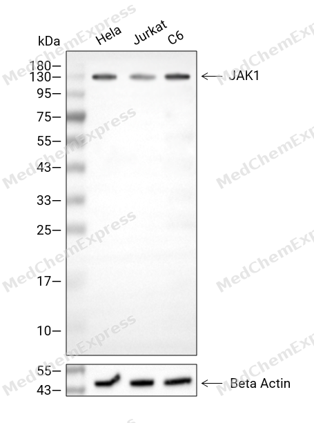 Western blot analysis of extracts from Hela (lane 2(20μg), Jurkat (lane 3(20μg), C6 (lane 4(20μg), using JAK1 Antibody. Proteins were transferred to a PVDF membrane and blocked with 5% BSA in TBST for 2 hour at room temperature. The primary antibody and Loading control antibody (Beta Actin, HY-P80438, 1/3000) was used in 5% BSA in TBST at 4°C overnight. Goat Anti-Mouse/Rabbit IgG-HRP Secondary Antibody (HY-P8004/HY-P8001, 1/10,000) was used for 1 hour at room temperature.
 -
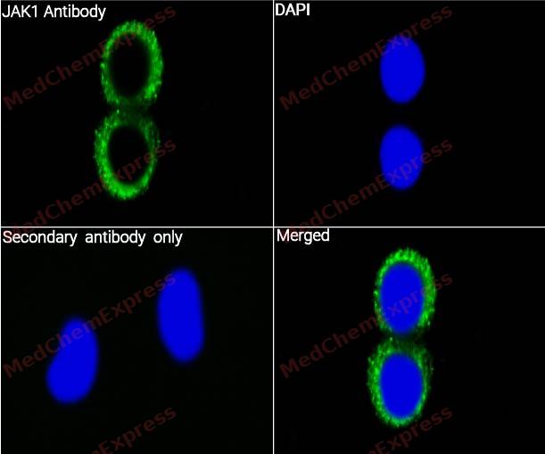 Immunocytochemistry analysis of NIH/3T3 cells labeling JAK1 with JAK1 Antibody (YA722) (HY-P80726) at 1/50 dilution. Cells were fixed in 4% paraformaldehyde for 15 minutes at room temperature, permeabilized with 0.1% Triton X-100 for 10 minutes at room temperature, then blocked with BSA for Immunol Staining for 10 min at room temperature. Cells were then incubated with JAK1 Antibody (YA722) (HY-P80726) at 1/50 dilution in BSA for Immunol Staining at 4 ℃,stay overnight. AF488-conjugated AffiniPure Goat Anti-Mouse IgG H&L(HY-P8005, Green) was used as the secondary antibody at 1/1,000 dilution. PBS instead of the primary antibody was used as the secondary antibody only control. The Nuclear counterstain was DAPI (Blue).
 -
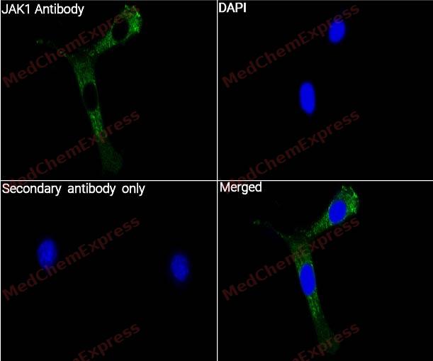 Immunocytochemistry analysis of jurkat cells labeling JAK1 with JAK1 Antibody (YA722) (HY-P80726) at 1/100 dilution. Cells were fixed in 4% paraformaldehyde for 15 minutes at room temperature, permeabilized with 0.1% Triton X-100 for 10 minutes at room temperature, then blocked with BSA for Immunol Staining for 10 min at room temperature. Cells were then incubated with JAK1 Antibody (YA722) (HY-P80726) at 1/100 dilution in BSA for Immunol Staining at 4 ℃,stay overnight. AF488-conjugated AffiniPure Goat Anti-Mouse IgG H&L(HY-P8005, Green) was used as the secondary antibody at 1/1,000 dilution. PBS instead of the primary antibody was used as the secondary antibody only control. The Nuclear counterstain was DAPI (Blue).


Background
-
Function
JAK1 is an essential Janus kinase that mediates biologic responses downstream of class II cytokine receptors, γc-containing receptors, and gp130-dependent receptors[1]. Mechanistically, the JAK/STAT pathway provides a rapid membrane-to-nucleus signaling module that drives transcriptional programs linked to inflammation, immunity, cancer, and autoimmune disease[2]. In inflammatory contexts, JAK1 transduces signals from γc cytokines, IL-6, interferons, and IL-10, connecting cytokine receptor activation to immune-cell regulation and synovial inflammation[3]. Disease models and human genetics support this role, because JAK1 gain-of-function variants drive immune dysregulation with atopic dermatitis, allergy, eosinophilia, and gastrointestinal inflammation[4][5]. Compared with related isoforms, JAK1 shows nonredundant signaling, and Jak1 has a dominant role over Jak3 in γc receptor signaling[1][6]. JAK2 more directly supports hematopoietic growth-factor pathways, whereas JAK3 is mainly linked to common γc receptor biology[3][6]. For experimental applications, selective JAK1 inhibitors help separate cytokine-driven inflammatory signaling from broader JAK blockade[7]. Chemical proteomics identified an allosteric cysteine in JAK1 C817, absent from JAK2 and JAK3, enabling inhibitors that block JAK1-dependent trans-phosphorylation and cytokine signaling[8].
-
Subcellular Localization
Endomembrane system; Peripheral membrane protein
-
Expression
Tissue_specificity:The expression level in primary colon tumors is higher than that in normal colon tissue. The expression level in metastatic colon tumors is comparable to that in normal colon tissue. -
Subunit
Interacts with IL31RA (PubMed:15194700). Interacts with IFNGR1 (PubMed:7615558). Interacts with JAKMIP1 (PubMed:15277531). Interacts with SHB (PubMed:12200137).
-
SwissProt ID
-
Synonyms
JAK1; JAK1A; JAK1B; Tyrosine-protein kinase JAK1; Janus kinase 1; JAK-1
-
Research Field
Cell Biology


Documentation
-
Data Sheet (262 KB)
-
SDS (251 KB)
- English - EN (251 KB)
- Français - FR (251 KB)
- Deutsch - DE (251 KB)
- Norwegian - NO (251 KB)
- Español - ES (251 KB)
- Swedish - SV (251 KB)
- Italian - IT (251 KB)
- Korean - KR (251 KB)
- Portuguese - PT (251 KB)
-
User Guide for Antibodies (1077 KB)
References
[1]. Rodig SJ, et al. Disruption of the Jak1 gene demonstrates obligatory and nonredundant roles of the Jaks in cytokine-induced biologic responses. Cell. 1998 May 1;93(3):373-83. [Content Brief]
[2]. Hu X, et al. The JAK/STAT signaling pathway: from bench to clinic. Signal Transduct Target Ther. 2021 Nov 26;6(1):402. [Content Brief]
[3]. Spinelli FR, et al. JAK1: Number one in the family; number one in inflammation? Rheumatology (Oxford). 2021 May 5;60(Suppl 2):ii3-ii10. [Content Brief]
[4]. Del Bel KL, et al. JAK1 gain-of-function causes an autosomal dominant immune dysregulatory and hypereosinophilic syndrome. J Allergy Clin Immunol. 2017 Jun;139(6):2016-2020.e5. [Content Brief]
[5]. Fayand A, et al. Successful treatment of JAK1-associated inflammatory disease. J Allergy Clin Immunol. 2023 Oct;152(4):972-983. [Content Brief]
[6]. Haan C, et al. Jak1 has a dominant role over Jak3 in signal transduction through γc-containing cytokine receptors. Chem Biol. 2011 Mar 25;18(3):314-23. [Content Brief]
[7]. Schwartz DM, et al. JAK inhibition as a therapeutic strategy for immune and inflammatory diseases. Nat Rev Drug Discov. 2017 Dec;16(12):843-862. [Content Brief]
[8]. Kavanagh ME, et al. Selective inhibitors of JAK1 targeting an isoform-restricted allosteric cysteine. Nat Chem Biol. 2022 Dec;18(12):1388-1398. [Content Brief]

 Powered by Bioz
